What is the essence of the EEG procedure?
An electroencephalogram records electrical signals from brain cells and allows us to identify epilepsy, trauma, neoplasms, inflammatory processes, and changes in blood vessels. Pathology is indicated by disturbances in the electrical activity of neurons, which are recorded using special sensors placed on the patient’s head.
Using an electroencephalogram, the doctor can:
- analyze the performance of the brain;
- identify foci of pathologies;
- assess the nature and extent of damage;
- confirm or clarify the diagnosis;
- monitor the effectiveness of the treatment.
Why is electroencephalography needed? Complete patient guide
Frequent awakenings at night, insomnia, enuresis, the need to detect epilepsy - these are just some of the pathologies that electroencephalography will help in diagnosing.
The neurologist at the Expert Kursk Clinic, Olesya Olegovna Bratchikova, told us about her.
— Olesya Olegovna, what is electroencephalography and how often is this research method prescribed?
Electroencephalography is a non-invasive method for studying brain function by recording its bioelectrical activity. “Non-invasive” means that there are no punctures, incisions, insertion of instruments into body cavities and organs, etc.
It is used everywhere, all over the world. It is widely used to diagnose and monitor the effectiveness of treatment for various conditions. Among them: loss of consciousness, differentiation of epileptic syndromes, various neurological conditions.
Why is MRI performed for epilepsy? Radiologist at MRI Expert Sochi, Zarema Bardudinovna Tseeva, tells
— What is the difference between daytime and nighttime EEG?
Daytime (otherwise known as routine) electroencephalography is performed during the daytime. Its duration does not exceed 20 minutes. Used to identify obvious disorders - for example, to diagnose epileptic syndrome.
It is used for medical examinations (screening studies), and for the delimitation of paroxysmal (accompanied by certain attacks) conditions. Daytime EEG provides no more than 30% of all possible information.
You can sign up for electroencephalography in your city here
Please note: the service is not available in all cities
Night EEG is the “gold standard” of this examination. It is used for a deep search for a problem: separating epileptic syndrome from non-epileptic syndrome, sleep disorders (walking and talking in sleep, periodic short-term pauses in breathing during sleep - for example, with snoring, etc.), enuresis, etc.
— What are the indications for electroencephalography?
An EEG can detect signs of epilepsy. This is her main indication. In this case, diagnosis is prescribed when epilepsy is suspected for the first time, when the treatment regimen is changed and its control.
Read material on the topic: Epilepsy: symptoms, causes, diagnosis and treatment
Electroencephalography is prescribed for enuresis; paroxysmal states; neuroses, anxiety, irritability; hyperactivity and learning disorders in children; delayed mental development, speech, stuttering; autism; frequent awakenings and excessive motor activity during sleep; episodes of walking and talking in a dream, etc.
— At what age is EEG monitoring performed for children?
Starting from 4 weeks.
— Are the indications for daytime and nighttime electroencephalography different or the same?
For a medical examination/screening of a healthy person (without complaints, burdened anamnesis), a daylong study is sufficient. If a person has a history of, for example, an episode of epilepsy, then an overnight study is necessary.
— In what cases does a patient need to undergo both daytime and nighttime electroencephalography?
In the presence of paroxysmal conditions, suspected epilepsy. Suppose a person had a history of an epileptic seizure, or the doctor himself observed it. In this case, a day examination is carried out. If epileptic activity is not detected during the daytime study, a night study must also be prescribed.
— Is it necessary to undergo EEG monitoring to obtain a certificate from the traffic police?
Yes, but not everyone. Since June 2015, according to the order of the Ministry of Health of the Russian Federation No. 344n, an EEG must be performed on any road user applying for a driving license of categories C, D, E. An EEG for drivers of other categories is performed according to indications determined by a neurologist at an appointment.
— How is electroencephalography performed? How long does diagnosis take?
A special cap with electrodes is placed on the subject’s head. A special gel is applied to the scalp at the point of contact with the electrode (for better contact and conductivity of the recorded impulses). Signals from the brain are transmitted through electrodes to a device called an electroencephalograph. It amplifies them and transfers them to a computer for further processing.
As a result, a special “curve” is formed - an electroencephalogram. Based on it, a conclusion is made about the state of the functional activity of the brain.
The daytime session lasts no more than 20 minutes. The study takes place in the so-called state of “relaxed wakefulness”. The man is relaxed, lying with his eyes closed, but not sleeping.
What are the reasons for insomnia? Olga Ivanovna Kuyantseva, a neurologist at Clinic Expert Voronezh, talks about the treatment of insomnia.
During diagnosis, functional tests are carried out to change the functional activity of the brain. The doctor asks the patient to close and open his eyes and breathe actively (3-5 minutes). Photostimulation is also performed: the subject looks at light flashes.
Night research is best done at home. It starts between 20 and 21 pm. Technical nuances are the same as during a daytime examination.
Two complete sleep cycles are recorded, which is approximately 2-3 hours after falling asleep.
— How to properly prepare for an electroencephalogram?
No special preparation is required for an EEG of the brain. The head must be clean; styling products must not be used when drying. On the eve of the study, avoid stress, overwork, and maintain a measured regimen. It is necessary to refrain from taking nervous system stimulants, strong tea, coffee, and alcohol.
Noisy games, watching TV, cartoons, working or playing on the computer are also undesirable for a child.
The presence of metal objects and jewelry on the body during diagnosis is not allowed.
In some cases, when conducting a sleep EEG to establish or exclude a diagnosis of epilepsy, as well as for the differential diagnosis of paroxysms, so-called sleep deprivation is recommended to the patient in preparation for the study. This means that on the eve of the procedure, the patient reduces the duration of night sleep, wakes up early - 3-4 hours earlier than his usual awakening time.
— Olesya Olegovna, how often can you undergo electroencephalography? Is this diagnostic method safe?
EEG is completely safe. You can undergo EEG monitoring as often as necessary for the doctor to make a diagnosis or monitor the patient’s condition.
— Does EEG have any limitations or contraindications?
No.
— In order to undergo an EEG in your clinic, do you need a doctor’s referral?
If a person undergoes a medical examination or is examined on his own initiative, in principle he can undergo the diagnosis independently, without a referral. It is enough to have an identity document.
If we are purposefully looking for some kind of pathology, determining the type of paroxysms, etc., then in this case it is advisable to have a doctor’s referral. From it we can understand the purpose of the survey, i.e. what the doctor needs.
Other questions:
Educational program on medical professions. When to contact a neurologist?
Holter (24-hour) ECG monitoring – complete instructions for the patient
Tension headache: causes, symptoms, diagnosis and treatment
For reference:
Bratchikova Olesya Olegovna
Graduate of the Faculty of General Medicine of Kursk State Medical University in 2004.
From 2004 to 2005, she completed an internship, and from 2005 to 2007, a clinical residency in the specialty “Neurology”.
I took a course in epileptology and certification courses.
Currently working as a neurologist at Clinic Expert Kursk.
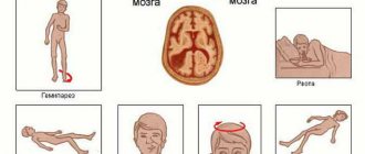
In what cases is EEG prescribed?
After a conversation with the patient and studying the medical history, the specialist decides to prescribe encephalography. Typically, indications for an EEG are frequent headaches, sleep problems, fainting, fatigue and chronic fatigue.
Deterioration in well-being may be a sign of disorders in the functioning of the brain. The above ailments often arise due to:
- vegetative-vascular dystonia;
- cardiac dysfunction;
- pathologies of blood vessels of the neck and head;
- inflammatory processes in meningitis and encephalitis;
- endocrine disorders;
- malignant or benign neoplasms.
For patients suffering from epilepsy who have undergone neurosurgical interventions and head injuries, EEG monitoring is mandatory.
Important aspects
All candidate drivers, as well as those who already have a license to drive a vehicle, undergo a medical examination.
In the latter case, you must obtain a certificate when changing your driver's license. The purpose of such a medical examination is to identify diseases for which it is prohibited to drive a vehicle. A car is a source of increased danger.
It can cause damage to property, as well as the life or health of citizens. Some diseases exclude the possibility of driving a vehicle.
Such ailments can cause serious and irreversible consequences. That is why the state obliges motorists to undergo inspections.
Without the appropriate certificate, they cannot obtain a license to drive a vehicle. We have already mentioned that epilepsy is one of the diseases that prevent you from driving.
This is due to the fact that patients experience seizures with loss of consciousness. The presence of epilepsy is detected by conducting an EEG.
This study is safe and painless for the patient. That is why everyone without exception can undergo it, including pregnant women.
In 2014, EEG was made mandatory for all driver candidates, as well as those motorists who change their license.
This caused a flurry of dissatisfaction due to the rise in price of the commission and the increase in the time it takes to complete it. In addition, equipment for performing EEG was not available in all hospitals.
This obligation has now been abolished. Today, only those drivers who apply for certain categories of driver’s license must undergo such a study.
How to prepare for an electroencephalograph examination?
Monitoring the electrical activity of the brain does not require complex training. During the examination, it is important to simply follow the doctor's instructions and remain calm. Flashes of light and noise are part of the procedure.
Three days before the examination, it is recommended to stop taking tranquilizers, anticonvulsants and sedatives. 24 hours before the examination, do not drink tea, coffee, energy drinks, or eat chocolate. The day before monitoring, wash your hair. Have a snack an hour before the examination. Before starting, loosen your hair and remove metal jewelry.
Rhythms in sleep
A separate category of types of rhythms that manifest themselves either in sleep conditions or in pathological conditions includes three varieties of this indicator.
- Delta rhythm. Characteristic of the deep sleep phase and for comatose patients. It is also recorded when recording signals from areas of the cerebral cortex located on the border with areas affected by oncological processes. Sometimes it can be recorded in children 4–6 years old.
- Theta rhythm. The frequency interval is within 4–8 Hz. These waves are triggered by the hippocampus (information filter) and appear during sleep. Responsible for high-quality assimilation of information and forms the basis of self-learning.
- Sigma rhythm. It has a frequency of 10–16 Hz, and is considered one of the main and noticeable oscillations of the spontaneous electroencephalogram, which occurs during natural sleep at its initial stage.
Based on the results obtained during EEG recording, an indicator is determined that characterizes a complete all-encompassing assessment of the waves - bioelectrical activity of the brain (BEA). The diagnostician checks the EEG parameters - frequency, rhythm and the presence of sharp flashes that provoke characteristic manifestations, and on these grounds makes a final conclusion.
How is an EEG performed?
The examination takes place in several stages.
Preparatory stage
- the patient enters the office, protected from light and sound;
- an encephalograph “cap” consisting of special sensors is put on it;
- The sensor wires are connected to a device that records the bioelectric impulses of the brain.
Diagnostic stage
- the encephalograph transmits data to the monitor in the form of a graph;
- the power of electric fields and its distribution in different parts of the brain are recorded;
- Functional tests are carried out: the patient is asked to blink, look at flashes of light, breathe less often or deeper, listen to a sharp sound.
Final stage
- the electrodes are removed from the patient;
- print out the results.
Process of studying the results
The analysis of the results obtained is carried out in parallel during the procedure, and during the recording of indicators, and continues after its completion. When recording, the presence of artifacts is taken into account - mechanical movement of electrodes, electrocardiograms, electromyograms, and induction of mains current fields. The amplitude and frequency are assessed, the most characteristic graphic elements are identified, and their temporal and spatial distribution is determined.
Upon completion, a patho- and physiological interpretation of the materials is made, and on its basis an EEG conclusion is formulated. Upon completion, the main medical form for this procedure is filled out, called a “clinical electroencephalographic report”, compiled by a diagnostician based on the analyzed data from the “raw” recording.
The transcript of the EEG conclusion is formed on the basis of a set of rules and consists of three sections:
- Description of the leading types of activity and graphic elements.
- Conclusion after description with interpreted pathophysiological materials.
- Correlation of indicators of the first two parts with clinical materials.
How long does the electroencephalogram procedure take?
A regular encephalogram (routine EEG or diagnosis of a paroxysmal state) takes from 20 to 30 minutes.
During the examination, a number of tests are carried out:
- rhythmic photostimulation;
- hyperventilation;
- load in the form of slow blinking.
If it is necessary to evaluate certain brain functions, the specialist adds additional tests, which he informs the patient about in advance. Such tests include:
- clenching your fingers into a fist;
- being in the dark;
- sleep deprivation for a certain period;
- night sleep monitoring.
Rhythms of a waking person
Brain activity recorded on the EEG in an adult has several types of rhythms, characterized by certain indicators and states of the body.
- Alpha rhythm. Its frequency remains in the range of 8–14 Hz and is present in most healthy individuals – more than 90%. The highest amplitude values are observed when the subject is at rest, in a dark room with his eyes closed. It is best identified in the occipital region. It is fragmentarily blocked or completely subsides during mental activity or visual attention.
- Beta rhythm. Its wave frequency fluctuates in the range of 13–30 Hz, and the main changes are observed when the subject is active. Pronounced fluctuations can be diagnosed in the frontal lobes under the obligatory condition of active activity, for example, mental or emotional arousal and others. The amplitude of beta oscillations is much less than alpha.
- Gamma rhythm. The oscillation interval is from 30, can reach 120–180 Hz and is characterized by a rather reduced amplitude - less than 10 μV. Exceeding the limit of 15 μV is considered a pathology causing a decrease in intellectual abilities. Rhythm is determined when solving problems and situations that require increased attention and concentration.
- Kappa rhythm. It is characterized by an interval of 8–12 Hz, and is observed in the temporal part of the brain during mental processes by suppressing alpha waves in other areas.
- Lambda rhythm. It has a small range - 4–5 Hz, and is triggered in the occipital region when it is necessary to make visual decisions, for example, when searching for something with open eyes. The vibrations disappear completely after concentrating your gaze on one point.
- Mu rhythm. Defined by the interval 8–13 Hz. It starts in the back of the head, and is best observed in a calm state. Suppressed when starting any activity, not excluding mental activity.
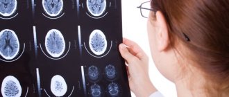
Interpretation of survey results
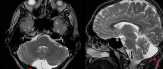
Even a qualified specialist is not always able to accurately name the cause of the patient’s poor health and immediately make a diagnosis. The doctor may be alerted to focal changes on the EEG. In this case, magnetic resonance imaging will be required to exclude a tumor or cyst.
When you contact a medical doctor, you will be able to undergo all examinations as quickly as possible, without delaying your visit to specialists.
If you need to do an EEG of the brain and you want to do it using modern expert-class equipment and in the shortest possible time, call the phone number listed on the website or leave a request in the feedback form.
Medical staff will answer your questions and conduct all necessary examinations within one business day. Let's take care of your health together!
Purpose of EEG video monitoring
One of the types of electroencephalography is EEG video monitoring. This is a long-term recording of electroencephalography, usually lasting several hours, which can be performed during sleep. The duration of the procedure in each specific case is determined by the attending physician and the personnel of the laboratory that conducts the examination.
EEG video monitoring is prescribed if a short standard procedure does not reveal pathologies, but they are present.
Also, this type of examination allows you to evaluate the EEG during wakefulness and sleep.
Many patients are interested in the question of whether they should necessarily sleep during the study. The answer to this question cannot be unambiguous, because it depends on the specific situation. So, for example, if the reason for the examination is a tic that bothers the patient while awake, there is no need to sleep during the examination.
At the same time, EEG video monitoring during sleep sometimes helps to identify conditions that neither the patient nor his loved ones may even be aware of.
The peculiarity of this procedure is that it can be carried out not only during the day, but also at night. In the event that sleep EEG is required, night monitoring is more rational. Not everyone can fall asleep without problems during the daytime.
At the same time, we should not forget that carrying out a multi-hour procedure in a completely isolated room can be extremely tiring for the patient, especially if we are talking about a child. Most pathologies can be detected during a relatively short recording of a conventional EEG.
Best materials of the month
- Coronaviruses: SARS-CoV-2 (COVID-19)
- Antibiotics for the prevention and treatment of COVID-19: how effective are they?
- The most common "office" diseases
- Does vodka kill coronavirus?
- How to stay alive on our roads?
Also, overnight EEG video monitoring is much more expensive.
Indications for an encephalogram
The doctor will definitely prescribe an electroencephalography if the patient has one or more alarming symptoms:
- frequent fainting;
- headache;
- disturbances in sleep phases, falling asleep, insomnia;
- head injuries;
- seizures;
- epileptic seizures;
- neuroses;
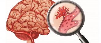
- acute cerebrovascular accident;
- degenerative diseases of the brain;
- neuroses;
- nausea and vomiting for no apparent reason;
- hypertension;
- suspicion of stroke, microstroke, cerebral infarction;
- suspected development of a brain tumor.
An encephalogram of the brain is an informative method for diagnosing infectious diseases of the brain and its membranes: encephalitis, meningitis. Electroencephalography may be prescribed during the treatment of these diseases in order to monitor its effectiveness.
An electroencephalogram is necessarily performed as part of preparing the patient for brain surgery.
Electroencephalography is widely used in pediatric practice. The diagnostic method is used for such conditions in children as:
- delayed mental, emotional and speech development;
- somnambulism;
- nervous tic;
- stuttering;
- head injuries;
- convulsive syndrome.
An encephalogram of a child’s brain is an important study in the diagnosis and treatment of children with diseases such as autism, Down syndrome, and cerebral palsy. With the help of an encephalogram, the diagnosis and its severity are clarified, how quickly the pathology progresses, and the effectiveness of treatment.
Due to the fact that the encephalograph does not emit impulses, but only records them, the procedure has no contraindications. The exception is when the patient has wounds on the head that prevent the installation of the electrodes of the device to obtain an encephalogram.
Decoding of electroencephalogram indicators
In order to decipher the EEG and not miss any of the smallest manifestations in the recording, the specialist needs to take into account all the important points that may affect the indicators being studied. These include age, the presence of certain diseases, possible contraindications and other factors.
Upon completion of the collection of all data from the procedure and their processing, the analysis is completed and then a final conclusion is formed, which will be provided for making a further decision on the choice of therapy method. Any disturbance in activity may be a symptom of diseases caused by certain factors.
Alpha rhythm
The normal frequency is determined in the range of 8–13 Hz, and its amplitude does not go beyond 100 μV. Such characteristics indicate a healthy state of a person and the absence of any pathologies. The following are considered violations:
- constant fixation of the alpha rhythm in the frontal lobe;
- exceeding the difference between the hemispheres by up to 35%;
- constant violation of wave sinusoidality;
- presence of frequency dispersion;
- amplitude below 25 μV and above 95 μV.
The presence of disturbances in this indicator indicates a possible asymmetry of the hemispheres, which may be the result of oncological tumors or pathologies of cerebral circulation, for example, stroke or hemorrhage. A high frequency indicates brain damage or TBI (traumatic brain injury).
A complete absence of the alpha rhythm is often observed in dementia, and in children, deviations from the norm are directly related to mental retardation (MDD). Such a delay in children is evidenced by: disorganization of alpha waves, shift of focus from the occipital region, increased synchrony, short activation reaction, overreaction to intense breathing.
Beta rhythm
In the accepted norm, these waves are clearly detected in the frontal lobes of the brain with a symmetrical amplitude in the range of 3–5 μV, recorded in both hemispheres. A high amplitude leads doctors to think about the presence of a concussion, and when short spindles appear, to the occurrence of encephalitis. An increase in the frequency and duration of spindles indicates the development of inflammation.
In children, the pathological manifestations of beta oscillations are considered to be a frequency of 15-16 Hz and a high amplitude present - 40-50 µV, and if its localization is the central or anterior part of the brain, then this should alert the doctor. Such characteristics indicate a high probability of delayed development of the baby.
Delta and theta rhythms
An increase in the amplitude of these indicators above 45 μV on a constant basis is characteristic of functional brain disorders. If the indicators are increased in all brain regions, then this may indicate severe dysfunction of the central nervous system.
If a high amplitude of the delta rhythm is detected, a tumor is suspected. Inflated values of the theta and delta rhythm recorded in the occipital region indicate a child’s lethargy and a delay in his development, as well as impaired circulatory function.
The most common diagnosed pathologies
Thanks to the electroencephalogram, diseases such as epilepsy or various types of traumatic brain injury (TBI) are quite easily diagnosed.
Epilepsy
The study allows you to determine the localization of the pathological area, as well as the specific type of epileptic disease. At the time of a convulsive syndrome, the EEG recording has a number of specific manifestations:
- pointed waves (peaks) - suddenly rising and falling can appear in one or several areas;
- the combination of slow pointed waves during an attack becomes even more pronounced;
- sudden increase in amplitude in the form of flashes.
The use of stimulating artificial signals helps in determining the form of epileptic disease, since they provide visibility of hidden activity that is difficult to diagnose with EEG. For example, intense breathing, requiring hyperventilation, leads to a decrease in the lumen of blood vessels.
Photostimulation is also used, carried out using a strobe (a powerful light source), and if there is no reaction to the stimulus, then most likely there is a pathology associated with the conduction of visual impulses. The appearance of non-standard vibrations indicates pathological changes in the brain. The doctor should not forget that exposure to powerful light can lead to an epileptic seizure.
Preparation for the procedure in adults
On the eve of the examination, tonic foods and liquids - tea, coffee, soda, chocolate, alcoholic drinks - are completely excluded from the diet.
The referral may indicate that an EEG will be performed with a test. In this case, the patient must abstain from sleep for a day or 1.5 days. The duration is set by the doctor based on the age of the subject.
Basic rules of preparation for adult patients on the day of examination:
- You can eat food 2 hours before attaching the sensors.
- Hair must be clean; do not use hairspray or gel in your hair.
- It is necessary to remove earrings and piercings before the procedure, but it is better to leave them at home.
- Smoking is allowed 2 hours before diagnosis.
- People with long hair should take a tissue or tissues with them to remove any remaining gel used to attach the electrodes.
There is no need to stop taking antiepileptic drugs. If it is necessary to undergo an examination and cancel it, it is carried out in a hospital with a round-the-clock stay and for special indications.
Decoding values in different age intervals
An EEG recording of a premature baby at 25–28 gestational weeks looks like a curve in the form of slow flashes of delta and theta rhythms, periodically combined with sharp wave peaks 3–15 seconds long with a decrease in amplitude to 25 μV. In full-term infants, these values are clearly divided into three types of indicators. During wakefulness (with a periodic frequency of 5 Hz and an amplitude of 55–60 Hz), the active phase of sleep (with a stable frequency of 5–7 Hz and a fast low amplitude) and quiet sleep with flashes of delta oscillations at a high amplitude.
Over the course of 3-6 months of a child’s life, the number of theta oscillations is constantly growing, while the delta rhythm, on the contrary, is characterized by a decline. Further, from 7 months to a year, the child develops alpha waves, and delta and theta gradually fade away. Over the next 8 years, the EEG shows a gradual replacement of slow waves with fast ones - alpha and beta oscillations.
Until the age of 15, alpha waves predominate, and by the age of 18, the BEA transformation is complete. Over the period from 21 to 50 years, stable indicators remain almost unchanged. And from 50, the next phase of rhythmicity restructuring begins, which is characterized by a decrease in the amplitude of alpha oscillations and an increase in beta and delta.
After 60 years, the frequency also begins to gradually fade, and in a healthy person, manifestations of delta and theta oscillations are noticed on the EEG. According to statistics, age indicators from 1 to 21 years, considered “healthy,” are determined in subjects 1–15 years old, reaching 70%, and in the range of 16–21 – about 80%.