When should you take a Reticulocyte (capillary blood) test?
- Assessment of erythropoiesis after bleeding or hemolysis;
- Assessing the ability of bone marrow to regenerate after taking cytotoxic drugs and/or bone marrow transplantation;
- Assessing the resumption of erythropoietin production after kidney transplantation;
- Doping control (for athletes when using erythropoietin);
- Diagnosis of various anemias;
- Assessment of bone marrow function in case of insufficient amounts of vitamin B12, iron, folate, copper and quality control of treatment.
Interpretation:
- Blood loss or destruction of red blood cells, hemolytic anemia (up to 300%), treatment of B12-deficiency anemia (reticulocyte crisis on days 5-8 of vitamin B12 therapy), effective therapy of iron deficiency anemia with iron preparations (8-12 days of treatment), thalassemia, malaria, others hematological diseases (polycythemia, cancer metastases to the bone marrow), acute oxygen starvation.
- Aplastic anemia, hypoplastic anemia, B12-deficiency anemia, autoimmune diseases of the hematopoietic system, alcoholism, metastases of cancer tumors in the bones, myxedema, kidney disease.
Sample result (PDF)
Detailed description of the study
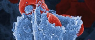
Reticulocytes are young red blood cells (erythrocytes). Reticulocytes are formed in the bone marrow from normoblasts that have lost their nucleus. It takes several days to ripen, usually 4-5. The maturation process begins in the bone marrow and then continues in the peripheral blood. The formation of red blood cells and their forms depends on the hormone erythropoietin, which is produced in the kidneys.
Reticulocytes are larger in size than red blood cells. The internal environment of the cell contains a basophilic network (reticulum), which is located in the form of threads and small grains. An important difference between reticulocytes and mature erythrocytes is their inability to fully transport oxygen and carbon dioxide, since the cells still do not have enough hemoglobin.
There are 5 different stages of maturity of young red blood cells:
- They have a core, the grain is located around the core in the shape of a corolla;
- Granular-mesh mass, which is represented by a ball;
- The grain size is represented by a dense network;
- Mass, which is represented by threads;
- There are individual grains.
It is important to note that about 80% of young forms belong to group 4-5.
In a healthy person, 0.2-1% of reticulocytes are determined in the blood. The percentage is calculated from the total number of red blood cells. With increased destruction of red blood cells (hemolysis) or bleeding, the number of immature forms will increase, so it is important to use the “reticular index” to assess the severity of anemia. Such a small concentration shows that young forms are able to exit into the peripheral blood and can then mature and turn into mature erythrocytes.
In infants in the first days after birth, the concentration of reticulocytes is higher than in an adult; they can be 10%. Gradually their number will decrease. Also, an increased number of young forms is observed due to the destruction of red blood cells with negative Rh factors of the mother and child. In case of blood loss or insufficient intake of iron from food, the concentration is also higher than normal, as the body independently tries to restore the level of red blood cells. There are conditions and/or diseases in which a decrease in the concentration of reticulocytes is observed. This can be due to damage to the bone marrow (after ionizing radiation, chemotherapy treatment or tumor damage), which leads to a decrease in the formation of reticulocytes and, as a consequence, a decrease in their number in the peripheral blood.
There are true and false reticulocytosis - an increase in the concentration of young forms. True is characterized by an increase in the number of immature red blood cells both in the peripheral blood and in the bone marrow. And if false, there will be an increased concentration of reticulocytes in the circulating blood and no increase in the bone marrow. This fact indicates the “washing out” of immature red blood cells from the bone marrow into the blood.
The concentration increases with accelerated erythropoiesis, and decreases with its slowdown. Erythropoiesis is the process of hematopoiesis during which red blood cells are formed. Determination of reticulocytes in capillary blood shows that the bone marrow is capable of regeneration and evaluates the hematopoietic system.
IN THE LABORATORY THEY PERFORM:
- General blood test with leukocyte differentiation (blood smear microscopy)
- Complete blood count with differentiation of leukocyte formula + reticulocytes
- Reticulocyte count (automatic RET analysis)
- Manual platelet counting
- Fluorescent platelet analysis (automatic counting)
- General blood test with differentiation of leukocyte formula
- Microscopic examination of the leukocyte formula (microscopy of a blood smear)
- Determination of ESR using the Westergren method
A general blood test with differentiation of the leukocyte formula, platelet and reticulocyte counting are performed on an automatic hematology analyzer XN 1000 manufactured by Sysmex Corporation, Japan.
Interpretation of clinical blood test indicators
- number of leukocytes
(white blood cells, WBC) with leukocyte formula.
White blood cells are cells that help the body fight infection. They are able to identify foreign agents (bacteria, viruses) in the body and destroy them. There are 5 different types of leukocytes: eosinophils, basophils, neutrophils, lymphocytes and monocytes, which are included in the leukocyte formula (the percentage of different forms of leukocytes in the blood serum and the calculation of their number per unit volume). All white blood cell differential parameters, including immature granulocytes (IG), are determined using fluorescence flow cytometry and provide an expanded list of clinically relevant parameters in each analysis. The sensitivity of the assay is especially important in the detection of inflammatory and infectious diseases. The total number of leukocytes is usually increased during an acute infectious process caused by bacteria. If there are too few white blood cells, the body becomes more susceptible to various infections. It also helps in monitoring therapy - the increased white blood cell count will begin to decrease, demonstrating the effectiveness of the treatment. Norm: (4-9)x109/L - number of
red blood cells (RBC).
Red blood cells are cells that contain hemoglobin. A general blood test allows you to determine whether there is a sufficient number of red blood cells in the blood, what their shape, size and hemoglobin content (MCV, MCH, MCHC) are. If the number of red blood cells detected by a general blood test is reduced, it means that the patient has anemia, which can manifest itself as weakness, fatigue and shortness of breath. Less common is an increase in the total number of red blood cells (erythrocytosis, or polycythemia). Norm: women - (3.7-4.7)x1012/L, men - (3.9-5.1)x1012/L - hemoglobin
content (Hb).
Hemoglobin is an iron-containing protein that has the ability to carry oxygen from the lungs to tissues and organs, and carbon dioxide from tissues and organs to the lungs, from which it is exhaled. Hemoglobin determines the main function of red blood cells; the color of these blood cells depends on its content. Norm: women - (120-150)g/L, men - (130-170)g/L - Hematocrit
(Hct) determines the volume of blood that red blood cells occupy in the bloodstream.
This indicator is expressed as a percentage. An increase in hematocrit occurs if the number of red blood cells increases or the volume of the liquid part of the blood decreases, which happens when there is excessive loss of fluid from the body (for example, with diarrhea). A decrease in this indicator is observed, on the contrary, with a decrease in the number of red blood cells (for example, due to their loss, destruction or decrease in their formation) or with overhydration - when a person receives too much fluid (for example, with excessive administration of intravenous solutions). Hematocrit reflects not only the number of red blood cells, but also their size. If the size of red blood cells decreases (as in iron deficiency anemia), the hematocrit will also decrease. Norm: women - (33-46)%, men - (38-49)% - Red blood cell indices
measure red blood cell size and hemoglobin content and include mean erythrocyte volume (MCV), mean erythrocyte hemoglobin content (MCH), mean erythrocyte hemoglobin concentration (MCHC), and erythrocyte distribution by size (RDW).
The determination of the above indicators is an integral part of the general blood test and is not performed separately. Norm: MCV - (80-98)fL, MCH - (27-37)pg, MCHC - (300-360)g/L, RDW - (11.5-14.5)% - platelets
(platelet count, PC, PLT) are cells that play a significant role in blood clotting.
If a person has a low platelet count, they are at increased risk of bleeding and bruising. Thrombocytopenia is a condition characterized by an abnormally low platelet count: below the normal platelet count in adults. In severe thrombocytopenia, when the platelet count is less than 20 x 109 /l, spontaneous bleeding (non-traumatic) may develop. Ignoring the symptoms of severe thrombocytopenia can have serious consequences for the patient, so obtaining reliable platelet count results is critical when making clinically important decisions. However, obtaining an accurate platelet count, especially from thrombocytopenic patient samples, is a rather challenging laboratory task. The XN 1000 analyzer from Sysmex offers an affordable solution to platelet counting - a fluorescent platelet assay that allows you to determine not only traditional indicators, but also specific markers, such as the immature platelet fraction, which is a more accurate marker of platelet production and reflects the percentage of immature platelets from their platelets. total amount, and is also an established parameter by which doctors determine the cause of thrombocytopenia, based on the etiology of various congenital and acquired thrombocytopenic conditions. Norm: (150-450)x10 9 /L - reticulocytes
(Retic count, RET) are young forms of red blood cells formed in the bone marrow and found in small quantities in the blood, allowing one to assess the functional state of red hematopoiesis.
An increased number of reticulocytes is a criterion for the activation of hematopoiesis in the bone marrow during hemolytic anemia, after blood loss, and a reduced number is characteristic of hypoplastic anemia. Automated reticulocyte analysis provides exceptional capabilities for comprehensive analysis of erythropoiesis. Reticulocyte counting uses a patented fluorescence flow cytometry method, the accuracy of which is recognized throughout the world. This analysis displays a wide range of reticulocyte parameters - both qualitative and quantitative - which helps in forming a complete picture of the state of the red germ of hematopoiesis; operational control of iron and/or erythropoietin therapy also becomes available, which gives the patient a greater chance of recovery, in the differential diagnosis of anemia , in monitoring the effectiveness of therapy. Along with the general count of reticulocytes, the analyzer ensures their separation into various fractions depending on the degree of maturity and, as a consequence, their erythropoietic activity. Additional insight into the quality of immature red blood cells can be obtained by assessing the level of reticulocyte hemoglobinization, which is an advanced clinical parameter useful in the treatment of patients with iron deficiency and anemia. In addition, RET analysis provides information about the content of hypo- and hyperchromic red blood cells, as well as red blood cell fragments, in the sample. Norm: (1.03-1.85)% - erythrocyte sedimentation rate (ESR)
.
An increase in ESR is a nonspecific test indicating the presence of an inflammatory process. With a reduced number of red blood cells in the blood, ESR increases regardless of the nature of anemia. A decrease in ESR is observed with erythrocytosis of various etiologies. The reaction accelerates in women during pregnancy and fasting. The increase in ESR is based on changes in the concentration of various plasma proteins associated with changes in their electrical charge, but other factors may also play a role: the size and shape of blood cells, changes in the lipid composition of plasma, etc. The determination of ESR is performed on an automatic analyzer for determination of erythrocyte sedimentation rate of the ROLLER series - model 20PN, made in Italy. The Roller 20 PN analyzer determines the erythrocyte sedimentation rate in venous and capillary blood, provides a result within 20 seconds by measuring the erythrocyte aggregation rate, which overcomes the limitations and dependence on variable factors of methods for determining ESR based on the phenomenon of sedimentation. Internal and external quality controls ensure accuracy, precision and reproducibility of patient results. The new patented technology is based on a microcapillary photometric method for measuring the rate of red blood cell aggregation under pathological conditions. Norm: women 15-50 years old - (2-20) mm/hour; over 50 years – (2–30) mm/h; men 15-50 years old – (2-15) mm/hour; over 50 years – (2-20) mm/hour
Important Notes
Material for research Capillary blood.
Children under 7 years of age: venous blood/capillary blood (for special indications). Children over 7 years of age and adults: venous blood. Capillary blood collection for research is carried out only for children under 7 years of age (for special indications)! According to GOST R 53079.4-2008, indications for taking capillary blood are possible: in newborns, in patients with very small or hard-to-reach veins, with large-area burns, and in severely obese patients.
What else is prescribed with this study?
Clinical blood test with leukocyte count and ESR (with microscopy of a blood smear to detect pathological changes) (venous blood)
3.9.1. Ven. blood 1 day
720 ₽ Add to cart
Leukocyte formula with mandatory “manual” microscopy of a blood smear (venous blood)
3.5.1. Ven. blood 1 day
360 ₽ Add to cart
Westergren ESR (venous blood)
3.3.1. Ven. blood 1 day
250 ₽ Add to cart
What do the test results mean?
The results must be interpreted by your doctor and in conjunction with the results of other tests, such as a complete blood count. In general, the reticulocyte count (absolute number or percentage) is a reflection of recent bone marrow activity. The results may indicate the presence of a disease or condition that causes an increased need for new red blood cells and whether the bone marrow can cope with the increased load on it. Sometimes the results may indicate excess red blood cell production.