Detailed description of the study
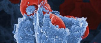
Rheumatoid factor (RF) is a group of antibodies to human tissues that are formed as a result of disorders in the immune system. The name of these antibodies explains their frequent detection in rheumatoid arthritis. However, RF may be elevated in other rheumatic and inflammatory diseases. Detection of RF is an important part of the diagnostic search when determining the causes of joint inflammation and confirming the presence of rheumatoid arthritis. This disease of an autoimmune nature develops mainly in women over 40-50 years of age. The reasons for it have not yet been sufficiently studied. The role of genetic and hormonal factors in combination with the adverse effects of the external environment is assumed. The formation of various antibodies to joint tissues leads to inflammation in them. Mostly small joints of the hands and feet are affected, inflammation is accompanied by redness and pain, and impaired motor activity. An important diagnostic sign of rheumatoid arthritis is stiffness in the limbs after waking up, which goes away within an hour. Chronic inflammation of the joints ultimately leads to their destruction and deformation of the hands and feet. RF is detected in more than 75% of patients with rheumatoid arthritis. Its titer weakly correlates with disease activity, but patients with high titers tend to have more severe rheumatoid arthritis. Sjögren's syndrome is a disease in which there is also an increase in RF levels. It is based on progressive damage to the salivary and lacrimal glands due to their erroneous destruction by the human immune system. Violation of salivary secretion is manifested by dry mouth, destruction of the lacrimal glands - dry eyes. There is insufficient hydration of the skin and mucous membranes. In addition, high RF titers can be found in a number of other diseases, which include: systemic lupus erythematosus, polymyositis, tuberculosis, syphilis, viral hepatitis, infectious mononucleosis, subacute septic endocarditis, autoimmune hepatitis and other autoimmune liver lesions. An increase in RF titer is sometimes observed among healthy people over 60 years of age, as well as during pregnancy and after blood transfusions (especially in the case of frequent blood transfusions). For a more accurate diagnosis of rheumatoid arthritis, it is recommended to determine not only the RF, but also the antibody titer to the cyclic citrulline-containing peptide. The use of both tests increases diagnostic sensitivity.
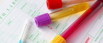
How to properly prepare for a rheumatic test test
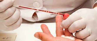
It is recommended to carry out the examination in the morning on an empty stomach. It is allowed to drink only regular non-carbonated water.
The time interval between the examination and the last meal should be more than 8 hours.
It is advisable to donate blood for rheumatic tests even before the patient begins taking medications. If this is not possible, it is advisable to postpone the examination. Only after 2 weeks after the end of the course of treatment and the patient has finished taking medications, a laboratory examination is prescribed. This measure is necessary to ensure that the survey results are as reliable as possible.
If the course of treatment is long and it is impossible to interrupt it, and the examination must be carried out urgently, then the name of the medications that the patient is taking must be indicated.
The day before the examination, try to exclude:
- all kinds of stress (physical and mental);
- fried and fatty foods;
- drinking alcohol, tea and coffee.
If you do not adhere to all the rules described above, there is an extremely high probability of obtaining an erroneous result.
There are diseases for the diagnosis of which it is recommended to undergo a rheumatic test. The most common of them:
- lupus erythematosus;
- discoid lupus;
- rheumatism and rheumatoid arthritis;
- sepsis;
- malignant neoplasms;
- scleroderma;
- burns;
- myocardial infarction;
- chronic kidney disease;
- liver diseases (hepatitis, intoxication, cirrhosis);
- pancreatitis;
- prolonged fasting.
References
1. Chaltsev, B. D., Vasiliev, V. I., Palshina S. G. [etc.]. Clinical guidelines for the diagnosis and treatment of Schörgen's disease with anticentromere antibodies. Scientific and practical rheumatology, 2019. 2. Rheumatoid arthritis. Clinical recommendations. approved Ministry of Health of Russia, 2021. - URL: https://legalacts.ru/doc/klinicheskie-rekomendatsii-revmatoidnyi-artrit-utv-minzdravom-rossii/ (access date: 02/11/2021). 3. Croia, C, Bursi, R, Sutera, D, One year in review 2021: pathogenesis of rheumatoid arthritis. Clinical Experience in Rheumatology, 2021.
Blood test for uric acid levels
Uric acid is the final breakdown product of purines. Every day a person receives purines through food, mainly meat products. Then, with the help of certain enzymes, purines are processed to form uric acid.
In normal physiological quantities, uric acid is needed by the body; it binds free radicals and protects healthy cells from oxidation. In addition, just like caffeine, it stimulates brain cells. However, high levels of uric acid have harmful consequences, in particular, they can lead to gout and some other diseases.
Testing uric acid levels makes it possible to diagnose uric acid metabolism disorders and related diseases.
1
Rheumatological examination
2 Rheumatological examination
3 Rheumatological examination
When to conduct an examination:
- with the first attack of acute arthritis in the joints of the lower extremities, which arose without obvious reasons;
- with recurrent attacks of acute arthritis in the joints of the lower extremities;
- if you have relatives in your family who suffer from gout;
- for diabetes mellitus, metabolic syndrome;
- with urolithiasis;
- after chemotherapy and/or radiation therapy for malignant tumors (and especially leukemia);
- with renal failure (the kidneys excrete uric acid);
- as part of a general rheumatological examination necessary to determine the cause of joint inflammation;
- with prolonged fasting, fasting;
- with a tendency to excessive consumption of alcoholic beverages.
Uric acid level
The level of uric acid is determined in the blood and urine.
Uric acid in the blood is called urecemia , and in urine - uricosuria . An increased level of uric acid is hyperuricemia , a decreased level of uric acid is hypouricemia . Only hyperuricemia and hyperuricosuria are of pathological significance.
The concentration of uric acid in the blood depends on the following factors:
- the amount of purines entering the body with food;
- synthesis of purines by body cells;
- the formation of purines due to the breakdown of body cells due to disease;
- the function of the kidneys, which excrete uric acid in the urine.
Under normal conditions, our body maintains normal uric acid levels. An increase in its concentration is one way or another associated with metabolic disorders.
Normal levels of uric acid in the blood
Men and women may have different concentrations of uric acid in the blood. The norm may depend not only on the gender, but also on the age of the person:
- in newborns and children under 15 years of age – 140-340 µmol/l;
- in men under 65 years old – 220-420 µmol/l;
- in women under 65 years of age – 40-340 µmol/l;
- in women over 65 years of age – up to 500 µmol/l.
If excess of the norm occurs for a long time, then crystals of uric acid salt (urate) are deposited in joints and tissues, causing various diseases.
Hyperuricemia has its own symptoms, but can also be asymptomatic.
Reasons for increased uric acid levels:
- taking certain medications, such as diuretics;
- pregnancy;
- intense loads in athletes and people engaged in heavy physical labor;
- prolonged fasting or eating foods containing large amounts of purines;
- some diseases (for example, endocrine), consequences of chemotherapy and radiation;
- impaired metabolism of uric acid in the body due to a deficiency of certain enzymes;
- insufficient excretion of uric acid by the kidneys.
How to reduce uric acid levels
Those who suffer from gout know how much trouble an increased concentration of uric acid can cause. Treatment of this disease must be comprehensive and must include taking medications that reduce the concentration of uric acid in the blood (xanthine oxidase inhibitors). It is recommended to drink more fluids and reduce the consumption of foods rich in purines.
It is also important to gradually lose excess weight, since obesity is usually associated with increased uric acid. The diet should be designed so that the amount of foods rich in purines is limited (red meat, liver, seafood, legumes). It is very important to give up alcohol. It is necessary to limit the consumption of grapes, tomatoes, turnips, radishes, eggplants, sorrel - they increase the level of uric acid in the blood. But watermelon, on the contrary, removes uric acid from the body. It is useful to consume foods that alkalize urine (lemon, alkaline mineral waters).
Diagnosis of anemia
CDC "Health Laboratory" diagnoses anemia using blood tests. On this page you will find a set of studies that you can take separately or together.
Anemia, or anemia, is a group of syndromes that have a common symptom: a decrease in the level of hemoglobin in the blood, usually with a simultaneous decrease in the number of red blood cells. Assessing the following indicators increases the accuracy of diagnosing anemia using blood tests:
- LVSS - ability of serum to bind iron; one of the main indicators used to diagnose anemia using a blood test ; changes with disturbances in microelement metabolism; in the case of iron deficiency anemia, the LVSS increases; if complex tests show a low level of serum iron, this indicates anemia, which is associated with chronic diseases, cancer, infections,
- vitamin B12 - important for the normal formation and maturation of red blood cells; The risk of cyanocobalamin deficiency is highest in vegetarians, since the vitamin comes with animal products, a decrease in concentration affects the decrease in the amount of folic acid,
- B9 - folic acid is also important for hematopoiesis: it stimulates the formation of erythro-, leukocytes and platelets, the compound enters the blood along with green vegetables, wholemeal bread and is partially formed by intestinal microflora, the reserve of acid in the liver is small, a month of stopping consumption is enough for a deficiency to occur source products, anemia develops after four months.
As part of the diagnosis, you can take a blood test for transferrin, ferritin, and erythropoietin. Choose tests according to the recommendations of your doctor. Only a specialist evaluates the results obtained. Diagnosing anemia using a blood test can be difficult, so the doctor evaluates clinical symptoms, comprehensive research data, and medical history.
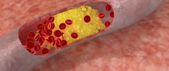
Lipid metabolism indicators
Indications for laboratory testing of lipid metabolism are the diagnosis of the following diseases:
- atherosclerosis and its complications,
- IHD - coronary heart disease,
- myocardial infarction,
- stroke.
Increased concentrations of lipids in the blood significantly increase the risk of developing vascular complications in atherosclerosis. The level of substances largely depends on the quantities in which they are supplied with food. Therefore, to correct lipid status, a low-fat diet is often prescribed.
Analyzes allow you to determine the content of the main lipids - cholesterol and triglycerides. For transport in the blood, they bind to proteins - apoproteins. Such combinations are called lipoproteins. There are several types, differing in the level of triglycerides, cholesterol and apoproteins.
As part of a laboratory study of lipid metabolism, the concentration and ratio of all indicators of lipid status are determined:
- cholesterol,
- triglycerides,
- high and low density lipoproteins,
- apolipoproteins A1 and B.
A blood test is performed for diagnosis. Biomaterial is given on an empty stomach in the morning. Before the study, you must refrain from eating for 12 hours. In the morning you can only drink water. The blood collection procedure is painless and takes only a few minutes.
The study is carried out over 3 days. The results are available on our website after filling out a special form. You can also pick up the report in person or use a courier delivery service. The cost of the study is indicated on the website. Collection of biomaterial is paid separately.