Brain meningioma (ICD 10 code – D32.0) is a neoplasm that originates from the arachnoid (arachnoid) membrane of the brain. Brain meningioma has a clear outline morphologically and looks like a horseshoe-shaped or spherical node, most often fused with the dura mater of the brain.
At the Yusupov Hospital, qualified specialists use the most advanced technologies and time-tested effective methods to treat meningioma: radiation therapy, stereotactic radiosurgery, high-quality removal of brain meningioma. Recovery after surgery is carried out in the rehabilitation department of the Yusupov Hospital under the careful supervision of competent rehabilitation doctors and attentive medical staff.
Meningioma: what is it?
As a rule, meningioma is benign in nature, however, like any other tumor localized inside the skull, benign meningioma of the brain is considered relatively malignant, accompanied by symptoms associated with compression of the brain matter. Malignant brain tumor (meningioma) is a less common disease characterized by aggressive growth and a high recurrence rate after surgical treatment.
Most often, brain meningioma is localized in the area of the foramen magnum, cerebral hemispheres, pyramid of the temporal bone, wings of the sphenoid bone, tentorial notch, parasagittal sinus and cerebellopontine angle.
In most cases, meningioma is located in a capsule. The tumor is not characterized by the formation of cysts; it can be small, just a few millimeters, or reach large sizes - over 15 centimeters in diameter. If a meningioma grows towards the brain, a node forms, which over time begins to compress the medulla. If a tumor grows towards the bones of the skull, over time it grows between the cells of the bone and causes thickening and deformation of the bone. The tumor can grow simultaneously towards the bone and brain, then nodes and deformation of the skull bones are formed.
Make an appointment
Reasons for development
The direct causes of the development of meningiomas have not been reliably studied to date. However, there are a number of factors that can provoke their occurrence:
- Most often, brain meningioma is diagnosed in mature patients, after 40 years;
- It is known that women are more susceptible to developing brain meningioma than men. This is due to the fact that tumor growth is greatly influenced by female sex hormones;
- the occurrence of various tumors in the brain is often associated with high doses of ionizing radiation;
- the influence of such negative factors as chemicals and toxic substances, trauma, exposure to a mobile phone and others;
- A significant role in the development of meningioma belongs to genetic diseases, one of which is neurofibromatosis type 2, which causes multiple malignant meningiomas.
Symptoms and signs
Meningioma of the brain (ICD 10 - D32.0) is characterized by relatively slow growth, so it can develop asymptomatically for a long time.
One of the first symptoms is a headache - dull, bursting or aching. It is distinguished by its diffuse nature and localization in the area of the back of the head, forehead or temples.
The occurrence of other symptoms is associated with the localization of the tumor (compression of certain brain structures). Such symptoms are called focal.
Brain meningioma may be suspected if the following symptoms are present:
- paresis of the limbs (severe weakness, decreased sensitivity, the appearance of pathological reflexes);
- loss of visual fields and other visual disorders (decreased visual acuity, double vision). A characteristic symptom is ptosis - drooping of the upper eyelid;
- deterioration of hearing function;
- reduction or complete loss of smell, olfactory hallucinations;
- epileptiform seizures;
- psychoemotional disorders, behavioral disorders - these symptoms most often manifest themselves as meningioma of the frontal lobe of the brain;
- thinking disorders;
- impaired coordination and gait;
- increased intraocular pressure;
- nausea and vomiting that does not bring relief.
If the outflow of cerebrospinal fluid is disrupted, hydrocephalus and cerebral edema occur, as a result of which patients develop persistent headaches, dizziness, and mental disorders.
Diagnostics
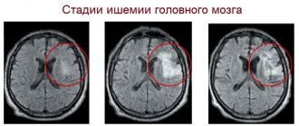
The most informative and accurate methods for diagnosing meningioma are computed tomography (CT) and magnetic resonance imaging (MRI). As a rule, these studies are carried out with contrast. CT and MRI can determine the size of the tumor, its location, the extent of damage to surrounding tissues and possible complications.
Magnetic resonance spectroscopy (MRS) is used to determine the chemical profile and nature of the neoplasm. Positron emission tomography (PET) allows identifying foci of recurrent meningioma.
An auxiliary method that allows you to determine the nature of the blood supply to the tumor is angiography. This study is often used in the process of preoperative preparation.
Make an appointment
MRI
MRI is considered the leading diagnostic method, since it allows detailed visualization of the vascularization of the tumor, determining the extent of damage to the venous sinuses and arteries, and identifying the relationship between the tumor and its surrounding structures. In the majority of meningiomas (65%), a dural tail is detected during the study, which allows for more accurate diagnosis of the disease. MRI is used more often than other methods to diagnose a tumor, however, the method has one significant drawback - frequent cases of false negative diagnoses in the presence of foci of hemorrhage and calcinitis.
Kinds
There are 11 types of benign meningiomas:
- meningothelial meningiomas – 60%;
- transitional meningiomas – 25%;
- fibrous meningiomas – 12%;
- rare types of meningiomas – 3%.
A brain tumor can be located in different areas of the brain:
- convexital tumor – 40%;
- parasagittal – 30%;
- basal location of the tumor – 30%.
Meningioma of the brain of the frontal lobe
Meningioma of the frontal region forms very often, in most cases it does not bother the patient for a long time. If the meningioma is located in the right frontal lobe, symptoms will appear on the opposite side of the body.
The reasons for the development of meningioma of the frontal region are various: traumatic brain injury, inflammatory disease of the meninges, genetic predisposition, food high in nitrates, neurofibromatosis and other reasons. Radiation exposure is considered a proven cause of tumor development; all other causes are considered risk factors.
Meningioma of the frontal region can cause blurred vision, headache, paresis of the facial nerves, arm muscles, lethargy and other symptoms.
Anaplastic meningioma
Anaplastic meningioma is a grade 3 brain tumor; tumor recurrence occurs in all patients within three years after treatment.
Parasagittal meningioma
Parasagittal meningioma is located in the occipital, parietal or frontal part along the longitudinal midline. Often this tumor is accompanied by a pathological increase in the content of bone matter in the bone tissue. Parasagittal meningiomas, growing in the frontal part of the head, cause:
- increased intracranial pressure;
- development of congestive optic discs in the fundus;
- severe nausea and vomiting, headache;
- epileptic seizures.
Parasagittal meningioma of the parietal region of the head is characterized by sensory disturbances and epileptic seizures. Meningioma of the occipital region is characterized by increased intracranial pressure and hallucinations.
Atypical meningioma
Atypical brain meningioma is a grade 2 tumor; tumor recurrence occurs in 30% of patients within 10 years after treatment.
Meningioma falx
A tumor growing from the greater falciform process of the brain is called falx meningioma. Over time, the tumor grows into the sagittal venous sinus, resulting in impaired venous circulation and intracranial hypertension. The growth of the tumor causes the following negative symptoms: epileptic seizures, impaired sensitivity and motor activity of the legs, pelvic disorders.
Stereotactic radiotherapy (radiosurgery)
The main advantage of this technique over radiation therapy is that it allows targeted treatment of the tumor from different angles. It is important that during the procedure only the tumor receives the maximum dose of radiation, and the surrounding tissues are not injured. Another important advantage of radiosurgery is its non-invasiveness. This treatment method is indicated in the following situations: when access to the tumor is difficult, the patient’s condition is serious, and there is a high risk of complications.
Compared to other treatment methods, radiosurgery does not require preoperative preparation, anesthesia, or long recovery after surgery. The prognosis for the patient depends on the size of the tumor. The technique is not used if the patient has been diagnosed with a tumor larger than 35 mm. In almost 100% of cases, this technique allows you to stop the growth of the tumor. In most cases, one course of treatment is sufficient, but sometimes a repeat course may be required. The risk of meningioma recurrence after stereotactic radiotherapy is very low. The risk of complications after this operation is much lower than with radiation therapy and surgery.
Treatment
Meningioma very often causes the development of edema of surrounding tissues, which affects the appearance of various negative symptoms. Steroid drugs are prescribed to relieve swelling. Treatment for meningioma depends on the type and size of the tumor, its location, the patient's health and age.
According to medical statistics, in 90% of cases, brain meningiomas are benign tumors, which are characterized by slow development and the absence of concomitant damage to vital organs.
Malignant tumors are characterized by rapid growth and the presence of metastases in any other organs of the human body.
Tumor removal
Removal of meningioma is not always carried out. Most often, a benign tumor is monitored. Surgery is required if the meningioma is malignant and increases in size.
The main treatment method for growing benign and malignant tumors is surgery to remove brain meningioma. It is very important to perform competent removal of the tumor. The consequences of incorrect surgical intervention, which damaged the surrounding brain tissue or venous sinuses, can be very dire. Such an operation can cause a significant decrease in the patient’s quality of life, so neurosurgeons often leave part of the cancerous tissue, constantly monitoring its growth.
Malignant meningiomas tend to recur, requiring repeated surgery.
Make an appointment
Consequences of the operation
Depending on the location of the tumor and its size, complications may develop after the operation: deterioration or loss of vision, partial or complete memory loss, paresis of the limbs, impaired concentration, changes in character, personality, cerebral edema, bleeding.
Radiation therapy
Treatment of brain meningioma without surgery involves the use of radiation therapy methods, which are used when it is not possible to effectively remove the tumor surgically. Pathological cells are destroyed under the influence of high doses of X-ray radiation. The use of standard radiation therapy is inappropriate for the treatment of patients diagnosed with large cerebral meningioma. However, treatment without surgery in such cases is ineffective.
When the tumor is localized in a place that is difficult for a neurosurgeon to reach, or when there are areas close to it, damage to which can lead to disruption of vital functions, specialists at the Yusupov Hospital give preference to stereotactic methods. This type of therapy can be used to treat large tumors. Stereotactic radiosurgery is based on targeted irradiation of a tumor formation with beams located at different angles.
Often, the stereotactic method is combined with surgical treatment - if there are contraindications for removing tumors in the usual way.
Chemotherapy is not used to treat benign brain meningiomas.
Radiation therapy
This technique is based on the fact that radiation is harmful to tumor cells. Traditionally, radiation therapy is carried out in several sessions, during which the tumor is always irradiated in the same position. This type of therapy takes a lot of time. There is a high risk of complications such as hair loss or radiation dermatitis. Typically, this technique is chosen for treatment when it is not possible to remove the tumor surgically. Due to complications and contraindications to radiation therapy, recently it has increasingly been replaced by another more innovative treatment method - stereotactic radiation therapy.
Relapses
If a benign, clearly circumscribed meningioma that has not grown into the surrounding tissue is detected in a patient, surgical intervention most often ensures a complete recovery.
However, it must be remembered that after removal of even benign meningiomas, relapses can occur. Recurrences of atypical meningiomas are recorded in almost 40% of cases, of malignant ones - in 80%.
The development of relapses within five years after surgery also depends on the location of the tumor.
Relapses are least likely to occur with meningioma localized in the cranial vault, more often in the area of the sella turcica and the body of the sphenoid bone. The most common tumors that recur are those affecting the sphenoid bone and cavernous sinus.
Surgery to remove brain meningioma
Surgical treatment involves complete or partial (for hard-to-reach meningiomas or in cases of invasion of large vessels) removal of the tumor using modern microsurgical instruments and an operating microscope under general anesthesia. After craniotomy, the specialist determines the damage to the skull bones and the possibility of returning the bone to its place.
If the bone tissue is overgrown by meningioma, it is necessary to perform cranioplasty, which allows you to close the defect in the skull bones. The neurosurgeon's tactics depend on the location of the tumor. After removal of the bone area, damage to the dura mater is determined. Convexital meningiomas are located immediately after the dura mater, which makes tumor removal easier.
Total removal of benign meningioma usually has no recurrence. If, due to inaccessibility, the neurosurgeon partially removes the tumor, the risk of regrowth remains, especially for Grade II and III tumors.