Many people have probably heard sad stories about someone’s sudden death due to a broken blood clot. Almost everyone understands that a “severed blood clot” is something life-threatening, but at the same time, not everyone knows what a blood clot is, when it forms and in general why and where it “breaks off”. Therefore, now we’ll talk about blood clots and their role in the body.
What are blood clots and why do they appear?
Thrombi are clots that form from connections between blood cells. Platelets, sticking together in chains, form lumps that are attached to the walls of blood vessels. In some situations, blood clots form due to disorders of the hematopoietic system, in others - as a result of damage to the inner wall of the vessel.
Large growths inside the vein prevent blood flow from passing through the obstructed area. As a result, congestion forms in the veins, leading to varicose veins. When a blood clot blocks the lumen, a heart attack occurs—the death of tissue that has not received oxygen through the bloodstream.
The formation of blood clots is influenced by several factors from a person’s daily life:
- Sedentary lifestyle. Lack of active mobility leads to stagnation of blood in the lower legs. Therefore, constantly sitting at a computer, as well as choosing an escalator instead of stairs, give the blood a reason to stagnate and form clots.
- Insufficient fluid intake. The quality of blood directly depends on what a person eats. With insufficient fluid intake, the blood becomes thick, which means it cannot fully perform its functions and puts a greater strain on the heart. It is easy to pump liquid blood through the system, but thick blood is much more difficult.
- Taking medications that affect the hematopoietic system. In medical practice, drugs are often used to treat certain diseases, one of the side effects of which is blood thickening. Therefore, such medications must be taken simultaneously with anticoagulants that prevent the formation of blood clots.
You can protect yourself from blood clots by regularly exposing your body to physical activity, drinking enough fluids, and including more plant-based foods rich in fiber in your diet.
What is this
Thrombosis is a condition in which blood clots form in blood vessels. If they do not dissolve naturally, they can cause harm. Such clots block blood flow and lead to serious complications. If the thrombus blocks more than three quarters of the lumen of the vessel, then oxygen starvation of the tissue begins. And if completely, it can lead to a heart attack, stroke or limb damage, depending on the location of the formation.
The first mention of thrombosis as a disease appears in 1271, but it was not until the early nineteenth century that scientists linked swelling of the legs with a blood clotting disorder. The phenomenon of thrombosis was understood and described in scientific articles and books by the German scientist and doctor Rudolf Virchow in the mid-nineteenth century. He also formulated the main reasons for its appearance, which are called Virchow’s triad in his honor.
How do blood clots occur?
If a blood clot has formed on the wall of an artery, its appearance can be described in the following stages:
- Some process damages the artery wall.
- The body notices the violation and begins to build protection against blood loss, forming a large number of special blood cells - platelets, which, attaching to the damaged area, form a kind of patch.
- In case of coagulation disorders or changes in the hematopoietic system, platelet formation does not stop on time and continues longer than expected. This causes too much growth to form on the wall. Or platelets, which are in small quantities in the blood, floating past in the bloodstream, stick to the resulting accumulation.
The causes of damage to the walls of blood vessels can be:
- mechanical disruption of the structure due to injury;
- infectious lesion;
- high levels of glucose molecules in the blood;
- immune system dysfunction.
If there are no factors that contribute to the formation of blood clots, any injury or other damage will not lead to a large accumulation of blood cells. Under the layer of platelets, the artery wall will heal and recover, and the crust will resolve over time.
There are several stages of thrombus formation:
- disruption of the structure of the inner surface of the artery;
- activation of blood clotting factors;
- platelet adhesion at the site of injury;
- the appearance of substances that trigger a chain of reactions that form fibrin threads, which contribute to thrombus formation;
- a kind of network of fibrin threads is formed, into which blood cells enter, creating a large clot;
- Over time, the clot thickens, forming a thrombus.
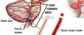
When a blood clot breaks off under the influence of any factors, it begins to move through the bloodstream. Once it hits the nearest bottleneck, the blood flow will be blocked. If a similar situation happens outside a medical facility, it is impossible to save the person.
Arterial thrombosis - symptoms and treatment
Arterial thrombosis is a sudden, acute cessation of arterial blood flow caused by a thrombus blocking a blood vessel. Thrombus (from ancient Greek - lump, clot) is an intravital blood clot that forms during diseases or injuries. Normally, the circulatory system does not contain blood clots. Their appearance in the vessel threatens the patient’s life.
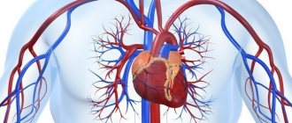
Arteries are blood vessels that carry oxygenated blood from the heart to the limbs, organs and tissues. Arterial thrombosis leads to a sudden cessation or deterioration of arterial blood flow in a limb or organ with a potential threat to its viability [1].
Arterial thrombosis in 40% of cases causes acute limb ischemia (lack of blood supply), and in 37% - embolism [2][3]. Embolism (from Greek - invasion) is the separation of a blood clot from the site of its initial formation, or the transfer of a pathological substrate with the bloodstream along the vascular bed with subsequent blockage of an arterial vessel. The pathological substrate can be solid, liquid or gaseous formations: drops of fat, gas or air bubbles, masses from a “burst” cholesterol plaque, pus, etc.
Arterial thrombosis and embolism cannot be considered independent diseases. They always arise as a consequence of other pathological conditions.
The causes of a blood clot in the lumen of a vessel are described by the German scientist Rudolf Virchow. They are united in the well-known triad:
- Damage to the vascular wall.
- Slowing blood flow.
- Violation of blood composition.
A thrombus is formed under the influence of all three factors with dominance of one of them.
Damage to the vascular wall is caused by:
- injuries - mechanical, thermal, electrical, etc.;
- inflammatory diseases of the arteries - arteritis, obliterating atherosclerosis with the development of atherothrombosis;
- severe infectious diseases - typhus, influenza, sepsis.
Slow blood flow occurs in the following cases:
- extravasal compression - compression of a vessel from the outside by a tumor, effusion of blood, an additional cervical rib, a bone fragment during a fracture, mechanical pressure during accidents, etc.;
- aneurysms (dilation of blood vessels);
- arterial spasms;
- acute circulatory failure;
- prolonged immobilization of limbs;
- oncological diseases.
to disturbances in blood composition [11]:
- blood diseases - leukemia, erythrocytosis, polycythemia;
- significant dehydration of the body;
- hereditary or acquired thrombophilia (pathological thrombus formation);
- systemic atherosclerosis;
- diabetes;
- hypertonic disease;
- malignant neoplasms;
- endotoxemia (accumulation of toxic breakdown products and bacterial activity in the blood and tissues);
- states of shock;
- taking medications - glucocorticosteroids, estrogens and gestagens (hormone replacement therapy and combined oral contraceptives), hemostatic agents, antifibrinolytic agents.
Emboli can be caused by heart disease or other causes.
Cardiac causes of embolism:
- cardiac ischemia;
- myocardial infarction;
- post-infarction cardiosclerosis;
- post-infarction aneurysms of the left ventricle;
- rheumatic heart defects;
- septic bacterial endocarditis;
- heart tumors (myxomas).
In these cases, a blood clot forms in the cavities of the heart. Then, under the influence of a hypertensive crisis, changes in heart rhythm and other reasons, its defragmentation occurs. The thrombus rushes with the blood flow, blocks a section of the arterial bed of a smaller diameter or a fork in the area of the division of the vessel.
Noncardiac causes:
- aortic aneurysm;
- ulcerated arterial plaques;
- pneumonia;
- lung tumor.
In rare cases, paradoxical embolism is possible - migration of a blood clot from the venous system through the right side of the heart to the left. This occurs with congenital heart disease and embolism with foreign objects (for example, a bullet or shot) [4].
The incidence of acute limb ischemia is one case per 6000 people annually [12]. With age, the incidence increases sharply. In the vast majority of cases, the pathology affects people over 60 years of age [8]. Ischemia, which occurs as a complication of thrombosis, more often affects men than women.
Smoking, an inactive lifestyle and poor nutrition lead to the development of atherosclerosis, hypertension and diabetes. At the same time, the risk of developing arterial thrombosis also increases.
The individual risk of vascular pathology includes:
- age younger than 50 years in the presence of diabetes mellitus and one of the risk factors for atherosclerosis: smoking, dyslipidemia (impaired lipid ratio in the blood serum), hypertension, hyperhomocysteinemia (increased level of the amino acid homocysteine in the blood);
- age 50-69 years and presence of diabetes mellitus or smoking;
- age 70 years and older [8].
Thrombosis factors
An increased risk of blood clots is caused by:
- Genetic inheritance to predisposition.
- Diseases that force you to limit physical activity, for example, bed rest.
- High degree of blood clotting.
- Arrhythmia, cardiomyopathy and other diseases that disrupt the strength and rhythm of blood flow throughout the system.
- Varicose veins.
- Liver dysfunction.
- Alcoholism.
- Smoking.
- High body mass index.
- Age-related changes in hormone levels in both men and women.
Some of these factors cannot be influenced, for example, genetic predisposition. However, you can protect yourself from the serious consequences of blood clots by maintaining an active and healthy lifestyle.
Then you will find out...
2/3 of venous thrombosis are asymptomatic or with mild symptoms. A person is bothered by moderate pain in the leg and slight swelling, but not so severe as to force him to see a doctor. Of the three cases of thrombosis, one progresses, and two resolve spontaneously.
However, this does not always mean that resolved thrombosis has passed without a trace. Even in this case, irreversible changes occur on the wall and valve of the affected vein, which lead to the development of chronic venous insufficiency. With duplex scanning of blood vessels, doctors often see post-thrombotic changes in the deep veins of the leg in patients who have never been diagnosed with venous thrombosis.
Blood control and nutrition without “greens”. A doctor on how to live with thrombosis Read more
How to prevent pulmonary embolism?
To avoid pulmonary embolism, it is necessary to prevent and promptly treat chronic diseases. One of the causes of venous thrombosis is varicose veins. Any person who suspects he has this disease should visit a phlebologist and have the venous system of the lower extremities diagnosed. After diagnosis, you need to follow the doctor's recommendations. Also, patients planning major surgery should be assessed for the risk of deep vein thrombosis, and people at high risk of DVT may require prophylactic doses of anticoagulants before and after surgery.
Who develops pulmonary embolism?
Almost all cases of pulmonary embolism are caused by deep vein thrombosis. Thus, people who are more likely to get pulmonary embolism are people who are prone to deep vein thrombosis. Let's look at some risk factors for pulmonary embolism. Important factors are immobility, severe somatic pathologies and major surgical intervention (especially gynecological operations, operations on the pelvis and lower extremities). The risk of developing deep vein thrombosis or PE in hospital can be significantly reduced by activating the patient to walk as soon as possible. Medicines to help prevent deep vein thrombosis and PE are also prescribed to those patients who are at high risk.
Causes
The main reasons why a blood clot may form in the leg are increased blood clotting and a decrease in normal blood flow activity. There are also a number of specific reasons that provoke the occurrence and development of thrombosis:
- mechanical damage to the walls of veins or blood vessels (consequences of bruises, fractures, surgical operations);
- damage to the venous membrane by atherosclerotic plaques;
- increased blood clotting in cancer;
- hormonal disorders;
- congenital abnormal structure of the circulatory system.
Prognosis after treatment method
Arterial thrombosis is a complication of various diseases. This means that removing a blood clot (thrombectomy) is an intervention that eliminates complications. Thus, the prognosis after treatment depends on eliminating the causes of thrombosis. If this is thrombosis associated with narrowing of the arteries, then restoration of the lumen by angioplasty and stenting eliminates the causes of thrombosis and provides a good long-term prognosis. If thrombosis or thromboembolism is associated with diseases of other arteries or the heart, then treatment of these diseases is necessary to prevent repeated episodes of blood clot transfer.
What are the prospects for pulmonary embolism?
This depends on the type of pulmonary embolism and the presence of concomitant diseases.
If PE is treated correctly and promptly, the prognosis is good and most patients can make a full recovery.
The prognosis is worse if there is a serious disease that contributed to the embolism, such as advanced cancer. Massive PE is more difficult to treat and is life-threatening. PE is a serious condition and can have a high risk of death, but this is greatly reduced with early treatment.
The most dangerous time for complications or death is the first few hours after an embolism. In addition, there is a high risk of developing a new pulmonary embolism within six weeks of the first one. Therefore, treatment must be started immediately and continued for at least six months.
Wait, who's coming?
How to detect thrombosis at an early stage? More often it “starts” in the veins of the leg and is accompanied by quite tolerable local pain and swelling. In an unfavorable scenario, the blood clot increases in size, involving the veins of the thigh and pelvis. Gradually, massive swelling, redness of the skin, bursting pain, and swelling of the saphenous veins appear. From the moment the first symptoms appear until you see a doctor, it usually takes 5-10 days. The exception is iliofemoral thrombosis, which makes itself felt immediately - with pronounced swelling and noticeable pain.
“Silent” thrombosis (with a complete absence of symptoms) can occur in bedridden patients (after surgery or paralysis) or if a blood clot forms in the pelvic veins (usually this occurs during pregnancy and childbirth, in the postpartum period, when taking oral contraceptives). In such situations, the first sign of disease may be pulmonary thromboembolism “without a clear source.”
Article on the topic
There is a cork in the vessel. Where did the child get thrombosis?
Possible complications during treatment
The following complications are possible when using a Fogarty probe:
- Rupture of the inner wall of the artery with its dissection and secondary thrombosis
- Tearing off the probe head and leaving it in the lumen of the artery
- Perforation of an altered artery wall with a probe with internal bleeding
- Complications associated with access (hematoma, lymph accumulation, wound suppuration)
Such complications are quite rare. Their frequency decreases as clinical experience with thrombectomies accumulates. In our clinic, we have not observed such complications over the past five years.
Complications of Rotarex thrombectomy:
- Dissection of the arterial wall due to rough passage of the probe
- Transfer of pieces of blood clots to small arteries below the site of thrombosis
- Formation of hematoma along the artery
Such complications are usually identified during the operation and are usually corrected immediately.
Symptoms
Clinical manifestations of a stationary thrombus are much less pronounced. The patient is rarely bothered by:
- tachycardia;
- shortness of breath when sitting.
In case of mobility in the left atrium, the following are observed:
- excruciating attacks of palpitations, when heart tremors shake the entire chest (what clinicians call “frantic”);
- release of cold sticky sweat;
- sharp pallor with cyanosis of lips and fingers;
- fainting states.
During an attack, the pulse in the radial artery is not detected or is sharply weakened. During auscultation, previously heard noises may disappear. A spherical thrombus or one attached to a “leg” is characterized by symptoms of deterioration in a sitting position, when it descends and clogs the venous opening.
Bringing the patient into a supine state significantly improves well-being.
The duration of attacks of loss of consciousness varies individually: from a few seconds to two hours.
Cardiac surgeons pay attention to the role of irritation of the atrial appendage zone in the reflex spasm of cerebral vessels, since subsequent removal of the blood clot shows that due to its size it could not block the blood flow.
Signs of thrombosis in a patient with heart disease may include:
- persistent heart failure that responds poorly to treatment;
- severe shortness of breath, cyanosis of the arms and legs, tachycardia, yellowing of the skin;
- formation of pulmonary hypertension.
Preparing for treatment
In the case of acute thrombosis, surgery to remove blood clots should be performed according to urgent indications in order to save the limb. The urgency of the operation depends on the degree of circulatory failure and the duration of the disease. Preparation for such an intervention is minimal.
- General blood and urine analysis
- Biochemical blood test (urea, creatinine, electrolytes)
- ECG
- Echocardiography - to identify the causes of thrombosis
- Ultrasound of the arteries of the affected limb and possible aneurysms.
- Installation of a urinary catheter
- Installation of an intravenous line catheter
- Cleansing enema
- Shaving surgical access sites
Frequently asked patient questions about pulmonary embolism (PE)
What are the symptoms of pulmonary embolism?
The main symptoms to suspect pulmonary embolism are cough, shortness of breath, chest pain and hemoptysis.
How to avoid pulmonary embolism?
In order to prevent the development of pulmonary embolism, it is necessary to prevent venous thrombosis: promptly treat varicose veins and other vein pathologies, avoid prolonged immobilization for various diseases and surgical treatment, use anticoagulants in case of increased risks of thrombosis.
Who is more likely to develop pulmonary embolism?
Pulmonary embolism, as a rule, affects patients with severe cardiac and pulmonary pathology, cancer patients, and patients in various surgical hospitals after long-term surgical interventions.
What should you do if you suspect pulmonary embolism?
At the slightest suspicion of pulmonary embolism, you must call an ambulance. Before the arrival of medical personnel, the patient must be laid on a flat surface and ensure free access of air.