In this article
- Causes of exophthalmos
- Symptoms of exophthalmos
- Types of exophthalmos
- Stages of development of exophthalmos
- Diagnosis of exophthalmos
- Treatment of exophthalmos
- Orbital decompression for exophthalmos
- Prognosis and possible consequences of exophthalmos
Exophthalmos is a forward displacement of one or both eyes. Various factors can provoke it. The form of the course and the method of treatment depend on them. Often you need the help of several doctors: an ophthalmologist, an endocrinologist, a therapist. Exophthalmos can cause visual impairment. Let's take a closer look at this disease.
Exophthalmos is not always a pathology. It can be a symptom of a disease or a natural condition. But in all cases we are talking about the protrusion of one or both eyes. This syndrome is also called bulging eyes, protrusion and proptosis. It was first described in the 18th century by the Irish physician R.J. Graves, who considered exophthalmos as one of the signs of endocrine ophthalmopathy.
Causes of exophthalmos
Proptosis is caused by various factors. In women it usually occurs against the background of endocrine system disorders, in men - after head injuries. The causes can be both ophthalmological and systemic pathologies. Among the first:
- orbital inflammation;
- traumatic injury to the orbit;
- eye socket diseases;
- dysfunction of the ciliary muscle;
- tumor processes;
- intraocular hemorrhages;
- congenital glaucoma;
- high degree of myopia;
- paralysis of the eye muscles.
Inflammation of the orbit can occur due to panophthalmitis, infectious lesions of the lacrimal sac, purulent processes on the eyelids and nose, dental diseases, ear infections, etc. Complicated forms of these diseases can lead to exophthalmos.
If the primary disease develops on one eyeball, proptosis of one eye occurs. Bilateral pathological processes cause binocular proptosis. Mechanical injuries, tumors and damage to veins, as a rule, become the causes of unilateral exophthalmos. Congenital glaucoma most often affects both eyeballs, which is why bulging eyes occur bilaterally.
Proptosis can be a consequence of diseases and pathological conditions that are not initially associated with the organs of vision:
- thyroid dysfunction;
- circulatory disorders;
- blood diseases;
- incorrect structure of the skull;
- hydrocephalus, etc.
These causes cause bilateral exophthalmos, when both eyes protrude. Proptosis does not always develop evenly. At first, symptoms may appear in only one eyeball. This is even more noticeable than the binocular form of the syndrome.
The factors for the development of exophthalmos in children and adults may differ. In children, thyroid problems occur less frequently. Their proptosis may be a consequence of congenital glaucoma, abnormal skull structure, progressive myopia, or trauma.
Diagnosis and treatment of bulging eyes
In the initial stages, you can independently determine exophthalmos, for example:
- A white, often with blood vessels, gap between the upper eyelid and the iris is clearly visible
- If you look down, the tunica albuginea stands out strongly and the eyes protrude
- The skin on the eyelid becomes much darker than usual
Because bulging eyes are most often associated with other diseases, so a full diagnostic examination is prescribed to make a correct diagnosis. In this case, specialists of other profiles, for example, an endocrinologist, may be involved. Most often, when the general condition of the body improves, the bulging eyes disappear.
Therefore, you should not self-medicate, but rather consult a specialist. At the Eye Clinic of Dr. Belikova, the cause of the disease is diagnosed and treatment is prescribed in a timely manner. In our clinic, consultations are conducted by doctors and candidates of medical sciences, doctors of the highest and first categories.
Symptoms of exophthalmos
Clinical symptoms of bulging eyes depend on its etiology. One or another of its varieties is characterized by corresponding characteristics. A common symptom is bulging eyes. As a rule, this is noticeable from the outside. The eyeball can move not only forward, but also slightly to the side, as with strabismus. Some people complain of pain when moving their eyes.
Other ophthalmological signs are also observed:
- swelling of the mucous membrane;
- dry cornea;
- tearfulness;
- diplopia;
- photosensitivity;
- redness of the sclera;
- failure to close eyelids;
- decreased visual acuity.
Any of these symptoms should be a reason to visit an ophthalmologist. A signal can also be systemic signs of a primary disease, against which exophthalmos develops: weakness, fatigue, insomnia, frequent dizziness, rapid weight loss, increased sweating, tachycardia, nervousness, anxiety, etc.
Treatment of endocrine ophthalmopathy
Options for therapeutic measures aimed at correcting endocrine ophthalmopathy are determined depending on the degree of dysfunction of the thyroid gland, the form of the disease and the reversibility of pathological changes. A prerequisite for successful treatment is the achievement of a euthyroid state (normal levels of hormones T4 free, T3 free, TSH).
The main goals of treatment are hydration of the conjunctiva, prevention of the development of keratopathy, correction of intraocular pressure, suppression of destruction processes inside the eyeball and preservation of vision.
Since the process develops against the background of an underlying autoimmune lesion of the thyroid gland, it is recommended to use drugs that suppress the immune response - glucocorticoids, corticosteroids
. Contraindications to the use of these drugs may include pancreatitis, gastric ulcers, thrombophlebitis, tumor processes and mental illness. In addition, plasmapheresis, hemosorption, and cryoapheresis are used.
Indicators for hospitalization of a patient include such signs as a sharp limitation of the movement of the eyeballs, diplopia, a corneal ulcer, rapidly progressing bulging eyes, and suspicion of optic neuropathy.
Correction of thyroid function is mandatory
thyrostatics or hormones. If there is no effect from the use of drugs, they resort to thyroidectomy - removal of the thyroid gland, followed by hormone replacement therapy. Currently, the opinion is becoming increasingly widespread that the thyroid gland must be completely removed at the first symptoms of ophthalmopathy, since after removal of the thyroid tissue in the blood, the titer of antibodies to the TSH receptor decreases significantly. A decrease in antibody titer improves the course of ophthalmopathy and increases the likelihood of a significant regression of its symptoms. The sooner thyroidectomy is performed, the more pronounced the improvement in eye condition is.
As a symptomatic treatment
endocrine ophthalmopathy, drugs are prescribed that normalize metabolic processes in tissues - actovegin, proserin, vitamins A and E, antibacterial drops, artificial tears, ointments and gels for moisturizing. The use of physiotherapeutic methods of treatment is also recommended - electrophoresis with aloe, magnetic therapy on the eye area.
Surgery
endocrine ophthalmopathy includes three types of operations - relieving tension in the orbit, operations on the muscular system of the eyes and eyelids.
The choice in favor of one type of surgical intervention or another depends on the symptoms of the pathological process. Orbital decompression
, for example, is indicated for optic neuropathy, severe bulging eyes, corneal ulcers and subluxation of the eyeball. With its help, an increase in the volume of the orbit is achieved by removing one or more orbital walls and excision of the periocular tissue.
Oculomotor muscles
are subjected to surgical treatment for persistent double vision and strabismus, if they are not corrected conservatively.
Surgical intervention on the eyelids
consists of a group of plastic and functional operations, the selection of which is carried out based on the form of the developed disorder (drooping, swelling of the eyelids, retraction, etc.).
Types of exophthalmos
Proptosis can be true or false. The first is characterized by acute or chronic damage to the eye tissues. In this case, inflammation is not always observed. Mandatory treatment is required. Imaginary bulging eyes are a consequence of asymmetry of the eye sockets, congenital anomalies in the structure of the skull, or severe myopia. False exophthalmos does not cause an inflammatory process. The protrusion of the eyes is within normal limits. There is no need to treat such proptosis. But the patient is at risk, and therefore he needs to visit the ophthalmologist more often.
Based on the characteristics of the clinical picture of eye protrusion, the following types of exophthalmos are distinguished:
- Constant. Occurs with endocrine disorders and after removal of the thyroid gland. Characterized by rapid progression.
- Intermittent. Develops against the background of damage to the ocular vessels. Exophthalmos moves from one eye to the other when the body or head is tilted.
- Throbbing. It is a consequence of eye injury, aneurysm or thrombosis. The protrusion occurs in time with the pulse. Often accompanied by migraine.
Based on the causes of eye protrusion, there are:
- Edema exophthalmos. It occurs due to endocrine and autoimmune pathologies or due to increased production of thyroid-stimulating hormone in the body. Progresses rapidly. The patient's intraocular pressure rises, severe pain is felt in the orbits, and visual acuity is greatly reduced. There is a risk of ulcers forming on the cornea.
- Thyrotoxic exophthalmos. It develops due to excessive production of thyroid hormones, with hyperthyroidism and against the background of hormonal imbalances. Often, such bulging eyes are temporary, and they are detected in women. Exophthalmos in thyrotoxicosis is accompanied by tachycardia and tremor. Often the syndrome goes away on its own, once the hormonal levels are restored.
- Hypothalamic-pituitary exophthalmos. It occurs when the hypothalamic centers are irritated, which happens with autonomic, metabolic and endocrine disorders. The syndrome appears suddenly and progresses very quickly. It is characterized by symptoms such as conjunctival edema, optic nerve palsy and increased ophthalmotonus. There are disorders of the psyche, nervous and reproductive systems.
- Intermittent exophthalmos. Its main features are similar to pulsating. But it occurs when overexertion occurs and when the head is tilted forward and not to the side. The eyes bulge when the jugular vein in the neck is pinched. A pulsation is felt in the eyeball.
- Exophthalmos with diffuse toxic goiter (Graves' disease or Graves' disease). The bulge is moderate, develops slowly without any consequences. The patient retains eye mobility, there is no pain, diplopia or severe discomfort. But there are other signs: strabismus, lag of the upper eyelid when looking down, a decrease in the frequency of natural blinks.
Binocular proptosis often develops. Sometimes unilateral exophthalmos occurs, which subsequently becomes bilateral.
Endocrine ophthalmopathy - what is it?
Endocrine ophthalmopathy
is a disease of the retrobulbar tissues and muscles of the eyeball of an autoimmune nature, which occurs against the background of thyroid pathology and leads to the development of exophthalmos, or bulging eyes, and a complex of eye symptoms. This disease was first described by R.J. Graves in 1835. That is why some authors call the pathology Graves' ophthalmopathy. Until recently, it was believed that endocrine ophthalmopathy was a symptom of an autoimmune disease of the thyroid gland - diffuse toxic goiter. Currently, endocrine ophthalmopathy is considered an independent disease.
Both endocrinologists and ophthalmologists study and treat patients with this pathology. According to medical statistics, the disease affects about 2% of the total population, and women suffer from it 6-8 times more often than men. Endocrine ophthalmopathy most often manifests itself in two age periods - at 40-45 and 60-65 years. In addition, the literature describes cases of this disease occurring in childhood in girls aged 5-15 years. In 80% of cases, Graves' ophthalmopathy accompanies diseases that cause disturbances in the hormonal function of the thyroid gland, and only in a quarter of cases occurs against the background of euthyroidism - a state of normal functioning of the thyroid gland.
Stages of development of exophthalmos
The clinical picture is determined by the stage of development of the disease and depends on the degree of displacement of the eyeball in the orbit. There are three in total:
- First: eye diameter - 21-23 mm. There may be no significant symptoms. Upon external examination, the protrusion is not noticeable. It can only be detected using equipment.
- Second: eye diameter - 24-26 mm. The first signs of bulging eyes appear. A person is bothered by diplopia, strabismus develops, and the mobility of the eyeballs is limited.
- Third: eye diameter is more than 27 mm. The eyelids do not close completely. Part of the sclera is always open and is not wetted by tear fluid. Dryness, burning, irritation, and redness of the sclera appear. Compression of the optic nerve disc occurs. Visual acuity decreases.
Exophthalmos is congenital, in which the convexity of the eyeballs corresponds to the norm, that is, no more than 2 mm. The pathological process becomes noticeable already at the second stage, and sometimes at the first.
Causes and mechanisms of development of endocrine ophthalmopathy
In 90-95% of cases, endocrine ophthalmopathy develops against the background of diffuse toxic goiter. Moreover, eye damage can be observed both at the height of the underlying disease and 10-15 years after its treatment, and sometimes long before it.
Endocrine ophthalmopathy is based on damage to the soft tissues of the orbit, associated with dysfunction of the thyroid gland of varying degrees of severity. The reasons that trigger the development of endocrine ophthalmopathy have not yet been clarified. The triggering factors for pathology are considered to be retroviral or bacterial infections, exposure to toxins, smoking, radiation, insolation and stress on the body.
The autoimmune nature of the disease is confirmed by the mechanism of its development, in which the patient’s immune system perceives the fiber surrounding the eyeball as a carrier of thyroid-stimulating hormone receptors, as a result of which it begins to synthesize antibodies against them (antibodies to the TSH receptor, abbreviated as AT to rTSH). Having penetrated the fiber of the orbit, antibodies cause immune inflammation, accompanied by infiltration. At the same time, fiber begins to actively produce substances that attract fluid - glycosaminoglycans.
The result of this process is swelling of the eye tissue and an increase in the volume of the extraocular muscles, which create pressure in the bony base of the orbit, which subsequently causes specific symptoms of the disease (primarily exophthalmos, protrusion of the eyeball anteriorly with the appearance of the symptom of “bulging eyes”). Over time, the inflammatory process subsides, and the infiltrate degenerates into connective tissue, i.e. a scar is formed, after the formation of which exophthalmos becomes irreversible.
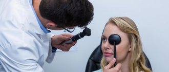
Diagnosis of exophthalmos
Protrusion of the eyes and the degree of displacement are detected using exophthalmometry, during which the distance between two parallel planes is measured: one passes through the apex of the cornea, the other through the lateral wall of the orbit. An exophthalmometer resembles a regular ruler, but has two frames. Two mirrors are attached to it at an angle of 45°. The ophthalmologist attaches the device to the outer sides of the eye sockets - closer to the temples, moves the frames and takes measurements.
The degree of exophthalmos is not the only indicator that needs to be identified. It is important to determine which parts of the eye are affected by the pathology. Typically, the following instrumental methods are prescribed for protrusion:
- Ophthalmoscopy. With the 3rd degree of bulging eyes, pathological changes in the fundus may occur. There is a risk of compression of the optic nerve head. He looks pale and puffy. Local hemorrhages are observed.
- Biomicroscopy. It can be used to detect ulcers and other damage to the cornea.
- Tonometry. Intraocular pressure does not increase in all types of exophthalmos. But this method is one of the mandatory ones in ophthalmology and is used during any examination. In addition, secondary complications of bulging eyes can be determined by ophthalmotonus.
- OCT. Allows you to visualize periorbital tissue, exclude or confirm swelling, neoplasms in the orbit and hemorrhages.
- Ultrasound of the eyeball in B-mode. Using this method, you can determine the degree of exophthalmos, evaluate the orbital tissue, and find out whether the disease is progressing or regressing.
The most important thing with exophthalmos is to find its cause, that is, the primary pathology. You need to undergo a comprehensive examination. The patient donates blood and urine for analysis, his hormone levels are determined, an ultrasound of the thyroid gland, MRI and other methods are prescribed. After the diagnosis is made and the type of exophthalmos is determined, treatment begins. As a rule, it is carried out not only by an ophthalmologist. You may need the help of an endocrinologist, otolaryngologist, neurologist and even a neurosurgeon.
How is endocrine ophthalmopathy diagnosed?
The diagnosis of “endocrine ophthalmopathy” is made on the basis of a set of instrumental and laboratory research methods carried out by an endocrinologist and an ophthalmologist.
Endocrinological examination
involves determining the level of thyroid hormones, identifying antibodies to gland tissues, and ultrasound examination of the thyroid gland. If ultrasound reveals nodes more than 1 cm in diameter in the structure of the gland, a puncture biopsy is indicated.
Examination by an ophthalmologist
consists of viziometry, perimetry, convergence studies. It is necessary to conduct an examination of the fundus of the eye - ophthalmoscopy, and determine the level of intraocular pressure - tonometry. If it is necessary to clarify the diagnosis, MRI, CT, ultrasound of the orbit and biopsy of the extraocular muscles can be performed.
Treatment of exophthalmos
The treatment method is determined by the causes of the pathology. So, with thyrotoxicosis, exophthalmos is treated with steroid drugs. They relieve inflammation and stabilize hormone levels. If bulging eyes develop due to a diseased thyroid gland, for example, with hyperthyroidism, radioactive iodine is prescribed. Sometimes pulse therapy with Prednisolone is used for exophthalmos. Edema is eliminated with medications and radiotherapy.
Exophthalmos may result from inflammation in the eye or orbit. In such cases, antibacterial and anti-inflammatory drops are prescribed, which help reduce inflammation and its toxicity. In some cases, antibiotics are used, intravenously. Vitamins are used to maintain general immunity.
Edema exophthalmos, which often occurs due to hormonal imbalances, hypothyroidism and hyperthyroidism of the thyroid gland, is treated by a neurologist, endocrinologist and therapist. First you need to restore the functioning of the thyroid gland. This is done with the help of drug therapy. The choice of a specific drug is determined by the primary pathology.
Pulsatile exophthalmos is treated with orbital radiotherapy. In addition, a pressure bandage is placed on the eye to cause thrombosis of the ophthalmic vein. This helps stop the development of bulging eyes. In severe cases, the carotid artery is ligated. These procedures can only be performed by a doctor.
Intermittent exophthalmos requires surgical treatment. During the operation, the surgeon ligates the internal or external carotid artery or places a clip on it inside the skull. Oncological diseases in which the patient has to undergo a course of chemotherapy are also treated surgically. Surgery is necessary if the optic nerve is severely damaged. Excess fatty tissue is removed from the eye socket to reduce pressure on the fundus. Severe damage to the cornea may require stitching of the eyelids. Special ointments are prescribed that help restore tissue of the cornea.
Endocrine ophthalmopathy - classification
There are several types of classifications of endocrine ophthalmopathy. In domestic medicine, the most common classification is according to V.G. Baranov, according to which the degrees of endocrine ophthalmopathy
accompanied by certain clinical manifestations.
— 1st degree
characterized by slight bulging eyes (up to 16 mm), moderate swelling of the eyelids, without dysfunction of the extraocular muscles and conjunctiva;
— 2nd degree
accompanied by moderately severe exophthalmos (up to 18 mm), significant swelling of the upper and lower eyelids, as well as the conjunctiva, and periodic double vision;
— 3rd degree
. It is characterized by severe exophthalmos (up to 21 mm), the inability to completely close the eyelids, erosions and ulcers on the cornea, limited mobility of the eyeball and signs of optic nerve atrophy.
Also in practice, the classification of endocrine ophthalmopathy by A.F. is often used. Brovkina, based on the severity of ocular symptoms, and including three main forms
diseases: thyrotoxic exophthalmos, edematous exophthalmos and endocrine myopathy.
Orbital decompression for exophthalmos
With exophthalmos, orbital decompression may be required, which helps reduce pressure on its walls. After the operation, the volume of space in the orbit increases, thereby reducing the degree of bulging eyes. Parts of one, two or three orbital walls are removed.
Orbital decompression is performed for compressive optic neuropathy, keratopathy, spontaneous eye prolapse, severe pain and to improve aesthetic appearance.
To relieve the symptoms of bulging eyes, it is recommended to adhere to the following rules:
- use moisturizing drops, ointments and gels;
- when going outside, take sunglasses with you;
- eliminate salt, select food products more carefully;
- sleep with your head on a raised surface (no more than 15 cm).
Use anti-inflammatory eye drops only for the first three days. They help eliminate inflammation and redness of the conjunctiva. But subsequently the opposite effect may occur due to vasodilation. Buy all medications only with a prescription. Treatment of exophthalmos with so-called folk remedies is unacceptable. At best, they will not affect the condition of the eyes, at worst, they will cause complications.
Treatment of exophthalmos can take several years. Under no circumstances should you abandon therapy at the first signs of recovery. The risk of relapse is quite high.
Symptoms of endocrine ophthalmopathy
Thyrotoxic exophthalmos
manifests itself clinically in the form of slight true or false protrusion of the eyeballs, retraction of the upper eyelid, due to which there is a widening of the palpebral fissure, slight trembling of the closed eyelids and insufficient convergence. No morphological changes are detected in the retrobulbar tissues. The range of movements of the periocular muscles is not limited, the fundus of the eye is unchanged.
For edematous exophthalmos
Characteristic is bilateral damage to the eyeballs, occurring more often at different time periods, with an interval of up to several months. During this form of endocrine ophthalmopathy, three stages are distinguished.
1. Compensation stage
. The onset of the disease is characterized by a number of specific symptoms, namely, in the morning there is a slight drooping of the upper eyelid, which disappears in the evening. The palpebral fissure closes completely. Over time, partial drooping of the eyelid is transformed into persistent retraction (contraction) due to spasm and prolonged increased muscle tone, which leads to contracture of the Müller muscle and the superior rectus muscle of the eye.
2. Subcompensatory stage
. The outer canthus and the area along the lower eyelid are affected by white chemosis, intraocular pressure increases and swelling of the periocular tissues of a non-inflammatory nature develops. Bulging eyes grows very quickly, the palpebral fissure ceases to close completely. The vessels of the sclera expand, become convoluted and form a figure resembling a cross. It is this symptom that gives rise to the diagnosis of edematous exophthalmos. When the eyeballs move, an increase in intraocular pressure is observed.
3. Decompensatory stage
. Characterized by a sharp increase in symptoms. A large degree of bulging eyes develops, the palpebral fissure does not close at all due to swelling of the eyelids and periocular tissue. The eye is immobilized. The development of optical neuropathy is observed, turning into atrophy of the optic nerve. Due to compression of the ciliary nerves, keratopathy and erosive and ulcerative lesions of the cornea develop. If the necessary treatment is not carried out, this stage of edematous exophthalmos ends with fibrosis of the orbital tissue and a sharp deterioration in vision due to corneal cataract or optic nerve atrophy.
Endocrine myopathy
most often affects both eyes, usually occurs in men against the background of a hypothyroid or euthyroid state. The onset of the pathological process is manifested by double vision, the intensity of which tends to increase. Then exophthalmos joins. Swelling of the periocular tissue is not observed in this form of endocrine ophthalmopathy, but the rectus oculomotor muscles thicken, which leads to disruption of their function and limitation in the abduction of the eyes outward, downward and upward. The infiltrative stage of this form of endocrine ophthalmopathy is very short-lived, and tissue fibrosis is observed after just a few months.
Protruding eyes in Graves' ophthalmopathy must be differentiated from false protruding eyes, which can occur with inflammatory processes in the orbit, tumors, and a significant degree of myopia.
Prognosis and possible consequences of exophthalmos
The prognosis depends on the severity of the disease, the timeliness of contacting a doctor, the correctness of the prescribed method of treatment and the individual characteristics of the patient. As a rule, it is possible to eliminate both the primary pathology and bulging eyes. Difficulties may arise with the malignant nature of exophthalmos, hydrocephalus and other serious illnesses.
As for complications of an ophthalmological nature, due to non-closure of the eyelids, increased intraocular pressure and insufficient nutrition of the eye with exophthalmos, there is a risk of:
- keratitis;
- neuritis;
- optic nerve atrophy;
- retinal hemorrhages.
Protruding eyes can lead to severe visual impairment, especially during atrophic processes. One or both eyes may lose the ability to move in their sockets. Complete loss of visual function is possible.
Forecast and prevention of exophthalmos
The prognosis for the development of eye protrusion depends on the stage of the disease and severity.
When treatment is started in the early stages of the disease, the chances of completely eliminating the pathology and restoring vision, as well as the patient’s appearance, are maximum. Against the background of complications and with advanced forms of pathology, the prognosis is not favorable; in such situations, complete loss of vision or proptosis of the eye is possible. If there is a predisposition to the development of pathology, as well as in the early stages of its development, preventive measures are required to prevent the progression of the disease. The most effective are the following:
- Rejection of bad habits. Consumption of alcohol and nicotine can provoke damage to the vascular system of the eyes, which in turn will lead to the development of complications of protrusion.
- Changing your diet. To enrich the body with vitamin complexes, it is recommended to avoid eating heavy foods in favor of fresh vegetables and fruits.
- Maintain eye hygiene.
- Prevention of stressful situations, which can also lead to protruding eyes.
- Prevention of injuries and mechanical damage to the eyeballs. Compliance with safety regulations at enterprises.
The most important factor in the prevention of eye disease is timely treatment of somatic diseases, the presence of which can provoke the development or complications of exophthalmos.
Prognosis of endocrine ophthalmopathy
The prognosis of endocrine ophthalmopathy depends on the timeliness of treatment. If the disease is diagnosed in the early stages and the correct treatment plan is developed, long-term remission of the disease can be achieved and severe irreversible consequences can be prevented. According to statistics, a third of patients experience clinical improvement, and two-thirds experience stabilization of the process. In 5%-10% of cases, further progression of endocrine ophthalmopathy is possible.
After treatment, ophthalmological monitoring is required after six months, as well as constant monitoring and correction of thyroid function by an endocrinologist. Patients with Graves' ophthalmopathy should be registered at the dispensary.