Blepharitis is an inflammatory disease that affects the edges of the eyelids. The disease is one of the most common causes of discomfort and eye irritation.
Every third patient who sought help from an ophthalmologist for eye inflammation was diagnosed with blepharitis. Of course, such a common pathology causes a lot of discussion in ophthalmological circles. What should the patient do? How to understand in what cases it is necessary to contact a specialist?
Causes of blepharitis
Why does this inflammation occur? Often this is a consequence of age-related changes, the presence in patients of chronic diseases of the gastrointestinal tract, seborrheic dermatitis, rosacea, diabetes mellitus, Sjogren's syndrome, rheumatoid arthritis, psoriasis, atopic dermatitis, rosacea and hormonal changes. Systemic intake of retinoic acid drugs (for the treatment of acne), hormone replacement therapy used by women during menopause, antihistamines, and antidepressants also contributes to the development of blepharitis. Violation of local immunity, the presence of immunodeficiency states and frequent use of cosmetics (especially high-density foundation) are provoking factors for this pathology.
Also, according to Russian and foreign publications, a common cause of inflammation is uncorrected or incorrectly corrected refractive errors (farsightedness, astigmatism, less often myopia). Therefore, when correcting vision, it is very important to find a competent specialist who selects glasses according to clinical standards.
Blepharitis - what kind of disease?
Blepharitis is an inflammation of the eyelids that affects the outer edges of the eyelids. This concept combines a whole group of pathologies with similar symptoms. In most cases, blepharitis is chronic, but there is also an acute form of inflammation.
Depending on the location of the lesions, blepharitis is divided into anterior marginal, posterior marginal and angular. In the first case, the inflammatory process affects the edge of the eyelids along the eyelash growth line, in the second it spreads to the meibomian glands. With posterior marginal inflammation of the eyelids, the conjunctiva and cornea can often be affected. Experts note that most often these two types of blepharitis appear in parallel. In the angular form of blepharitis, the damage affects the corners of the eyes.
Most often, eyelid inflammation occurs in adults over 40 years of age, but sometimes children also suffer from it. Blepharitis must be treated, because its long course can lead to complications such as keratitis, conjunctivitis, decreased visual acuity, or chalazion.
It is worth noting that the disease is quite difficult to treat, and in rare cases, complete recovery can be achieved. But it is quite possible to reduce the risk of complications and the frequency of exacerbations with properly selected therapy.
Symptoms of blepharitis
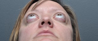
The most common symptoms of blepharitis are redness of the edges of the eyelids and the eye itself, itching of varying severity, burning, lacrimation, foreign body sensation, and increased eye fatigue. With a long course, the disease provokes ulceration of the edges of the eyelids, changes in the shape of the eyelids, loss of eyelashes and the wrong direction of their growth, inflammation of the cornea. All this can lead to significant and irreversible vision loss. That is why it is necessary to consult an ophthalmologist in time, because timely prescribed treatment will ensure a quick recovery. Otherwise, the process will become chronic, and treatment will take months or even years.
Classification
Based on anatomical characteristics, they are distinguished:
- Anterior marginal blepharitis (only the ciliary edge of the eyelid is affected).
- Posterior marginal blepharitis - damage to the edges of the eyelids is caused by inflammation of the meibomian glands located in the thickness of the eyelids (meibomian blepharitis).
- Combined.
In accordance with the etiology, non-infectious and infectious blepharitis are distinguished. According to the nature of the course, they are divided into acute, subacute and chronic blepharitis.
According to the clinical course, various forms of the disease are distinguished:
- Simple.
- Seborrheic (scaly).
- Demodectic.
- Ulcerative ( ostiofolliculitis ).
- Allergic.
- Acne ( rosacea-blepharitis ).
- Mixed.
Clinically distinguished:
- superficial blepharitis (the inflammatory process is localized in the area of the eyelid edge);
- deep (subcutaneous tissue, muscle and other tissues of the eyelid are involved in the pathological process).
Types of blepharitis
Let's look at the types of blepharitis in more detail. The anatomy of the eyelids will help us.
The eyelids belong to the auxiliary organs of the eye, which are mobile structural formations that cover our organ of vision. They perform the function of protecting the eyeball from negative environmental factors. By making blinking movements, the eyelids contribute to the uniform distribution of tear fluid over the ocular surface, thereby moisturizing it. Each eyelid consists of two plates - the outer (skin and muscles) and the inner (cartilaginous plate and delicate tissue - conjunctiva). The meibomian glands are located in the thickness of the cartilage; they are located in parallel columns and open with excretory ducts near the posterior edge of the eyelids. These are very important glands that produce a viscous lipid secretion that is part of tears. Along the front edge of the eyelid there are eyelashes that border the top and bottom of the eyes. Next to the eyelashes are the Zeiss sebaceous glands, which secrete secretions into the hair follicle of the eyelashes.
By location, all blepharitis is divided into anterior (in the area of eyelash growth) and posterior (in the area of the exit of the meibomian gland ducts). Let's look at them in more detail.
Anterior blepharitis
- is a local manifestation of skin pathology (seborrhea, rosacea), with the addition of a staphylococcal infection.
Seborrheic
(or scaly) blepharitis
. With this type of inflammation, there are very typical symptoms - the presence of a large number of small dry scales, like dandruff, on the edges of the eyelids and eyelashes. Symptoms are usually more pronounced in the morning, but if the patient also suffers from dry eye syndrome, the burning sensation and foreign body sensation may continue throughout the day. Most often, the disease affects both eyes. If the process continues for a long time in one eye, this may be an alarming symptom of a tumor process and requires immediate consultation with a specialist!
Ulcerative (or staphylococcal) blepharitis
.
The cause of this type of disease is bacteria. The most common are Staphylococcus epidermidis
and
Staphylococcus aureus
. Although the exact mechanism of development of ulcerative blepharitis is unclear, three convergent pathways likely underlie the pathophysiology:
1) direct bacterial infection; 2) hypersensitivity to exotoxin (a component of the bacterial cell wall); 3) delayed immune hypersensitivity reaction (i.e., the human body’s resistance to the effects of this bacterium).
As a result of an immune reaction, hard scales and crusts, the so-called muffs, form around the bases of the eyelashes. The eyes become red and dry eyes occur due to disruption of the stability of the tear film.
With a long process, a secondary infection may occur. In such cases, stye of the eyelid, inflammation of the cornea or conjunctiva of the eye occurs. Due to constant ulceration, the edges of the eyelids become scarred and eyelashes fall out or begin to grow incorrectly. All this leads to cosmetic defects that require the intervention of cosmetologists, dermatologists and even plastic surgeons.
Posterior blepharitis—characterized by inflammation of the posterior margin of the eyelids and has a variety of etiologies, including meibomian gland dysfunction, infectious and allergic conjunctivitis, and systemic conditions such as rosacea, eczema, and atopic dermatitis.
Meibomian gland dysfunction (MGD) is a chronic, diffuse anomaly of the glands, manifested by blockage of the duct outlets, a change in the amount of gland in their structure. As a result, the excretion and production of lipid secretions are disrupted, the tear film is not protected from evaporation and is thinned. This provokes dryness of the ocular surface, the negative impact of environmental factors on the eye increases, damage to the cornea and other structures of the eyeball occurs, discomfort and increased visual fatigue appear. A foamy white secretion accumulates in the corners of the eyes, the edges of the eyelids turn red, thicken, itching and cloudiness are felt before the eyes. An infection can join the accumulation of secretions inside the gland, which provokes the development of inflammation of the eyelid. All this significantly reduces the quality of life.
A separate form of blepharitis is demodectic blepharitis. It is quite specific and requires different treatment. Therefore, this pathology should be given a separate article.
At the appointment, the ophthalmologist primarily relies on the patient’s complaints, concomitant diseases and clinical picture. After a conversation with the patient, the edges of the eyelids are always examined using a special microscope, the condition and stability of the tear film and the presence of discharge are assessed. Special tests may be required to evaluate tear fluid. In some cases, examination is not enough, and to clarify the diagnosis, smears from the conjunctiva are cultured on special media and eyelashes are examined for the presence of a pathogen.
Pathogenesis
The pathogenesis of blepharitis is based on a violation of the secretory function of the meibomian glands, leading to a “thickening” of the secretion, which creates a favorable environment for the entry/reproduction of various pathogens (viruses, bacteria, Demodex mites, fungus) and the development of the inflammatory process at the edges of the eyelid.
The inflammatory process of the eyelid margins is a central pathophysiological link. According to various authors, chronic blepharitis is a consequence of changes in the properties of lipids in the secretion of the meibomian glands, caused by lipases of the bacterial flora with subsequent blockage of the openings of the excretory ducts of the glands. Clogged ducts promote the growth and development of bacteria and mites, which trigger the inflammatory process. The constant presence of bacterial flora in the conjunctival cavity contributes to the development of a chronic process with subsequent narrowing and obliteration of the excretory ducts of the meibomian glands.
Treatment of blepharitis
Although the etiology and clinical forms of blepharitis can vary significantly, treatment methods are the same in most cases.
First of all, it is necessary to eliminate the causes of the disease. Refractive errors are corrected and unfavorable factors (dust, chemical fumes, infectious foci of inflammation) are removed.
Non-drug therapy
.One of the most important and frequently used treatment methods is proper eyelid hygiene.
Eyelid hygiene
— ensures normal and proper functioning of the glands, normalizes metabolic processes in the skin of the eyelids and promotes the formation of a stable tear film. Protection of the eyelids, especially the marginal part, from the effects of negative environmental factors, various parasites and infections is the basis for the prevention and treatment of blepharitis.
All therapeutic eyelid hygiene can be divided into two parts:
1) Applying a warm compress to the eyelids; 2) Eyelid massage;
A warm compress normalizes metabolic processes, does not allow the lipid secretion in the gland ducts to harden, which prevents blockage of the ducts and, as a result, prevents the provocation of inflammation inside the glands.
When applying a compress, it is recommended to use cotton cosmetic pads, previously soaked in hot water and wrung out until damp. The discs are applied to closed eyes for 1-2 minutes.
After the secretion has become more liquid, a light massage of the eyelids is performed. This allows you to remove excess secretion into the eye cavity, stimulate blood circulation and lymphatic drainage, which will improve metabolic processes.
In case of severe blepharitis and at the beginning of treatment, it is recommended that the massage be performed by a specialist, since it is necessary to maintain sterile conditions and carry out manipulations using a special instrument - a glass rod, as well as instill anesthetics. After the removal of the primary symptoms and with positive dynamics of the condition, the patient performs the massage independently.
During self-massage, a special gel is used, which allows you to cleanse the eyelids of toxic substances, scales and crusts that occur against the background of inflammation, and also moisturize the skin of the eyelids, which will reduce discomfort and itching.
Oddly enough, even increasing the number of blinking movements will help in the treatment of blepharitis, especially if the patient works at the computer!
conclusions
- Demodectic blepharitis is the most common eye reaction of the body to waste products of a parasite that lives on the skin of the face;
- The disease is more susceptible to the elderly, children with diseases of the gastrointestinal tract and lungs, as well as patients with systemic diseases;
- In some cases, the cause of inflammation of the eyelids may also be a visual disturbance that cannot be corrected with glasses or contact lenses;
- The course of treatment takes from two weeks to a month. Therapy is prescribed by an ophthalmologist;
- For preventive purposes, it is recommended to carefully monitor personal hygiene, see a dermatologist, and, if possible, eliminate all risk factors.
Drug therapy
You should immediately pay attention to the fact that all pharmacological drugs are prescribed by the attending physician only after an examination and taking into account the individual tolerance of the drug components.
Self-medication in cases of blepharitis brings only temporary relief, the disease does not go away and in the future it only complicates recovery. It is necessary to understand for what purpose and what result needs to be achieved using this or that drug.
When treating staphylococcal blepharitis, an important aspect is the choice of antibacterial drug. Broad-spectrum antibiotics (tetracyclines, fluoroquinolones) are most often used. Preference is given to ointment forms, as they allow simultaneously moisturizing and healing the wound surfaces of the eyelid margins.
Non-steroidal anti-inflammatory drugs are used to reduce the inflammatory response. They suppress the inflammatory response in tissues, reducing redness.
In case of severe burning, itching, swelling of tissues, it is recommended to take orally and locally use (instillation) antiallergic drugs. These drugs reduce discomfort from the clinical manifestations of inflammation, thereby alleviating the patient’s condition.
Next, tear replacement therapy is used - its goal is to replenish the tear deficiency, protect the ocular surface from the effects of negative factors, moisturize and reduce the symptoms of a foreign body in the eye. To do this, use drops with gel textures containing hyaluronic acid.
During treatment, you should limit visiting the pool, sauna and baths, and taking warm baths.
In case of a prolonged inflammatory process, you may need to consult a gastroenterologist, dermatologist (for the prevalence of seborrhea, atopic dermatitis, rosacea on the surface of the skin in the face), a nutritionist (for nutrition correction - exclusion from the diet of spicy, salty, acidic foods, as well as foods with high sugar and gluten content).
If a patient is diagnosed with blepharitis, you need to understand that the duration of treatment and the speed of getting rid of this disease directly depends on following the specialist’s instructions. Usually, when the process is chronic and a late visit to the doctor, improvement occurs very slowly, and the risk of exacerbation of the disease is high. The most difficult treatment is for staphylococcal blepharitis, which can lead to the appearance of styes, chalazions, inflammation of the conjunctiva and cornea of the eye.
When conducting scientific research and clinical trials, experts came to the conclusion that a universal approach to treatment is ineffective. A combination of treatment strategies was most effective in chronic blepharitis, and these methods required an individual approach to each patient. Drug treatment in combination with hygiene and eyelid massage is the most modern model in the treatment of this pathology.
Pathologies
Inflammatory diseases of skin folds are acute and chronic. It can affect the upper, lower eyelid, or both eyes at the same time.
Acute
Among the most common acute ones:
- Barley. External (purulent inflammation of the hair follicle) or internal (meibomian gland). Caused by Staphylococcus aureus disease.
- Furuncle. Purulent necrotic inflammation of the hair follicle, sebaceous glands and surrounding connective tissue. Localized on the upper eyelid, in the eyebrow area. Rarely on the edge of the eyelid in the area of the outer canthus. The causative agent is staphylococcus.
- Dacryoadenitis is inflammation of the upper eyelid. Complications after common infections (flu, sore throat, etc.)
- Dermatitis. Occurs as a result of allergies, gastrointestinal disorders, autoimmune diseases. To the symptoms listed above are added itching, peeling, and rashes in the form of blisters.
Less common are eyelid abscess and phlegmon.
Chronic
Chronic diseases include:
- Blepharitis is inflammation of the edges of the eyelids. Caused by skin bacteria. Demodectic blepharitis - caused by mites of the genus demodex. There is redness and swelling of the edges of the eyelids. Itching. Eyelash loss. With ulcerative blepharitis, there are ulcers and purulent crusts.
- Chalazion is a proliferative inflammation of the meibomian gland and the cartilage around it. It can occur both after barley and independently.
Blepharitis and chalazion are often combined with conjunctivitis.
How to prevent the disease?
A good prevention of blepharitis is special eye exercises. It can be carried out when working at a computer and during prolonged visual stress.
- look left and right 10 times, then up and down 10 times. - Close your eyes tightly and then open both eyes wide. Repeat this 15 times. -Rub your palms together (don’t forget to wash your hands before this exercise!) and gently place your warm palms on your closed eyelids, hold for 15-20 seconds.
Do not forget to carry out preventive hygiene and eyelid massage 2-3 times a week, and clean the rooms weekly (this will prevent dust from accumulating and it will be much easier on your eyes). Pay attention to your health. At the first symptoms of malaise and redness of the eyes, consult a specialist! A timely examination and prescribed treatment will give the fastest positive effect!
Take care of your eyes!
Tests and diagnostics
Diagnosing blepharitis is not difficult. It is based on an ophthalmological examination of the patient with a slit lamp. Instrumental diagnostic procedures (visometry, refractometry, eye biomicroscopy) can effectively identify refractive errors and assess the condition of the eye structures and tear film.
If demodectic blepharitis is suspected, a laboratory examination of the eyelashes is performed to determine the presence of the causative mite. To identify the infectious pathogen, a bacterial culture of a smear from the conjunctiva is performed.
Clinical manifestations of demodectic and allergic blepharitis
The causes and symptoms of this disease are directly related to the demodex mite, which affects the eyelids. In a person with this pathology, the eyes begin to itch very much, and a discharge appears that “glues” the eyelashes together. The edges of the eyelids become thicker and redden. At the base of the eyelashes, an accumulation of sebaceous secretions and cellular particles appears, which is shaped like a collar or muff. Mites can usually be detected at an ophthalmologist's appointment after microscopy.
Allergic blepharitis is usually accompanied by inflammation of the connective membrane of the eye. The cause of the disease may be the individual characteristics of the body - for example, high sensitivity to certain medications, cosmetics, dust, wool. In the allergic form of the pathology, there is not purulent, but mucous discharge, lacrimation, swelling and itching of the eyelids, and a burning sensation in the eyes. As a rule, symptoms appear on two organs of vision at once.


