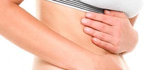
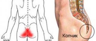
Causes of pain in the gallbladder
Pregnancy
The appearance of pain and discomfort in the gallbladder area is due to the development of cholestasis in pregnancy. Symptoms occur in the 3rd trimester of pregnancy and disappear in the first days of the postpartum period. Pain is caused by stagnation of bile in the biliary tract (BDT). In addition to pain, yellowness of the skin and mucous membranes and severe itching are noted. The urine becomes dark in color and the stool becomes gray.
Biliary dyskinesia
Functional disorders, which include gallstones, are the most common cause of pain in the gallbladder in children, young and middle-aged people. The nature and duration of the pain syndrome is determined by the type of motor impairment. With the hypermotor variant, the pain is cramping, strong, reminiscent of biliary colic. They appear 20-30 minutes after finishing a meal. Pain is often caused by psycho-emotional stress.
With the hypomotor variant of dyskinesia, aching or nagging pain in the right hypochondrium is disturbing. It begins 40-90 minutes after eating and is provoked by fatty foods and alcohol consumption. In contrast to hypermotor JP, the pain syndrome is longer lasting - up to several hours. It is accompanied by nausea, vomiting, a feeling of heaviness in the abdomen, alternating constipation and diarrhea.
Chronic cholecystitis
With this pathology, periodic pain is observed, localized on the right in the hypochondrium. They are provoked by eating large amounts of fatty foods, which leads to increased secretion of bile and increased load on the gallbladder. In women, symptoms worsen before menstruation. In chronic cholecystitis, the pain is dull or aching, not intense, combined with bitterness in the mouth, nausea, and stool instability.
Acute purulent cholecystitis
This disease is characterized by severe cramping pain in the gallbladder area that appears suddenly. To alleviate the condition, a person lies on his side and pulls his knees towards his stomach. A painful attack is accompanied by fever, sweating, and tachycardia. When inflammation passes into empyema of the gallbladder, pain reaches maximum intensity, body temperature rises to 40°C.
Cholangitis
In the acute form of inflammation of the bile ducts, sharp, severe pain is observed in the right hypochondrium. For them, irradiation is specific to the right shoulder, neck, and interscapular region. The symptom appears suddenly in combination with febrile fever and jaundice. There is no clear connection between pain and dietary errors or other typical factors. At the same time, nausea and vomiting, diarrhea, and weakness occur.
Chronic cholangitis is characterized by aching pain in the projection of the gallbladder. They are felt periodically, accompanied by heaviness in the hypochondrium on the right and a feeling of fullness in the epigastrium. The pain syndrome is not intense and does not require analgesics. In most cases, the pain disappears on its own after a few hours. The addition of general symptoms is typical: prolonged low-grade fever, increased fatigue.
Cholelithiasis
Pain syndrome is the main manifestation of the disease. A typical attack of cholelithiasis is called biliary colic. It develops with the abuse of fatty foods and emotional experiences. Suddenly, intense pain appears above the gallbladder and in the epigastrium. It radiates to the scapula, lower back, and precordial region. Often the pain is so strong that the person rushes about in bed and cannot find a position in which the pain subsides.
The attack lasts several hours. The pain is cramping in nature and is not relieved by conventional analgesics. For cholelithiasis, it is typical to increase body temperature simultaneously with the onset of pain. Clinical symptoms are supplemented by severe nausea and vomiting, and stool disorders. Chills and pale skin are noted.
Parasitic infestations
Pain in the right hypochondrium can be a sign of giardiasis. With this pathology, a mild pain syndrome is observed. There is no connection between pain and diet disorders or physical overexertion. The symptom is combined with unstable bowel movements, flatulence, and nausea. With parasitic infections, intoxication syndrome is also pronounced.
Postcholecystectomy syndrome
Symptoms of the disease occur after surgery to remove the gallbladder. Postcholecystectomy syndrome is characterized by recurrent pain attacks that develop for no apparent reason. The pain has a different character - cutting, spastic, dull, aching. The pain is accompanied by dyspeptic symptoms: belching, bitterness in the mouth, nausea, bloating. Diarrhea occurs with copious foul-smelling feces.
Gallbladder tumors
Benign polyps of the organ are characterized by scant symptoms. Periodically, dull pain is felt at the location of the gallbladder, which is not associated with food intake. In a small number of patients, the disease manifests itself as sharp pain and spasms in the right hypochondrium, which are provoked by eating fatty foods or physical activity.
In case of gallbladder cancer, pain syndrome is expressed at an advanced stage. There is a mild dull pain over the organ, which appears for no apparent reason. At first it bothers you sporadically, and then it becomes constant. The clinical picture is characterized by a progressive decrease in body weight, lack of appetite, and prolonged low-grade fever.
Cholelithiasis
Gallstones are nothing but the presence of deposits in the gallbladder or bile ducts. Gallstones are more likely to affect women, obese people, older adults, diabetics, and patients taking oral contraceptives or other estrogen medications.
Gallstones
Gallstones can occur at the same time as duct stones because deposits in the gallbladder are tiny and can travel with bile into the bile ducts, blocking their narrow lumen. In addition to hepatic colic, jaundice indicates urolithiasis, which is additionally accompanied by a change in the color of the stool and unpleasant itching of the skin.
Diagnostics
A careful collection of medical history and eating habits plays a major role in determining the cause of pain. During a physical examination, a gastroenterologist checks for cystic symptoms that indicate an inflammatory process. The diagnostic search involves a comprehensive laboratory and instrumental examination of the biliary system. The following methods are used:
- Ultrasound of the gallbladder.
According to sonography, the size and contours of the gallbladder and the condition of the bile ducts are assessed. During the examination, stones and adhesions may be noticed. To study the contractile activity of the organ, a test with a choleretic breakfast is indicated. - ERCP.
The technique of reverse contrasting of the bile ducts is necessary for detailed visualization of their condition, finding small stones that were not visible during sonography. ERCP is performed using an endoscope, so it is both a diagnostic and therapeutic method. - Duodenal sounding.
Obtaining several portions of bile is necessary for its microscopic and bacteriological analysis. An increased number of leukocytes and mucus are found in the bile. During bacterial culture, a mixed bacterial flora is usually found. - X-ray techniques.
A plain radiography of the abdominal cavity is performed to identify complications - calcification of the gallbladder wall, free gas under the diaphragm. To clarify the diagnosis, cholecystography, MSCT of the abdominal organs, and dynamic scintigraphy of the hepatobiliary system are recommended. - Laboratory research.
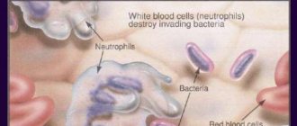
In the hemogram for inflammatory diseases of the gallbladder, leukocytosis and increased ESR are detected. A biochemical blood test is performed to look for signs of cholestasis (increased alkaline phosphatase and cholesterol levels) and to assess the content of direct bilirubin.
Hepatic colic and diet
A properly selected diet can not only cure hepatic colic, but also relieve the symptoms of cholelithiasis. The diet should be low-fat and easily digestible.
Patients should eliminate or limit the consumption of foods such as cheese, lard, mayonnaise and cream. In addition, frying should be avoided; it is better when you cook the dish in the oven or steam.
Patients who are obese should reduce their body weight, and those who additionally suffer from hypertriglyceridemia should reduce their levels of simple sugars and disaccharides.
Weight loss in obesity
Treatment
Help before diagnosis
For mild pain, indicating a chronic process, it is permissible to limit yourself to non-drug methods. The patient is given a detailed diet plan, which involves the exclusion of animal fats, extractives, and alcohol. The diet depends on the preservation of the biliary function of the bladder. To increase the volume of bile, mineral waters are prescribed. In case of an acute pain attack, you should go to the hospital as soon as possible.
Conservative therapy
Drug treatment is carried out for uncomplicated forms of gallbladder disease, without the risk of organ destruction. At the initial stage, antispasmodics and analgesics are used to relieve painful symptoms. Then etiopathogenetic therapy is selected, which may include several groups of drugs:
- Antibiotics
. For exacerbation of cholecystitis and cholangitis, broad-spectrum antibacterial drugs are used. To accelerate the elimination of bacterial toxins and endotoxins, treatment is supplemented with infusion therapy. - Choleretic agents
. After stopping the acute process, drugs are prescribed that improve the flow of bile. They are divided into 2 groups: cholekinetics, which stimulate the contractile activity of the gallbladder, and choleretics, which increase the volume of the water component of bile. - UDHC
. The use of ursodeoxycholic acid is one of the methods of non-surgical treatment of cholelithiasis. With long-term use (up to two years), the active substance dissolves stones and helps normalize the biochemical composition of bile.
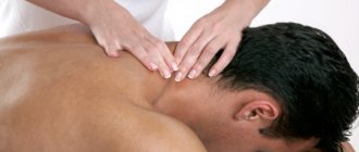
After the elimination of acute inflammation, physiotherapeutic techniques are prescribed. For chronic cholecystitis, it is advisable to use reflexology, SMT therapy, and electrophoresis. It is effective to use decoctions of medicinal plants that have hepatoprotective and choleretic properties. Spa treatment at balneological resorts is recommended.
Cholecystectomy. Gallbladder stones
Lithotripsy
For calculous cholecystitis, methods of non-invasive crushing of stones are widely used. In gastroenterology, extracorporeal shock wave and contact lithotripsy are more often performed. This treatment is characterized by the absence of tissue trauma and a short rehabilitation period. Lithotripsy is only required for stones up to 2 cm in size.
Complications of hepatic colic
Ignoring the disease of hepatic colic can lead to unpleasant consequences requiring hospitalization. Among the complications of untreated hepatic (biliary) colic, we note:
- acute pancreatitis;
- acute cholecystitis;
- inflammation of the bile ducts;
- in rare cases, secondary cirrhosis of the liver may occur;
- chronic inflammation of the walls of the gallbladder.
Acute pancreatitis and acute cholecystitis are diseases for which it is necessary to immediately hospitalize the patient, as they are life-threatening! Therefore, it is very important that patients do not ignore hepatic colic and in any case consult a gastroenterologist. This will help to recognize the disease in a timely manner and prevent possible complications.
ONLINE REGISTRATION at the DIANA clinic
You can sign up by calling the toll-free phone number 8-800-707-15-60 or filling out the contact form. In this case, we will contact you ourselves.
If you find an error, please select a piece of text and press Ctrl+Enter
Bile ducts
The bile ducts, into which the bile produced by hepatocytes enters, begin blindly and are directed to the periphery of the hepatic lobule. Here they open into larger interlobular bile ducts, which, merging and gradually enlarging, form the common hepatic duct emerging from the porta hepatis (Fig. 3). Since bile is produced in the liver around the clock and enters the intestines only during digestion, a reservoir for storing bile is needed. The gallbladder serves as such a reservoir.
Diseases of the biliary system
Gallstone disease (hereinafter referred to as cholelithiasis) is a chronic disease of the gallbladder, which can be asymptomatic, exacerbating only with the development of inflammation and blockage of the bile ducts by stones.
GSD already occurs in 10% of the world's population3. Gallstones consist of substances that are poorly soluble in bile, in most cases cholesterol. There are various reasons for the formation of stones: genetic, demographic, dietary, and also due to various concomitant diseases or taking certain medications. The structure of once formed stone constantly changes, anhydrous cholesterol accumulates inside it4.
- Risk factors are age (40-69 years)3, heredity, predisposition, female gender.
- The development of this disease in women is associated with hormonal changes during pregnancy and childbirth, which contribute to the formation of stones5.
During menopause, due to a decrease in sex hormones, the contractility of the gallbladder weakens5.
The connection between cholelithiasis and the nature of nutrition has been proven. Excessive saturation of bile with cholesterol is promoted by foods high in animal fats (fatty meat, milk, cream, lard), simple carbohydrates (fast food, sweets, baked goods). Stone formation is provoked by frequent consumption of spicy seasonings and a deficiency in the diet of vitamins (especially vitamin E)5.
The tendency to stone formation occurs with increased reabsorption of bile acids. This occurs under conditions of slow movement of food through the intestines, which is facilitated by a lack of dietary fiber (vegetables, fruits)5. One of the main factors in the development of cholelithiasis is obesity. Moreover, the risk of stone formation is directly proportional to excess weight. If with a body mass index (BMI) from 25 to 30 the risk of cholelithiasis increases by 2 times, then with a BMI over 35 it increases by 20 times6.
Sudden weight loss also has a negative impact on stone formation. With a sharply limited unbalanced diet, the circulation of bile acids is disrupted, which leads to the formation of stones5.
There is information about the connection between lack of physical activity and the process of stone formation. Thus, in people who spent 40 hours a week watching TV, cholelithiasis was detected 2 times more often. The disease is much less common in people engaged in physical labor5.
Structure of the liver
Liver and gallbladder
The performance of numerous functions is associated with the peculiarities of the internal structure of the liver. The dense membrane covering the liver under the peritoneum goes deep into the organ and divides it into prismatic lobules with a diameter of about 1.5 mm. The number of such hepatic lobules in humans reaches 500 thousand; they are the structural and functional unit of the liver (Fig. 2). In the lobule, liver cells (hepatocytes) are grouped in the form of radial plates, between which there are wide blood capillaries (sinusoidal), converging to the liver). Inside the radial plates, between two adjacent rows of hepatocytes, slits are formed, called bile ducts: bile produced by hepatocytes enters them.
Each liver cell is in contact with the wall of the blood capillary on one side, and with the lumen of the bile duct on the other. This structure allows hepatocytes to work in two directions: secrete bile into the bile ducts and send glucose, proteins, fats, vitamins, urea, etc. into the blood. Raw materials for the production of bile and numerous substances also enter through the capillaries with arterial and venous blood. As already mentioned, arterial blood arrives in the liver through the branches of the hepatic artery, and venous blood through the branches of the portal vein. In the wide capillaries of the hepatic lobules, arterial blood mixes with venous blood and flows very slowly, which promotes the exchange of substances between blood and hepatocytes. The capillary wall also contains special cells - stellate macrophages, which perform a protective function. They can capture from the blood and destroy various foreign particles, microorganisms, and damaged cells. Blood saturated with waste products of hepatocytes flows from the capillaries into the central vein of the lobule, and from there into larger veins, through which it is carried out of the liver and enters the inferior vena cava, i.e. returns to the general bloodstream.