Nystagmus is the name given to rapid, repetitive eye movements that occur regardless of a person’s conscious commands. A similar condition sometimes occurs normally if a person has to watch a rapidly moving object, cold water gets into his ear, or his body rotates in space (for example, on a carousel). Some diseases of the nervous system, eye or inner ear can also lead to the appearance of this symptom. The pathology is very easy to diagnose, but it is difficult to treat.
The mechanism for the development of nystagmus is a violation of a peripheral organ (eyeball, nerves of the extraocular muscles, labyrinth in the peripheral part of the vestibular apparatus and the nerve coming from it), or the brain.
The long-term existence of nystagmus, even when it appears as a result of damage to the central nervous system and vestibular apparatus, leads to an inevitable decrease in visual acuity, since the brain does not receive normal images of objects from the eyes.
Causes
Nystagmus is usually divided into congenital and acquired:
Congenital pathology is quite rare. It is inherited or occurs as a result of birth injuries and intrauterine infections, giving impetus to the development of incomplete atrophy of the optic nerve, strabismus, retinal dystrophy, farsightedness, astigmatism, and myopia.
Acquired nystagmus can develop due to:
- Multiple sclerosis.
- Tumors in the brain.
- Brain contusion.
- Inflammation of the inner ear.
- Encephalitis.
- Temporal bone cracks.
- Stroke.
- Tumors of the nerve that carries information from the vestibular and auditory analyzers (vestibular-cochlear).
- Exposure to drugs and certain toxins.
- Taking a number of medications (lithium compounds, carbamazepine, barbiturates).
- Some ophthalmological diseases.
Pendulum-like eye movements often cause damage to the brain, namely: the cerebellum, brain stem, pons, midbrain, sella turcica, pituitary gland, rhomboid fossa, medulla oblongata.
At the same time, the direction of involuntary eye movement may indicate the localization of the lesion:
- With horizontal movements, as a rule, the inner ear or middle sections in the rhombencephalon are affected.
- With vertical or diagonal movements, the problem lies in the area of the upper sections in the diamond-shaped fossa.
- During rotational movements, the lower parts of the rhombencephalon are often affected.
- With converging movements, we are talking about pathologies of the midbrain.
When diagnosing, to more accurately determine the level of damage, the neurologist must also evaluate the speed of eyeball movements and their amplitude.
Forecast for the development of the disease
With adequate treatment of the underlying pathology that caused visual impairment, nystagmus of the eyeball can be eliminated almost completely. Visual functions are restored, quality of life does not suffer.
To prevent the appearance of nystagmus, it is necessary to promptly diagnose and treat pathologies of the brain, eyes and inner ear. If nystagmus appears as a complication, medication dosage adjustment is required in patients taking sleeping pills and anticonvulsants.
Author of the article: Marina Vitalievna Degtyareva, ophthalmologist, ophthalmologist
Types of nystagmus
According to the direction of oscillatory eye movements, the disease is divided into:
- Vertical nystagmus, if the eye moves up and down (vertically);
- Horizontal nystagmus, when movements occur along a horizontal axis;
- Rotatory nystagmus, with rotational movements around the sagittal axis;
- Convergent nystagmus, in which there is rapid movement of the eyes towards each other;
- Diagonal nystagmus - movement along a diagonal.
The direction in which the nystagmus is directed must be judged by its rapid phase.
If the eye movements are of the same type, the nystagmus is called associated, if different - dissociated.
Nystagmus is also distinguished by the location of the lesion:
1. Vestibular. Its causes are due to pathologies of the part of the brain that receives information from the vestibular apparatus, as well as a disease of the labyrinth itself (peripheral vestibular apparatus). Such nystagmus can be caused by rotation of the body, as well as a caloric test (pour cold or warm water of a certain temperature into the ear). It can also occur spontaneously, accompanied by nausea and dizziness. 2. Central. Such nystagmus develops with traumatic, dystrophic, tumor or inflammatory lesions of the cerebellum, structures in the posterior cranial fossa, subcortical and cortical centers for the regulation of eye movements.
There are also other classifications.
Diagnostics
When examining a patient, a doctor can immediately determine the presence of nystagmus by uncontrolled oscillatory eye movements. But his main task is to understand the cause of this pathology. To do this, it is necessary to conduct a number of studies:
- interviewing the patient or parents about whether the patient has a congenital or acquired abnormality, which could have caused it (perhaps there was a head injury, inflammation of the inner ear, or something else);
- checking the eyes for refractive errors;
- examination of the retina, fundus of the eye;
- checking for malfunctions in the optic nerve and the apparatus responsible for moving the eyes.
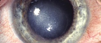
Healthy fundus
During the test, various techniques are used, which depend on the type of nystagmus:
- spontaneous nystagmus is determined during the initial examination by a neurologist. The patient, in a lying or sitting position, follows the doctor's hammer (various positions are necessary, because the disease sometimes manifests itself only in one position or when changing it).
- experimental (can be both physiological and pathological) - checked by special provoking methods: rotatory is determined using the Barany chair, color and pressor test.
- Hardware studies are also carried out: Electroencephalography (EEG), Magnetic resonance imaging (MRI), Echoencephalography of the brain.
- When testing optokinetic nystagmus, a special drum with black and white stripes is used. It is rotated horizontally and vertically in front of the patient.
How is electroencephalography performed?
These types of tests are needed to correctly diagnose pathologies of the nervous system, as well as to determine the presence of vision in infants.
It is possible to accurately diagnose pathology using video nystagmographs, but it is very expensive and therefore not available to everyone.
Symptoms of nystagmus
Nystagmus is characterized by movements of the eyeball that occur spontaneously or after some provocation. Movements are pendulum-like (back and forth) and last the same amount of time in the horizontal and vertical planes or diagonally. If the eye moves faster in one direction than in the other, then such nystagmus is defined as jerky.
There is also a mixed type, in which when a person looks forward, pendulum-like movements occur, but when looking in any direction, jerky movements occur.
Nystagmus of the eyeball - what is it?
With nystagmus, the eyeballs repeat the same vibrations, and this process cannot be controlled by any effort. Nystagmus occurs even in healthy people after rapid rotation of the body, or when following a rapidly moving object with the gaze. However, most often the basis of this pathology is damage to the central nervous system, disease of the inner ear, and visual impairment.
If nystagmus occurs due to a dysfunction of some organ, it is necessarily accompanied by a decrease in visual acuity. The share of eyeball nystagmus among all eye diseases is 18%; among visually impaired children, from 20 to 40% of patients suffer from nystagmus.
The eyeballs make spontaneous movements due to increased tone on one side of the labyrinth of the inner ear. Normally, the signal transmitted from this vestibular analyzer reaches the eyeballs at the same speed. This synchronicity allows the eyes to make the same movements or remain at rest. With the disease, hypertonicity of the labyrinth of the inner ear disrupts the coherence of signals from the vestibular apparatus, and the eyeballs involuntarily oscillate in different directions.
If nystagmus appears when changing body position, it means that the pathology has spread to the semicircular tubules of the inner ear.
Nystagmus in children
Nystagmus in children has its significant differences:
1. The likelihood of a child having the disease exists if the baby does not fix his gaze by the 4th week of life. This type of pathology is congenital in nature and is caused by the influence of unfavorable factors on the child’s brain during the prenatal period of development or a genetic disorder of the innervation of the extraocular muscles. This condition has the following characteristics:
- Appears by 2-3 months, persisting for life;
- Not visible during sleep;
- It has a jerky character and a horizontal orientation;
- There is a direction of gaze in which nystagmus does not appear.
2. Early acquired nystagmus is caused by pathology of both eyes, which reduces central vision. In terms of symptoms, it is similar to congenital, but appears a little later. In this case, the child notices eye twitching and this greatly bothers him. 3. Nodule spasm is a pathological condition that accompanies nystagmus, which develops by 3-18 months. It may be of unknown nature (and disappears by the age of 3 years), and is often caused by some pathology (including tumors) of the brain or cranial nerves. In this case, nystagmus has a small amplitude, high frequency and develops in the horizontal plane (sometimes with vertical components), accompanied by nodding of the head. 4. Hidden, latent nystagmus, develops due to infantile strabismus, occurs without paresis of upward or downward gaze. Such nystagmus is absent with open eyes, but appears when the intensity of illumination of one eye decreases and has a horizontal direction. 5. Infantile nystagmus is often observed with albinism, a genetic disease characterized by the absence of pigment in the iris. Involuntary eye movements in children can also be caused by post-traumatic encephalopathy or be the first signs of Meniere's disease.
Surgery to eliminate nystagmus
Correction of nystagmus in the punctate form of this pathology consists of weakening the strong muscle on the side of the strong phase and strengthening the weak muscle on the side of the weak phase. This fixes the middle position of the relative rest of the eyes:
Operation stages:
- Bilateral symmetrical intervention (recession) on the muscles responsible for the slow phase.
- With a sharp decrease in nystagmus, the second stage is not carried out. If there is no effect, bilateral symmetrical intervention (recession) is performed on the muscles responsible for the fast phase.
When nystagmus is combined with strabismus, a smaller resection is performed on the side of the deviation, and a larger resection is performed on the side opposite to the deviation. The use of laser and radiotherapy methods allows for maximum preservation of the nerve endings and blood vessels of the eyes. After surgery, it is necessary to consolidate the results using conservative therapy methods.
According to medical statistics, successful rehabilitation is guaranteed in 78% of cases of surgical intervention. The patient gets the opportunity to have an even gaze with a confident fixation on the object, high visual acuity without the use of glasses, and the ability to perceive 3D format.
Treatment of nystagmus
Treatment for the condition depends on the type of pathology detected:
- Inflammation of the labyrinth or eye requires conservative treatment of these diseases;
- For albinism, wearing sunglasses or pierced glasses and tinted contact lenses is prescribed;
- In some cases, vision correction by surgical methods is required;
- Surgical removal of a brain tumor;
- Medications are prescribed to improve the nutrition of the retina and other structures of the organ of vision (vitamin complexes, vasodilators, drugs that reduce blood viscosity).
In the medical department, everyone can undergo examination using the most modern diagnostic equipment, and based on the results, receive advice from a highly qualified specialist. The clinic is open seven days a week and operates daily from 9 a.m. to 9 p.m. Our specialists will help identify the cause of vision loss and provide competent treatment for identified pathologies.
You can find out the cost of a particular procedure or make an appointment at the Moscow Eye Clinic by calling 8 8 (499) 322-36-36 (daily from 9:00 to 21:00) or using the online registration form.
Literature sources
Veselago O.V. Algorithms for diagnosing and treating dizziness. // Russian medical journal. 2012. 20. (10). 489-492.
Zamergrad M.V., Parfenov V.A., Yakhno N.N. and others. Diagnosis of systemic dizziness in outpatient practice. // Neurological journal. 2014. 19. (2). 23-29.
Lavrik S.Yu., Borisov A.S. Shprakh V.V., Neurological aspects of diagnosis and treatment of dizziness (literature review). Siberian scientific medical journal. 2021. T. 38. No. 1. P. 59-64.
Lavrik S.Yu., Borisov A.S., Shprakh V.V. The use of videonystagmagraphy in the diagnosis of dizziness. Methodological recommendations / Irkutsk State Medical Academy of Postgraduate Education. Irkutsk, 2021.
Brandt T. Vertigo. Its Multisensory Syndromes. London: Springer, 2000. 503 p. Cohn B. Can bedside oculomotor (HINTS) testing differentiate central from peripheral causes of vertigo? // Ann Emerg Med. 2014. 64. (3). p.265-268.