Muscle wasting is a type of muscular dystrophy that occurs as a result of metabolic disorders and a decrease in the supply of vital nutrients to muscle tissue. This is a dangerous disease that leads to muscle loss and disability. For the treatment of patients who have muscle wasting of the upper and lower extremities, all the necessary conditions have been created:
- European level of room comfort;
- Equipment and equipment from leading manufacturers in the USA and European countries;
- Multidisciplinary approach to treating the disease;
- Attentive attitude of medical staff towards patients and their relatives.
Complex cases of malnutrition of the legs, arms, torso and other parts of the body are discussed at a meeting of the Expert Council with the participation of candidates and doctors of medical sciences, doctors of the highest category. Leading specialists in the field of neurology analyze research results, collectively establish a diagnosis and determine the cause of muscle wasting. Neurologists take an individual approach to the choice of treatment method for malnutrition and direct all efforts towards eliminating the cause of the disease. Rehabilitation clinic specialists create an individual rehabilitation therapy plan that accelerates the restoration of muscle function.
Causes and types
What causes muscle wasting? The main cause of muscle wasting is insufficient supply of nutrients. A decrease in muscle mass can occur as a result of infectious diseases, exposure to toxins from burns, frostbite, chemical poisoning, and intoxication due to long-term compartment syndrome. Hypotrophy of the leg muscles in an adult develops when peripheral nerves or tendons are damaged. Muscle wasting occurs with paralysis.
There are several types of muscle wasting. Congenital hypotrophy of the leg muscles occurs due to pregnancy pathology (impaired blood supply to the fetus, infectious diseases of the mother, poor nutrition and bad habits of a pregnant woman). The acquired form of the disease develops after birth trauma, with unbalanced nutrition of the child, metabolic disorders, and dysfunction of the endocrine organs. Muscle wasting of the lower extremities in old age occurs when a person is insufficiently active.
The most common type is hip muscle wasting in adults. It develops when the function of the hip joint is impaired and coxarthrosis develops. Lower leg hypotrophy most often occurs in young children who do not receive a balanced diet and are often injured. Athletes who have stopped exercising are at risk of developing muscle wasting. Leg hypotrophy develops in office workers, cashiers, and people who spend most of the day at the computer. Muscle wasting is characteristic of damage to the peripheral nervous system.
Muscle wasting accompanies flaccid paralysis, which occurs with the paralytic form of poliomyelitis. Muscle atrophy develops gradually with: the following pathology:
- Hereditary degenerative diseases of the muscular system;
- Metabolic disorders;
- Chronic infections;
- Long-term use of glucocorticoids;
- Violations of the trophic functions of the nervous system.
Local muscle wasting occurs with prolonged immobility associated with a violation of the integrity of tendons, nerves or muscles, or joint diseases.
What is SMA?
The abbreviation SMA combines the three capital letters of the name of the disease - Spinal Muscular Atrophy. This is a rare inherited disease caused by a genetic defect in the SMN1 gene. It encodes a protein that is essential for the survival of large nerve cells that maintain muscle tone. Due to a decrease in the level of this protein, the function of neurons decreases and atrophy occurs, that is, weakening, reduction in muscle size.
SMA is inherited in an autosomal recessive manner—for this mutation to manifest, the defective gene must be inherited from both parents. The overall prevalence of the disease is approximately 1:10,000, but approximately one in 50 people are carriers of the defective gene1,2.
Signs
There are several stages of muscle wasting:
- The amount of subcutaneous tissue throughout the body decreases. A person loses up to 20% of body weight. He has pallor, weak appetite, muscle tone decreases;
- Subcutaneous tissue on the abdomen and chest practically disappears. The skin becomes gray, the muscles become flabby, and the liver increases in size. Mental health disorders and irritability occur;
- Severe wasting (cachexia) occurs when the patient loses 30% of muscle mass. The condition requires intensive therapy.
Common symptoms of muscle wasting include the following:
- Constant muscle pain;
- Weakness;
- Inability to perform normal movements;
- Significant loss of body weight.
If areas of muscle wasting are located symmetrically, this raises suspicion of myopathy or spinal amyotrophy. With progressive muscular dystrophy, relatively isolated wasting of the quadriceps femoris or biceps brachii is observed. If malnutrition is located in the distal limbs, we are talking about polyneuropathy (with impaired sensitivity and loss of reflexes in the distal limbs) or Steinert’s myotonic dystrophy.
Unilateral acquired isolated muscle wasting is always a consequence of damage to the root, plexus or peripheral nerve. Decisive for the topical diagnosis is the characteristic distribution of the process of malnutrition and sensory disturbances or prolonged inactivity of the muscle. Hypotrophy of the quadriceps muscle occurs with arthrosis of the knee joint and with sarcoma of the hip. Focal wasting of individual muscles or muscle groups, isolated and sometimes symmetrical, can slowly progress over many years. This is a sign of focal damage to the ganglion cells of the anterior horns or ischemia in the area of the blood supply of the artery.
Hypotrophy of the calf muscles often occurs. With progressive muscular dystrophy, sometimes in significantly hypotrophied muscles areas with intact muscle fibers are identified, which look like nodules. Doctors at the Yusupov Hospital distinguish them from a muscle roll, which is formed when the short head of the biceps brachii muscle is ruptured and is noticeable on the flexor surface of the shoulder.
Forms of muscular dystrophy
Muscular dystrophy is characterized by progressive symmetrical muscle atrophy. The disease in all its forms and manifestations takes a long time. Muscle weakness in this case is due to the lack of an important protein - dystrophin. It is important for humans as it connects the muscle fiber skeleton with the extracellular matrix.
The absence of dystrophin causes constant fatigue, gait disturbances, problems with swallowing and coordination of movements. In total, there are more than 30 forms of the disease. They differ in character, severity, and symptoms.
9 most common ones:
- Duchenne muscular dystrophy. This is the most common form of pathology among children. Mostly only boys suffer from it. In addition to problems with coordination of movements and walking, patients often fall, walk on tiptoes or wobble, and suffer from mental retardation. Usually detected at the age of 2-3 years. The muscles gradually change in size, become weaker, and cannot perform their functions. In most cases, by the age of 12-13 years the patient requires a wheelchair. The life expectancy of such people usually does not exceed 20 years.
- Becker muscular dystrophy. The symptoms in this case are approximately the same as in the previous form. But here the symptoms are more mild, and the progression of the pathology is slowed down. This form of dystrophy is diagnosed in childhood and adolescence and affects only men. The average life expectancy of such patients is only 10 years longer than with Duchenne disease.
- Myotonic disorders. This is the most common form of the disease among adults and is diagnosed in childhood or adolescence. Such disorders affect not only the muscles of the arms and legs, they negatively affect the heart, nervous and hormonal systems, vision, and gastrointestinal tract. The disease often progresses slowly.
- Humoscapulofacial myopathy. The pathology affects the muscles of the face, shoulder blades, arms and legs. The diagnosis is usually made in childhood and occurs in both boys and girls. The disease develops slowly, but is characterized by a sharp deterioration and periods when the person’s condition improves. Typically, most patients with this diagnosis are able to work and move independently, although they have problems with walking, chewing and swallowing.
- Erb-Roth muscular dystrophy affects both boys and girls. It is usually diagnosed in childhood. Most patients live to be 40 years or more. Symptoms first develop in the thighs and then spread to the arms and legs.
- Congenital pathology. This muscle dystrophy is diagnosed in newborn age. It occurs in both girls and boys. This form of the disease exists in two variants: myosin deficiency dystrophy and Fukuyama disease. Both of them are severe and have pronounced symptoms. The child remains disabled forever, suffering from seizures and pathological changes in the brain.
- Distal dystrophy. This form is a rare disease that manifests itself in adulthood. The progression of the pathology is slow, which gives people a chance to lead a normal lifestyle and live to old age. The muscles of the limbs suffer the most from it.
- Dystrophy of the muscles of the eyeball and pharynx. Usually diagnosed at the age of 40-50 years. Causes problems with swallowing and chewing food, weakness of the eye muscles and related symptoms. In the later stages it causes suffocation and spasms, and relapses of pneumonia.
- Emery-Dreyfus muscular dystrophy. The disease is quite rare, but different from other forms. It appears in adolescence among boys. It affects the limbs and causes pathological changes in the heart even in girls who are only carriers of the gene and do not suffer from severe symptoms. Usually such patients can live to adulthood.
It is noteworthy that this is a hereditary disease that can appear at any age. Most often, the first symptoms are detected during preventive examinations by specialists in early childhood. According to statistics, boys suffer from the pathology more often than girls.
The prognosis for such patients depends on many factors. Someone lives with the disease for years, only maintaining their condition and slowing down the progression of muscle atrophy. Others quickly lose the ability to lead a normal lifestyle, cannot walk independently and often need a wheelchair.
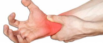
Hypotrophy of arm muscles
Hypotrophy of the muscles of the hand, forearm and shoulder develops as a secondary disease against the background of impaired innervation or blood circulation in a certain area of muscle tissue and as a primary pathology (with myopathy), when motor function is not impaired. The following causes of wasting of the muscles of the hand and other fragments of the upper limb are identified:
- Constant overexertion during heavy physical labor;
- Arthrosis of the wrist joint;
- Endocrine pathology (obesity, diabetes mellitus, acromegaly, thyroid diseases);
- Scar processes after injuries, systemic diseases (lupus erythematosus);
- Neoplasms of various origins;
- Congenital anomalies of the lower limb.
The main typical sign of the disease is the symmetry of the lesion (except for myasthenia gravis) and the slow development of the disease (except for myositis), wasting of the affected muscles and weakening of tendon reflexes with preserved sensitivity.
Hypotrophy of the arm muscles begins with the most distant (distal) parts of the upper limbs - the hands. Due to damage to the finger muscles, the hand takes on the appearance of a “monkey hand”. Tendon reflexes are completely lost. Sensitivity is observed, but sensation is retained in the affected limb. As the disease progresses, the muscles of the trunk and neck are involved in the pathological process.
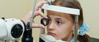
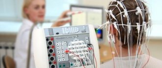
Making a diagnosis does not cause any particular difficulties for the doctors at the Yusupov Hospital due to the availability of modern equipment that allows them to perform electromyography and biopsy of the affected muscles. The patient is prescribed biochemical and general blood tests, and a urine test. The activity of muscle enzymes (mainly creatine phosphokinase) is determined in blood serum. The amount of creatine and creatinine in the urine is calculated. According to indications, the patient undergoes a computer or magnetic resonance imaging scan of the cervicothoracic spine and brain, and the level of hormones in the blood is determined.
Treatment
Neurologists at the Yusupov Hospital prescribe complex treatment to patients suffering from muscle dystrophy, aimed at eliminating the cause of the disease, influencing the mechanisms of development of the pathological process, and reducing the manifestations of the disease. To improve blood flow in peripheral vessels, angioprotectors (trental, pentoxifylline, chimes), low molecular weight dextran, and prostaglandin E preparations (vasaprostan) are used. After dilating blood vessels with no-shpa and papaverine, the supply of muscle fibers with oxygen and nutrients improves.
B vitamins (thiamine hydrochloride, pyridoxine hydrotartrate, cyanocobalamin) normalize metabolic processes and the conduction of nerve impulses. Biological stimulants stimulate the regeneration of muscle fibers and restore muscle volume: aloe, actovegin, plasmol. To restore muscle conduction, prozerin, galantamine, and armin are used.