A fracture is a violation of the integrity of a bone under the influence of an external force that exceeds the strength of the bone. With an incomplete fracture, a partial violation of the integrity of the bone occurs with the formation of a fracture, crack or holey effect of the bone tissue. When treating fractures, it is important not only to restore the integrity of the bone and its anatomical shape, but also to improve the function of the damaged parts of the body.
1
Treatment of fractures
2 Treatment of fractures
3 Treatment of fractures
Bone fractures can lead to complications such as:
- disruption of vital organs (heart, lungs and brain);
- the occurrence of paralysis as a result of injury to nerve cells by bone fragments;
- infection and the appearance of purulent inflammation in the area of the open fracture.
To avoid various complications, medical care for fractures and other injuries should be provided as quickly as possible. It is very important to transport the victim to a hospital or emergency room in a timely manner!
Causes of fractures
Bone fractures are divided into traumatic and pathological .
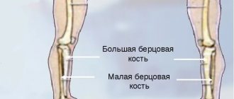
Traumatic bone fractures occur as a result of external mechanical influences. A broken leg and a broken arm most often occur when a person is hit or falls. A rib fracture, like a collarbone fracture, often occurs as a result of an unsuccessful jump and fall. A fracture of the heel bone (the so-called “skydiver’s injury”), as a rule, occurs when landing unsuccessfully on one’s feet from a height.
Others, pathological fractures , are caused by weakness and fragility of the bone itself. These include, for example, bone fractures in older women suffering from osteoporosis (a common injury in this disease is a hip fracture).
1 Treatment of fractures
2 Treatment of fractures
3 Treatment of fractures
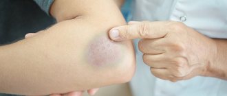
Complications
Like any disease, a bone injury can be complicated by many pathological conditions if treatment for a clavicle fracture is not started in time. Fortunately, complications from injury are quite rare because patients often seek medical attention immediately. However, possible complications due to poor-quality or untimely medical care cannot be excluded:
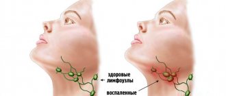
- Injury to a vascular or nerve formation. A displaced clavicle fracture can cause damage to a large vascular trunk or nerve formation, which leads to the formation of characteristic symptoms. In the case of damage to the nerve fiber, disturbances in the motor or sensory sphere in the injured area are observed as long-term consequences of the damage. The severity of neurological symptoms depends on the nature and extent of nerve damage, however, one should not forget about the likelihood of injury to nerve fibers. Damage to the vessel often leads to serious bleeding, especially if a sharp bone fragment has injured a large major vessel, resulting in serious blood loss.
- Damage to the pleura . A life-threatening condition for a person, pneumothorax, is formed when displacement during a clavicle fracture causes damage to the parietal pleura. Air enters the pleural cavity, which is accompanied by a characteristic clinical picture (shortness of breath, lack of air, lag of one of the halves of the chest when breathing). Pneumothorax requires prompt action by medical professionals and immediate treatment.
In most cases, fracture complications can be prevented thanks to competent and proper care for a clavicle fracture.
Types of fractures
Depending on the reasons that caused the fractures, different types of bone injuries can be distinguished.
Classification of bone fractures based on skin integrity:
Fractures can be closed (i.e., without deformation of the skin) and open (with damage to soft tissues and skin).
In addition, doctors distinguish between fractures with displacement of bone fragments and without displacement.
Classification of fractures by direction and shape:
- longitudinal fracture (fracture line parallel to the axis of the tubular bone);
- transverse (the fracture line is perpendicular to the axis of the bone);
- oblique (V-shaped), in which the fracture line is at an acute angle to the axis of the bone;
- helical fracture (rotation of bone fragments relative to their usual location);
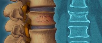
- wedge-shaped fracture (both bones are pressed into each other, most often found with a fracture of the spine);
- comminuted (the bone is broken into several parts);
- compression fracture (all fragments are small, there is no single fracture line);
- impacted fracture (with this type of compression fracture, one of the bone fragments is firmly embedded in the other).
First aid for an open fracture
Such an injury is extremely dangerous, as the wound can become infected. First aid for an open fracture begins with stopping bleeding using a pressure bandage or tourniquet if significant blood loss is observed. Antiseptic preparations are used to treat the skin around the wound. To transport the victim, it is necessary to carefully apply a splint without touching the wound.
Fracture treatment
In a hospital or other medical institution, an x-ray is taken, with the help of which the diagnosis, location, nature of the damage and the direction of displacement of bone fragments are clarified.
Then the doctor sets the bone fragments (repositions them). This is done only after pain relief. If the fracture is not clearly visible, an incision may be made in the patient's skin. The injury site is secured with plaster or other medical device.
In case of severe injuries, surgical treatment of the fracture is performed; bone fragments are secured using plates, nails and screws. The fracture site is then fixed (immobilized) to ensure proper fusion of the bones.
In some cases, bone traction is required. In this case, a steel pin is attached to the bone below the site of injury, and a weight is attached to the two ends of the pin.
It should be noted that the rate of bone healing depends on the patient’s age, type of fracture, degree of bone mineralization and the presence of concomitant diseases.
Currently, modern devices such as the Ilizarov apparatus and orthosis .
1 Treatment of fractures
2 Treatment of fractures
3 Treatment of fractures
Treatment of fractures using the Ilizarov apparatus
The Ilizarov apparatus is used to reliably connect bone fragments in open and comminuted complex fractures. The spokes passing through the bones of the damaged limbs are attached to rings, which are secured with special transition elements. If necessary, this allows you to compress or stretch certain areas of the bone.
Using this design, you can not only fix a fracture, but also influence the rate of bone fusion. In addition, the Ilizarov apparatus allows you to move with a broken leg.
The process of installing and removing the Ilizarov apparatus
Installation of the device is carried out under local or general anesthesia. Two wires are passed over the fracture through parts of the bones perpendicular to each other. And the ends of the spokes are secured to the bone using clamps. The entire time the design is worn, it is necessary to properly care for it and wipe the knitting needles with a disinfectant solution.
Removal of the Ilizarov apparatus, as a rule, is carried out in the same clinic where the installation took place, or any other medical institution in which the corresponding traumatologist works. Removal of the Ilizarov apparatus is carried out using anesthesia.
Treatment of joint and bone diseases using an orthosis
An orthosis includes several types of orthopedic devices that are used to treat joints. These can be corsets, bandages, orthopedic shoes, as well as orthopedic insoles.
Orthoses can be used in the following cases:
- fixation and unloading of the spine and joints;
- restoration of musculoskeletal function after various injuries (used to treat fractures, dislocations, sprains and bruises);
- correction of deformities of the musculoskeletal system (kyphosis, scoliosis);
- pain relief from arthritis, arthrosis, osteochondrosis, etc.;
- protection of the spine and joints during increased physical activity.
But most often, an orthosis comes to the rescue in cases where it is necessary to fix a damaged joint.
Types of orthoses
According to their purpose, orthoses can be divided into 3 large groups:
- orthoses for the joints of the lower extremities (ankle orthosis, knee orthosis, hip joint device, orthopedic shoes and insoles);
- orthoses for the joints of the upper extremities (shoulder brace (scarf or orthosis), wrist orthosis, finger braces and elbow pads);
- orthosis for the spine (postpartum and prenatal bandages, collar splints, corsets).
Orthoses come in soft , rigid , semi-rigid and splint types . Most often, the degree of rigidity determines its purpose. For example, a soft ankle orthosis (or knee orthosis) resembles a bandage that is used to prevent joint diseases.
The rigid device is somewhat similar to plaster; it is a rather complex structure made of plastic and metal inserts. Prescribed for injuries, fractures, after surgery, for dislocations, when it is necessary to immobilize the joint.
The splint is used to restore an arm or leg after surgery or injury. The splint differs from an orthosis in that it has a different design, in which there are no hinges.
1 Treatment of fractures
2 Treatment of fractures
3 Treatment of fractures
Arm fractures: what should be done and what should not be done?
The most important point is to immobilize the broken limb as quickly as possible so that a closed fracture does not become an open one. To immobilize the arms, it is customary to use splints, fixing them to the limb in a certain way. If a real tire is not at hand, you can use improvised materials - for example, a thick tree branch, plywood, boards, etc. In case of severe pain, the victim should be given a painkiller. The arm with the splint should be bent at the elbow and suspended, and in order to prevent the development of pain shock, the patient is warmed in all possible ways, from hot tea to blankets or heating pads.
If the patient has an open fracture, the main task is to stop the bleeding by applying a tourniquet above the wound surface. The time for applying a tourniquet should be no more than 30, maximum 40 minutes, otherwise necrosis of the limb may develop, since it practically does not receive blood flow. If there is no tourniquet nearby, any available means in the form of a piece of torn fabric, rope, or belt will do instead.
To prevent infection, the wound is treated with an antiseptic. It is unacceptable to pull the patient’s injured limb in an attempt to straighten the bone.
Treatment of fractures at the MedikCity clinic
If you need emergency and professional help, then contact the paid emergency room of the MedicCity clinic!
We work every day from 9.00 to 21.00. X-rays work for you in the same mode, and we perform MRIs around the clock!
Experienced traumatologists use in their work all the advanced methods of treating fractures, as well as modern, high-quality materials, in particular plastic plaster, which is lightweight, does not deform from water and is comfortable to wear.
Types of modern plastic plaster
One of the easiest is Scotchcast . The patient practically does not feel its weight, and the body breathes in it. Scotch tapes are available in various colors, which slightly “brightens up” not the easiest period in a person’s life. Among the disadvantages, it can be noted that the plaster cannot be exposed to water (it is put on with a special cotton-rag stocking, which serves as a layer between the rough material and the skin), and it can only be removed with the help of a specialist.
Softcast is a bandage made of a very flexible, elastic material, which allows it to be used for sprains and sprains. In cases of fractures, the bandage is worn together with adhesive tape. This plaster is made from fiberglass fabric impregnated with polyurethane resin and is therefore water resistant. In this case, the cast ensures the flow of air to the injured limb.
HM-cast is a synthetic mesh with large cells, made in the shape of a sleeve. It is very light, but you only need to wear it with a special synthetic stocking. This product can be used for water procedures; it is very light in weight and is available in various sizes, which makes it convenient for treating limb fractures of different locations. This cast allows X-ray penetration, allowing specialists to monitor bone healing without removing the cast.
Turbocast is the most common plastic gypsum with high strength. A cotton stocking is not worn under this cast, so you can take water procedures in the turbocast. Another plus is that this material has a working memory, so it can be heated and used repeatedly. A turbocast plaster cast looks aesthetically pleasing and is easy to use (it can be hygienically treated with a soap solution).
In our clinic you can replace the uncomfortable old plaster cast with a modern lightweight turbocast bandage. Follow our promotions! Very often there is a discount on the plaster replacement procedure.
If necessary, the doctor will quickly and professionally install an orthosis for you and remove the Ilizarov apparatus. If you need to remove an Ilizarov apparatus, you can find out the cost of the service by phone.
Improved technique
The Ladisten Orthopedics and Traumatology Clinic offers an alternative solution. Its founder, Dr. Veklich, improved the Ilizarov apparatus. The Veklich apparatus is installed in a minimally invasive way. It can be used to increase height or correct disproportion of limbs.
In case of bone injuries, surgical intervention to install the device cannot be avoided. Its design is almost identical to the Ilizarov apparatus, but instead of spokes, titanium rods with a diameter of 6 mm are used. The rod can be applied only on one side, reducing the risk of osteomyelitis significantly. It also does not come into contact with nerves and blood vessels. The entire structure is not too bulky and reliably holds damaged bone fragments.
Treatment with orthoses
ORDEKT orthoses are designed specifically for comfortable recovery after injuries to ligaments, tendons, muscle tissue, bone fractures, and rehabilitation after surgical treatment. You can learn about all the advantages of ORDEKT products made from low-temperature plastic on our website. If you have any questions, write us a message in the feedback form or call 8 800 500 8333. Our specialists will help you choose the ideal model for you.
Almost every sixth bone fracture in the human body is a fracture of the radius in a typical location. The method of treatment and rehabilitation of such injuries has been sufficiently developed. It is important to make a timely and correct diagnosis, differentiate the type of damage, and immediately begin treatment.
The article was checked by Strakhov Maxim Alekseevich - Candidate of Medical Sciences, Associate Professor of the Department of Traumatology-Orthopedics and Military Field Surgery of the Federal State Autonomous Educational Institution of Higher Education Russian National Research Medical University named after. N.I. Pirogov of the Ministry of Health of Russia, Associate Professor of the Department of Traumatology and Orthopedics of the Federal State Budgetary Institution Federal Scientific and Clinical Center of the Federal Medical and Biological Agency of Russia (Moscow).
Recovery
Physiotherapy is widely used in medicine for more active recovery.
Physiotherapeutic methods of rehabilitation after a clavicle fracture are actively used in medicine to accelerate the healing of the injury, as well as reduce pain and discomfort for the patient. In addition, physiotherapy has an anti-inflammatory, anti-edematous effect, stimulates metabolism in the area of damage and tissue regeneration.
The classification of physiotherapeutic procedures is based on the time of their implementation. Thus, there are methods of physiotherapy that are used only during the period of plaster immobilization, after removal of the plaster, and also regardless of whether the patient has a plaster cast.
During plaster immobilization, SUV irradiation in erythemal doses . At the local level, the procedure provides relaxation of muscle tissue in the damaged area, expansion of the capillary network, which stimulates blood flow in the irradiated tissue. Finally, tissue swelling is reduced, as well as the severity of pain due to a decrease in the sensitivity of pain receptors. The general effect of SUV irradiation in erythemal doses is manifested in stimulating the formation of vitamin D, which accelerates the process of callus formation. The second procedure, electrophoresis of painkillers, ensures the accumulation of painkillers in the subcutaneous fat and muscle tissue, which long-term reduce the intensity of the patient’s pain syndrome.
After the moment when a person's plaster cast is removed, it is possible to use new physiotherapeutic procedures. If the traumatologist has removed the plaster, then the callus has already formed quite well, which means that it is possible to put small loads on the bone to increase its strength. For this purpose, therapeutic massage, high-frequency magnetic therapy, amplipulse therapy, UHF therapy, ultrasound therapy, and remote shock wave therapy are used.
Therapeutic massage provides active and persistent dilation of blood vessels in the injured area. Active blood flow accelerates the transformation of formed callus into healthy functioning bone tissue. Therapeutic massage has a significant overall effect through a reflex effect on the vasomotor center located in the brain. Thanks to therapeutic massage, blood pressure in patients is normalized.
High-frequency magnetic therapy provides an analgesic effect by influencing nerve tissue. In addition, the tension in the patient's skeletal muscles decreases.
Amplipulse therapy creates an analgesic effect and leads to relaxation of muscle fibers. The physiotherapeutic method actively influences blood vessels, dilating them, which enhances tissue trophism. Thus, amplipulse therapy stimulates the reduction of swelling, the resorption of infiltrates, and also activates recovery processes in the injured area.
UHF therapy creates an active warming effect, which directly affects cellular metabolism. Thus, microcirculation processes are activated, the muscle layer of blood vessels relaxes, and the swelling of the damaged area decreases. As a general effect, UHF therapy reduces the tone of the sympathetic nervous system, increasing the tone of its parasympathetic department.
Ultrasound therapy actively stimulates blood circulation processes, as well as lymph circulation. At the local level, capillary permeability increases, which affects the intensity of wound healing.
Remote shock wave therapy activates the synthesis of special biologically active components. They provide vasodilation in the area of injury, which stimulates the processes of proliferation of cells and tissues in the injured area. Cell division ensures the direct process of repair of the bone and cartilage base.
Regardless of immobilization, electrophoresis of vasodilators, low-frequency magnetic therapy and mineral water intake can be performed. Low-frequency magnetic therapy becomes an excellent stimulating factor for reparative processes. In addition, this method has a good anti-inflammatory effect. Mineral waters restore the balance of electrolytes in the human body and provide the necessary minerals for the active healing of damaged bone and cartilage tissue.