Treatment of a spinal fracture is a complex process. Rehabilitation of a patient after an injury takes quite a long time, and sometimes a lifetime. Fractures in which the spinal cord is not damaged are divided into three degrees:
- The first is the height at which the vertebra is reduced by less than half;
- Second - the height is reduced by half;
- Third, a reduction in vertebral height by more than 50% is determined.
A spinal fracture may be accompanied by a disruption of the integrity of the spinal cord, which is responsible for transmitting impulses to peripheral nerve endings and back. When it is damaged, the connection is interrupted, and the organs and systems of the body begin to work incorrectly or shut down. A common occurrence after a spinal fracture is incomplete and complete paralysis. Diagnosis and treatment of spinal fractures using modern techniques are carried out by doctors at the Yusupov Hospital.
Traumatologists provide emergency care around the clock to victims with spinal fractures of any complexity. The Yusupov Hospital has created conditions for comfortable treatment of patients using conservative methods. Traumatologists are fluent in the techniques of all surgical interventions. In cases of spinal cord injury, patients are consulted by a neurosurgeon. Rehabilitation clinic specialists use innovative methods of rehabilitation therapy. Rehabilitation measures begin from the first day of the patient’s admission to the hospital. After discharge from the hospital, the patient receives individual recommendations for rehabilitation treatment developed by rehabilitation clinic specialists.
Why does a vertebra break?
Statistics show that older people with a history of osteoporosis are most susceptible to trauma to the vertebral bodies. Osteoporosis is a severe bone disease that leads to thinning and decreased bone density, making them fragile and vulnerable to even minor physical stress. For a patient with this disease in severe form, sometimes it is enough just to sneeze or go down a step to cause a fracture and flattening of the weakest vertebra. Women, especially mature and retired women, suffer from osteoporosis more often than men, and therefore the predisposition of the female audience to CP is higher.
But a compression fracture of the spine, rehabilitation and treatment for which is a mandatory measure, is not only the lot of pensioners. It may well occur in young patients, including children, who have a completely strong and healthy spinal column. Typically, a violation of the integrity of the vertebrae occurs due to intense external mechanical impact on the back area, the force of which exceeded the physiological potential of the strength of the bone structure, in other words, due to severe injuries of various types (sports, household, etc.). Let us summarize and add what common unfavorable factors cause compression-type defects of the vertebral bodies, these include:
- falling from height onto legs, back, buttocks;
- a powerful blow that came to the ridge;
- lifting very heavy objects with a jerk;
- squats with a barbell in bodybuilding;
- all kinds of sports emergencies, in particular falls from high apparatus;
- a heavy object falling from above onto your back;
- a sharp blow to the head on the bottom of a reservoir when diving;
- traffic accident;
- diseases of bone tissue, especially osteoporosis;
- tuberculous and tumor neoplasms pressing on the spine.
Any of the 33 elements included in the structure of the main part of the human axial skeleton can be damaged at any level. According to clinical observations, the lumbar or thoracic region is most often affected, but keep in mind that with a compression fracture of the spine, rehabilitation begins only after the completion of the main treatment process.
Therapeutic and rehabilitation measures should be planned exclusively by a doctor, strictly on an individual basis. The etiology and scale of destruction, the number of injured bodies, the degree of their compression (reduction in height), post-traumatic complications, the personal characteristics of the patient’s body and much more - all this is taken into account when drawing up a treatment and rehabilitation program.
Anatomy of the spine
The spine is one of the main organs of the human body, responsible for maintaining it in an upright position. It is formed by 33 vertebrae, between which are intervertebral discs. They are responsible for cushioning bone fragments under static and dynamic loads.
From the back of each vertebra there are arches shaped like a semicircle and forming the spinal canal. It is in it that one of the most important organs of the human body is located - the spinal cord.
The arches have 7 processes:
- 4 articular;
- 2 transverse;
- spinous.
The articular processes of adjacent vertebrae are connected in pairs to each other, thereby forming intervertebral joints. They are entrusted with the function of protection under heavy loads. Thus, openings are formed between 2 adjacent vertebrae, called foraminal. The nerve roots extending from the spinal cord pass through them. Additionally, the bodies, processes and arches are connected by ligaments that provide stability and strength to the entire spine.
Since the spinal cord runs a few millimeters from the spinal column, and its joints are densely penetrated by nerve fibers, spinal fractures are always dangerous. They require not only lengthy and sometimes complex surgical treatment, but also long-term rehabilitation. Moreover, sometimes, despite all the efforts of doctors, such injuries still lead to disability and death.
What is the danger of pathology?
It is important to understand that any pathology that affects a vital anatomical formation, with an illiterate approach or lack of appropriate therapy, can result in dire consequences for a person. Timely treatment and rehabilitation after a compression fracture of the lumbar, thoracic or cervical spine will help to avoid all negative complications. We will talk about them further.
The spinal system serves as a container for the most important component of the central nervous system - the spinal cord. Thanks to the spinal substance, musculoskeletal functions are ensured, in other words, the overall ability to move and maintain a stable body position.
The spinal canal, its nerve roots, arterial and venous vessels affected by pathological pathogenesis, which is more often observed with compression-comminuted fractures, with traumatic excesses with the formation of wedge-shaped deformities and instability of elements, is not a joke. The degree of damage to the spinal cord can vary - from concussions, compression and contusions, to its complete rupture.
The worst thing that a neglected clinic can threaten is partial or complete paralysis of the body.
In addition, without urgent medical care and rehabilitation after compression fractures in the spine, secondary degenerative and neurological diseases develop, which will be very difficult to combat. These include:
- segmental instability in the damaged area;
- osteochondrosis and intervertebral hernia;
- kyphosis of the spinal column, or non-physiological curvature;
- radiculopathy, paresis and other neurogenic disorders;
- spinal canal stenosis;
- vascular malformations and development of hematoma in the epidural space;
- chronic pain, paresthesia;
- persistent movement disorders;
- dysfunction of the pelvic organs.
Therefore, do not risk feeling discomfort and pain in your back, especially if they appeared immediately or after a certain time after an injury or unsuccessful movement. You can’t hesitate here, believing that nothing serious has happened. It is better to be examined once and make sure that everything is really in order, than to ignore a dangerous problem and pay for your indifference at the cost of your own ability to work.
Forecasts
In most cases, simple compression injuries can be successfully treated. The complete recovery process takes no more than 6 months. The patient may experience moderate back pain throughout the recovery period.
After completion of treatment, the following phenomena are observed:
- Persons who have suffered a compression injury, which is accompanied by neurological symptoms, may at times experience minor pain. This is due to the fact that the deformed cartilage affects the spinal cord and root;
- with the pathology in question accompanied by infections of bone tissue, the prognosis is also overwhelmingly favorable. Persons who have suffered a fracture due to purulent osteomyelitis can count on a high chance of recovery provided timely treatment;
- If the fracture occurred against the background of puncture tuberculosis, unfortunately, the prognosis is unfavorable. Such patients become disabled.
As for a fracture caused by latent osteoporosis, after proper therapy the patient’s condition stabilizes within 2 months. Complete recovery is possible only if the pathology is detected in a timely manner and therapy is started on time.
Symptoms and medical care
Let's start with the symptoms that are characteristic of this classification of traumatic lesions. The main symptoms include:
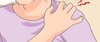
- acute local pain, it can radiate to the arms and legs;
- pain when palpating the diseased part;
- local swelling and hyperthermia of soft tissues,
- visible traces of injury on the skin (redness, abrasion, bruise, etc.);
- loss of sensitivity in the limbs, feeling of muscle weakness;
- asymmetry of the vertebral axis and visible deformities;
- reduction in range of motion or complete immobility of the problem area;
- difficulty breathing;
- girdle pain in the abdominal cavity;
- general malaise, sometimes fever.
In the early period, emergency drug therapy is recommended in parallel with an accurate determination of the nature and severity of the injury to determine the advisability of surgical treatment. Based on the results of a thorough diagnosis, the doctor decides whether to perform surgery or treat conservatively. Upon completion of the treatment cycle, rehabilitation always follows at a certain stage; after a fracture of the thoracic spine, etc., it is an extremely necessary measure.
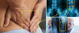
Diagnostics
In the Yusupov Hospital, if a spinal fracture is suspected, a comprehensive examination of the patient is carried out. It includes the following studies:
- Computed tomography – for a detailed study of the structure of all damaged vertebrae;
- X-ray of the spine - to identify a damaged vertebra;
- Myelography – makes it possible to assess the general condition of the spinal cord in the area of injury;
- Magnetic resonance imaging – determines the presence of soft tissue damage.
After receiving the research results, the attending physician analyzes them and makes a final diagnosis.
Make an appointment
Non-surgical treatment
If a form of damage is diagnosed in which it is possible to do without surgery, they are limited to non-surgical reduction of the fragments. Next, favorable conditions are created for bone consolidation by installing a fixing device (plaster cast, semi-corset, bandage), long-term bed rest for several weeks and sometimes months, as well as periodic use of hardware traction. Most of the time, a person is supposed to lie on his back, the mattress is replaced by a hard shield. Usually, standing and walking are allowed after about 2 months, and sitting even later, after about 4 months.
Additionally, calcium supplements are prescribed to stimulate bone repair processes, muscle relaxants and NSAIDs to reduce spasms and pain. Only after the final fusion and strengthening of the bone (after 2-3 months) is the patient allowed to become relatively normal; rehabilitation for a spinal fracture begins as soon as possible after fixing the body in an advantageous position.
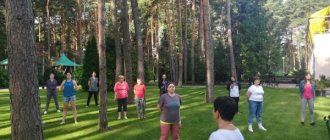
According to the canons of orthopedics, rehabilitation after a fracture of the lumbar spine, as well as other locations, is first based on performing simple exercise therapy exercises in a lying position. Early exercise therapy is necessary to prevent muscle atrophy and circulatory disorders. In addition, at the initial stage, great importance is paid to breathing exercises to prevent congestion in the lungs.
Somewhat later, more active training is added to the rehabilitation program after a fracture of the cervical spine, lumbar or thoracic region of a compression nature. Approximately halfway through the course, physiotherapy, light massage and acupuncture are prescribed. In most cases, the recovery of a victim with unsevere forms of deformities that do not require surgery is based on this principle.
Vertebroplasty
This procedure is most useful for reducing pain. It also strengthens damaged bone, allowing patients to recover faster. Using a needle, under X-ray control, a cementing substance, polymethyl methacrylate (PMMA), is injected into the area of the vertebral fracture. The cement hardens within 15 minutes. This fixes the bone and prevents further destruction and reduces pain in more than 80% of patients.
Surgical techniques
Let's say a specialist has diagnosed a lumbar, cervical or thoracic spinal fracture, rehabilitation is described at the end, saying that there will be a surgical treatment. What surgical techniques are used to correct the shape, height and anatomical integrity of the vertebra? Today, in orthopedics and traumatology, unique gentle methods are used that involve closed surgery, that is, during a particular procedure, the surgeon does not make large incisions and does not expose the damaged element at all. Such loyal stabilizing procedures include vertebroplasty or kyphoplasty.
- Vertebroplasty involves injecting bone cement into the vertebral body through a syringe. First, local anesthesia is administered, and after the analgesic effect occurs, a special guide needle is inserted into the cavity of the defective object. A syringe filled with a semi-liquid mixture based on acrylates is connected to the conductor wire. Next, the specialist squeezes a unique solution into the vertebra, where it fills all the voids and cracks, replenishing the height of the body. After 10 minutes, the cementoplastic mass “sets”, turning into a hard material similar to bone, only much stronger. The advantage of the procedure is that the element that was corrected with cement also acquires “immunity” to a similar type of destruction. After such a microinvasive session, there is no need to wait painfully for the spinal fracture to heal; treatment and rehabilitation are carried out in a short time.
- Kyphoplasty is a procedure similar to the previous tactics, but its principle is based on placing a special balloon through a probe inside the vertebra, after which it is inflated. Inflating the balloon helps to very well align the geometry and height of the distorted body. Next, cement is injected into the intravertebral structures in the same way as in vertebroplasty. Thus, this session copes equally successfully with the reconstruction of the destroyed area. Plus, it not only raises a certain link to its normal level and restores its strength resource, but also prevents its destruction in the future, and also corrects abnormal curvatures of the column.
Both techniques are especially widely used when a compression fracture of the thoracic spine is diagnosed of osteoporotic etiology; rehabilitation, by the way, is very easy to tolerate and does not require lying in bed for months. It is enough to lie down after the manipulations for several hours, a maximum of a day, and calmly go home on your own feet. It is worth noting that these technologies are also highly effective in restoring broken vertebrae located at the level from the coccyx to the ribs, as well as in the neck area.
The two technologies are of particular value for older people, since thanks to the use of a high-tech cement composition, signs of osteoporosis in the corrected part are completely eliminated. Moreover, they involve a very gentle intervention with minimal risks of side effects, and also do not require the use of aggressive types of anesthesia.
Types of fractures
Injuries can be uncomplicated or complicated when the spinal cord is affected. Injury may be accompanied by deformation of discs and nerve endings. Depending on the number of vertebrae affected, isolated and multiple fractures are distinguished.
If all parts of the vertebra are simultaneously affected and displacement is possible, then this is an unstable fracture. When one section is damaged, it is a stable fracture because the spinal column remains stable.
Compression injuries are characterized by flattening of the vertebrae—the vertebral body becomes smaller. Depending on the compression, there are several degrees of complexity.
Physical rehabilitation
Rehabilitation for a spinal fracture is the next stage of the treatment process of paramount importance. After providing first aid, even if it involved minimally invasive cementoplasty, the patient must strictly observe a specific physical regimen for a certain period, attend physiotherapeutic sessions, undergo therapeutic exercises, etc. It cannot be otherwise if you have suffered such a serious injury.
Complicated injuries and those provoked by osteoporotic processes require a very careful selection of recovery methods. Therefore, it would be more advisable to take care in advance where to undergo rehabilitation after a spinal fracture; today there is no particular shortage of good specialized institutions.
Prescribing any restorative tactics to yourself is strictly prohibited! Rehabilitation of patients with a spinal fracture is being developed exclusively by the treating doctor together with a rehabilitation specialist. Our recommendations are given in general form and are for informational purposes only, so before using them, consult with a specialist about the possibility of using them for your specific medical problem.
We will talk about that important period when complete bone consolidation has already been achieved, since some patients at this stage stop working on their spine, but in vain. It is after 2-3 months that all emphasis should be directed toward developing and strengthening the musculoskeletal corset, which has become considerably weakened after prolonged immobilization and long-term unloading. In addition, thanks to the unique physical organization, the work of the gastrointestinal tract, cardiovascular system, respiratory system, reproductive, urinary system and many other important components of the body, which are entirely dependent on the health of the spine, will be stabilized.
Medicines
Most patients in the acute stage of fractures receive analgesics. With pain control, the patient can get up and move around easily and avoid problems caused by immobilization.
Corseting
Most patients are prescribed the use of a rigid corset, which is selected individually. A corset can dramatically reduce the range of motion and fix a broken vertebra. Patients who wear a special brace are allowed to move, but strenuous activities such as lifting and bending are significantly limited.
Motor activity control
. Bed rest is usually recommended for several days, since prolonged immobilization leads to weakening of the muscular corset, and in the presence of osteoporosis, to further bone loss and progression of osteoporosis.
Correction of osteoporosis
. Bone-strengthening drugs such as bisphosphonates (Actonel, Boniva, and Fosamax) help stabilize or reverse bone loss. This is a critical part of the treatment and helps prevent further fractures when axial loads are placed on the vertebrae.
Thoracic region
The main tasks of rehabilitation for compression fractures of the thoracic spine are solved through exercise therapy. By performing certain exercises, physiologically normal mobility is ensured not only of all central segments, but also of the entire back, muscle tone is restored, pain in the scapulothoracic zone is eliminated, and posture is normalized. Here are examples of some useful exercises. The average repetition frequency is 10 times.
- Take a standing position. Place your feet hip-width apart, hands on your waist. As you inhale, bend back, slightly raising your shoulders and pushing them back; as you exhale, round your back, pointing your shoulders forward.
- Now we complicate the task a little by doing the same thing, but with the body turning to the right/left: we turn to one side and spread the shoulder girdle, return to the starting position and round the back, turning the shoulders forward.
- Let's move on to a new task. The position is the same, only the arms are extended along the body. Perform lateral bends: bend to one side, the arm of the bending part slides down the side of the limb, while simultaneously pulling the opposite arm towards the armpit. Similarly, we tilt the body in the other direction. Each side – 10 repetitions.
- Get on all fours with your straightened upper limbs resting on your palms. As you inhale, we bend your back, your pelvis moves back, your head drops down. As you exhale, arch your back upward (like a cat), smoothly throwing back your head, while tightening your stomach.
- We partially change the previous pose - the support in the hands now falls on the forearm area. We lift our right hand off the floor, take hold of the shoulder, slightly turn our chest to the right and make 5 springy movements with this glenohumeral part with the emphasis “up”. We work in a similar way with the left side.
- Lie on the floor with your stomach down, resting on your forearms, chest open. When the thoracic fracture has healed, the “sphinx” pose has a beneficial effect on this line of the spine; ask your orthopedist whether your rehabilitation can include this type of warm-up. Method of execution: pull your head up towards the ceiling (feel your ribs straightening), stay in this position for a few seconds, then relax, lowering your head to the floor.
- Lie on your side (head on the floor), bend your knees (knees together), stretch both arms forward in front of you, clasping your palms. Next, with a sliding movement of the upper hand, as if opening, we pass it along the inner surface of the adjacent arm, then along the chest, and finally place it on the floor. At the moment of the so-called “opening”, turn your head in the direction of movement of the limb, as a result the shoulder blades lie on the plane. The pelvis and legs should remain in position at all times. p. Then “get together” again and repeat the task. After five times, turn to the other side and repeat the exercise by analogy.
- Lumbar part
Rehabilitation for compression fractures of the lumbar spine has similar goals. This is to restore the full functionality of the musculoskeletal system, make it resilient and strong, and balance your posture. What kind of exercise do physical therapy instructors and orthopedic doctors suggest their clients do?
- Lying on your back, perform simultaneous flexion of the knee and hip joints. Thus, the lumbosacral region and abdominal muscles are excellently trained.
- We do not change the IP. Raise your legs straight up. Extending the limbs to the sides followed by bringing them together. When your legs come together, you need to cross them slightly.
- The well-known exercise called “bicycle” brings considerable benefits. We won’t dwell on the technique; we think everyone knows how to simulate riding a bicycle in a recumbent position.
- Roll over onto your stomach, spread your arms to the sides. Lift your chest off the surface along with your arms, hold in this position for as long as your physical fitness allows, then lower and relax. Note that the feet do not come off the floor. If it is more convenient for someone, you can stretch your arms in front of you.
- The position remains the same, but your legs need to be spread slightly and your arms should be extended straight in front of you. Lifting the sternal complex (along with the arms) and legs from the support, lift them simultaneously with a bend in the back. Fix the pose and stay in it for as long as possible. Lower yourself, rest a bit, then repeat.
- Get on your knees. The pelvis and back are strictly vertical, hands on the belt. Walk on your knees in a straight line forward, then backward. We complicate the task: we begin to walk in a circle, first clockwise, and then in the opposite direction.
Physiotherapy for recovery
Physiotherapy is prescribed to the patient already at the second stage of treatment, when doctors allow him to gradually restore motor functions. Such procedures help restore muscles, bone tissue and nerves. During rehabilitation, the following methods are used:
- UHF is used to reduce pain, relieve swelling and improve blood circulation.
- Electrophoresis – helps to introduce medications into the body for better effect.
- Myostimulation is muscle contraction using electrical current pulses. Helps relieve stress, pain, and muscle tension.
- Paraffin-ozokerite sessions are the application of different types of energy to a diseased area of the body (in this case, thermal energy). The method helps improve muscle condition.
- Kinesiotherapy using the Exart apparatus is a treatment method consisting of physical exercises necessary to strengthen muscle groups. Performing these exercises on a special device helps get rid of joint pain and normalizes blood circulation.
- Carboxytherapy is a treatment with carbon dioxide, one of the new methods of therapy, very effective for degenerative diseases of the joints and spine.
- Ural rays are used to destroy harmful microflora.
- Balneotherapy – treatment with mineral waters improves blood circulation and reduces muscle tension.
- Magnetic therapy – relieves pain and speeds up recovery.
Cervical area
After removing the plaster, rehabilitation after a fracture of the cervical spine involves restoring the mobility of the corresponding segment, strengthening the muscular-ligamentous apparatus of the neck and upper shoulder girdle. At the same time, coordination abilities are improved, and complete adaptation to social and living conditions occurs.
Surprisingly, all of the above methods are optimally suited after such a spinal-motor body injury as a fracture of the cervical spine; rehabilitation with the inclusion of these techniques in the exercise therapy complex, naturally, must be approved by a specialist. As for the fundamental exercises, they are outlined below.
- First you need to warm up the glenohumeral part, which will increase blood flow in the neck. To do this, take a vertical position, place your hands on your shoulders. Perform calm rotational movements in the shoulder joints - 10 times back, and then the same amount forward. When finished, lower your hands and shake them.
- I. p. is the same as in the first exercise. No. 1. Now spread your upper limbs in different directions, straightening them, and place them on your shoulders again. Make sure that at the moment when you spread your arms, they remain on the same horizontal plane with your shoulders, that is, do not lift them up or lower them. After 10-15 repetitions, lower the limbs that worked hard and shake them a little, and at the same time rest.
- Cross your hands with your fingers, place them on your forehead (with the inside of your palms facing the frontal area). Apply resistance to your head with your upper limbs, continuously pressing your forehead on your palms for 5 seconds. Simply put, imagine that you want to lower your head, but an obstacle prevents you from performing this action. Please note that the head does not drop anywhere, but remains in place, but you feel tension in the back of the neck. On the count of five, relax and remove your hands from your forehead. We do 5-7 approaches.
- The next method of warming up is based on the same principle, but we place the “lock” on the back of the head, pressing the head backwards. Make sure that the spine does not bend and the head does not fall back. Work with this type of exercise, observing the time and frequency of sets according to method No. 3.
- Both for stretching, balance, strength of the neck muscles, and for a beneficial effect on the respiratory center and heart function, shoulder abduction is used. The person stands with his arms along his body. In practice, it looks like this: we tense the shoulder muscles and turn our shoulders forward as if you want to close them, then we open our shoulders, straighten up and try to bring our shoulder blades towards each other.
- We save the original position. Now simultaneously move your right hand along the side of the body to the armpit and turn your head to the right, while the shoulder rises and touches the chin. Now we will carry out an identical manipulation with the reverse side.
Such fruitful rehabilitation after a fracture of the cervical spine, or rather its biomechanics, will lead to an amazing state. Of course, training should be implemented systematically, and not just during this difficult period, but preferably throughout life.