There are contraindications. Specialist consultation is required.
Causes of occurrence Stages of development of pathology Signs Symptoms of degeneration into cancer Diagnostics Methods of surgical treatment
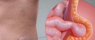
When talking about this disease, we mean all benign neoplasms, different in structure:
- nodes formed from glandular cells (nodular cyst of the thyroid gland);
- Colloid cysts are fluid-filled cavity formations.
If the thyroid cyst has dense inclusions and a complicated structure, then special studies (for example, ultrasound), tests and biopsy are needed, because such a condition may be a sign of malignant degeneration.
General information
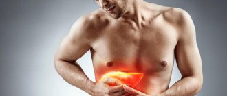
The number of patients with thyroid pathology increases annually and nodular formations occupy a leading place. Nodular diseases of the gland include cysts , colloid nodes , adenomas and malignant formations .
Patients with cysts make up a large group. Thyroid cyst, what is it? This is a cavity formation filled with liquid contents of a benign nature. Cysts are formed as a result of micro-effusions, dystrophy or hyperplasia of the main structural unit of the organ - the follicle. Typically, cysts do not affect the function of the gland and are asymptomatic for a long time. The course of cysts is benign and malignant cysts are very rare. Thyroid cysts are more common in women.
Simple formations are observed over time; when the size increases and complaints appear, the cyst is emptied during a biopsy . Most cysts in women recur—refill with fluid after aspiration. In this case, sclerotherapy is used. The ICD-10 code for “thyroid cyst” is D34 - this code combines all benign formations of the gland (cysts and nodes).
Publications in the media
Thyroid cancer accounts for 90% of all malignant tumors of this organ. Thyroid cancer is detected at autopsy in 5% of patients with no indication of thyroid disease. However, death from thyroid cancer is rare, which is explained by the characteristics of thyroid cancer: usually the tumor grows slowly, does not cause functional impairment, and rarely metastasizes. Incidence: 5.6 per 100,000 population in 2001.
The main predisposing factors are considered to be: long-term stimulation of gland tissue due to increased levels of TSH; ionizing radiation, especially at a young age; the presence of autoimmune processes.
• Heredity and thyroid cancer. Some thyroid carcinomas are hereditary, for example •• Papillary cancer (*188550, D10S170 gene mutation, 10q11–q12, Â) •• Follicular cancer (188470, Â) •• Medullary cancer (#155240, RET oncogene mutation, 10q11. 2, В). • Radiation exposure •• X-ray irradiation of the head and neck with therapeutic doses increases the incidence of thyroid cancer by 5-10 times. Radiation is carried out for various diseases (for example, enlargement of the thymus gland in a child, congenital hemangiomas of the head and neck, Hodgken's disease) •• The latent period between irradiation and the appearance of a tumor depends on the age at which the patient received radiation therapy ••• In those exposed to radiation in childhood age, the tumor was observed after 10–12 years ••• In those irradiated in adolescence, the tumor was observed after 20–25 years ••• If the gland was irradiated in an adult, the latent period before tumor formation is about 30 years.
Classification . The most common histological types of thyroid cancer are: papillary (79.9%), follicular (14.2%), medullary (3.7%), Hurthl cells (2.7%), undifferentiated (anaplastic - 1, 6%).
• Papillary cancer • Characteristics •• Characterized by slow tumor growth. Metastases in regional lymph nodes - in 50% of patients. Hematogenous metastases - less than 5% •• Tumor size is very variable: from hidden (less than 1.5 cm in diameter) to significant (affects one or both lobes) •• In 40% of patients, the tumor is multifocal in nature •• Tumors are well demarcated or are poorly delimited and grow into adjacent tissues • Prognosis •• Most favorable for hidden and well-encapsulated primary lesions localized deep in the parenchyma of the gland. In these cases, the 20-year survival rate of patients exceeds 90% •• The prognosis is unfavorable in the absence of a capsule and germination into surrounding tissues. 20-year survival rate less than 50% •• Poor prognosis also in patients over 40 years of age.
• Follicular cancer is often reported in areas where iodine deficiency is endemic •• Affects women 2 times more often •• The likelihood of the disease increases over the age of 40 years • Characteristics •• The tumor histologically resembles normal thyroid tissue, often functions as an endocrine gland, capturing iodine according to the TSH-dependent type •• The tumor grows slowly and is usually unifocal (represented by one node). Metastasizes by hematogenous route. It rarely affects the lymph nodes (the exception is tumors that grow into surrounding tissues, including the parathyroid glands) •• Sometimes cylindrical cells characteristic of papillary carcinoma are found in the tissues of follicular cancer. In such cases, the biological features of the tumor are similar to papillary cancer • Prognosis •• Follicular cancer is more malignant than papillary cancer; this tumor often metastasizes to the bones, lungs and liver. 10-year survival rate - 50% •• In the absence of metastases, the prognosis is good: 20-year survival rate >80% •• With tumor dissemination, 20-year survival rate after surgery <20%.
• Medullary carcinoma • General information •• Medullary carcinoma arises from the parafollicular cells (C cells) of the thyroid gland •• Most often occurs sporadically, but may be hereditary (20%). The sporadic form usually occurs as a single lesion. Hereditary form - an independent disease or a component of familial polyendocrine adenomatosis type II (Sipple syndrome - a combination of medullary carcinoma of the thyroid gland and pheochromocytoma) • Local (lymphatic vessels) and distant (hematogenous) spread is observed more often than with follicular carcinoma • Medullary carcinoma has a hyalinized stroma and stains like amyloid. One type of tumor is characterized by aggressive, rapid growth, rapid spread and early metastases; the other is slow growth and progression despite metastases • The tumor often produces calcitonin, less often other hormones • The prognosis is worse than for papillary or follicular carcinomas and depends on the stage of the tumor at initial diagnosis •• For stage 1 tumors, 20-year survival is 50% •• At stage 2, less than 10% of patients live longer than 20 years •• Death usually occurs from metastases to vital organs •• Familial polyendocrine adenomatosis can be completely cured by total thyroidectomy if diagnosis and treatment are carried out before the appearance of clinical signs of the tumor.
• Anaplastic carcinoma • General information •• Anaplastic thyroid carcinoma accounts for less than 10% of all thyroid tumors •• Affects patients over 50 years of age • Tumors usually arise from pre-existing well-differentiated thyroid tumors (eg, follicular) • Tumors are extremely malignant: rapidly grow into neighboring organs (trachea, esophagus) and early metastasize through lymphogenous and hematogenous routes. These tumors are usually inoperable at the time of discovery • Prognosis •• Fatal outcome occurs within a few months (regardless of treatment methods) •• If treatment is successful, a diagnostic error should be suspected (eg, there was lymphoma rather than anaplastic small cell carcinoma).
• Lymphosarcoma (less than 1% of all thyroid tumors) affects mainly women over 50 years of age • • Morphologically, tumors consist of small cells, so they are difficult to distinguish from anaplastic small cell carcinoma using conventional histological methods. Differentiation is possible with electron microscopy • The tumor can either primarily arise in the thyroid gland, or be part of a generalization of the lymphosarcomatous process • • Local tumor responds well to radiation therapy. When lymphosarcoma generalizes, systematic use of chemotherapy drugs is necessary • The prognosis depends on the cell type of the tumor and the nature of the lesion.
Clinical picture. The main symptom is the presence of a nodule in the thyroid gland. In some cases, the tumor causes hoarseness, symptoms of tracheal and esophageal compression (eg, dyspnea, dysphagia), or pain. • Age of the patient •• In children, malignant nodes are observed in 50% of cases •• Nodes that occur in a pregnant woman are usually benign •• In people over 40 years of age, the frequency of registered cancer nodes increases by 10% in each subsequent decade •• Benign nodes and thyroid cancer glands are more often observed in women •• Malignancy of the node occurs more often in men. • Features of nodular formation •• Consistency ••• Malignant tumors are characterized by dense nodes, but sometimes cancer degenerates into cysts and becomes soft ••• Soft nodes are often benign; long-term benign adenomatous hyperplasia can be combined with calcification of the node •• Infiltrative growth of the node into the surrounding tissue of the gland or adjacent structures (trachea, muscles) suggests malignancy. Sometimes thyroid cancer does not have signs of infiltrative growth and looks like a benign node •• The probability of malignancy with single nodes is 20%, with multiple nodes - 40% •• Signs of growth. If nodes suddenly appear or grow unexpectedly quickly, a malignant tumor should be suspected. Hemorrhage into a pre-existing node (eg, adenomatous hyperplasia) will also cause it to suddenly enlarge, but is almost always accompanied by pain. • Enlarged lymph nodes on the affected side suggest malignancy. In children, more than 50% of cases are first diagnosed due to enlarged cervical lymph nodes. • Condition of the vocal cords •• Paralysis of the vocal cord on the side of the node is always a sign of cancer that has infiltrated the recurrent laryngeal nerve •• Since vocal cord paralysis can occur without impairment of phonation, the glottis should be examined by direct laryngoscopy •• The examination should be repeated after surgery if hoarseness occurs .
Diagnosis • Testing the functions of the thyroid gland when cancer is suspected is of little value. Most malignant tumors of the gland do not have hormonal activity, as do nodes with adenomatous hyperplasia. Less than 1% of thyroid tumors are hormonally active. • Antibodies titer to thyroid tissue is increased in Hashimoto's thyroiditis. However, thyroid cancer can be combined with thyroiditis, so the detection of antithyroid antibodies does not exclude an oncological diagnosis. • Thyrocalcitonin content is increased in patients with medullary thyroid cancer. • Radioisotope examination of the gland is performed using either radioactive iodine or 99mTc •• Hot and cold nodes. Zones of isotope accumulation in normal gland tissue on a scanogram are called hot spots; nodes that have not accumulated the isotope are cold ••• Approximately 20% of cold nodes are tumors. About 40% of tumors can accumulate the isotope ••• Radioisotope scanning does not allow differentiating benign cold nodes from malignant ones •• Isotopes 123I and 125I give less radiation exposure than 131I because they have a shorter half-life. They have no advantages over 131I in the differentiation of tumors and benign nodes •• The thyroid gland is capable of accumulating 99mTc, but (unlike radioactive iodine) does not include it in the produced hormones ••• Nodules that are cold in relation to radioactive iodine will remain cold relative to 99mTc ••• Tumors can take up 99mTc due to abundant vascularization. In this case, hot nodes will appear on the scanogram ••• 99mTc gives less radiation exposure compared to 131I, but its use does not facilitate the differential diagnosis of malignant and benign lesions. • Ultrasound •• Gives an idea of the size, shape of the thyroid gland and the presence of nodes in its parenchyma. It is possible to identify nodes either as cysts, or as solid, or as complex formations (a combination of solid and cystic components) •• Able to identify simple cysts, which rarely turn out to be tumors, but with solid and complex nodes it does not allow to differentiate benign and malignant formations •• Allows to identify thyroid nodes that are not detected by palpation, and perform a targeted puncture biopsy of the node. • Puncture (aspiration) biopsy ••• The method allows you to obtain material for cytological studies; study individual cells and their clusters ••• The method is quite accurate and specific in the diagnosis of certain lesions of the thyroid gland, therefore, during operations for nodular goiter, urgent intraoperative histological examination is necessary. Does not cause complications. However, cytological puncture is informative only in highly specialized oncology centers •• Trephine biopsy ••• With a special needle (for example, Vim-Silverman or Tru-Cut), a column of thyroid tissue is obtained and its histological examination is carried out ••• The method allows you to most accurately determine the nature tumors, however, due to the large size of the needle, it is inconvenient to biopsy small nodes; relatively high incidence of complications (bleeding) •• Core needle biopsy ••• Aspirate the node tissue through a tube attached to the needle ••• Fewer complications than with trephine biopsy.
TNM classification is applicable only for cancer ••• T1 - tumor up to 1 cm in greatest dimension, limited to thyroid tissue ••• T2 - tumor more than 1 and less than 4 cm in greatest dimension, limited to thyroid tissue ••• T3 - tumor more than 4 cm in greatest dimension, limited to the tissue of the thyroid gland ••• T4 - a tumor of any size, spreading beyond the capsule of the thyroid gland ••• N1 - there is damage to regional lymph nodes by metastases ••• M1 - there are distant metastases (except peritoneal).
Grouping into stages depends on the age and histological structure of the tumor • In the case of undifferentiated cancer, all tumors are classified as stage IV • Medullary cancer • Stage I: T1N0M0 • Stage II: T2–4N0M0 • Stage III: T0–4N1M0 • Stage IV: T0– 4N0–1M1. • Follicular and papillary cancer before 45 years of age •• Stage I: T1–4N0–1M0 • Stage II: T0–4N0–1M1. • Follicular and papillary cancer over 45 years of age •• Stage I: T1N0M0 • Stage II: T2–3N0M0 • Stage III ••• T4N0M0 ••• T1–4N1M0 • Stage IV: T0–4N0–1M1.
Treatment. The main method of treatment is surgical. All operations are performed extrafascially. Preoperative radiation therapy is indicated for patients with medullary or undifferentiated thyroid cancer, less often for locally advanced, well-differentiated tumors •• The extent of intervention is determined by the histological type of tumor, its aggressiveness and prevalence. • For anaplastic carcinoma, treatment is mainly palliative. Surgery is used to relieve obstruction, and chemotherapy can delay death. • For papillary, follicular and medullary carcinomas, a combination of surgery, thyroid hormone therapy and radioactive iodine is usually used. With a single node limited to one lobe, the optimal method is complete removal of the lobe and isthmus of the thyroid gland along with the anterior part of the opposite lobe •• Urgent histological examination of the removed node is necessary (before completion of the operation) ••• In some cases, the diagnosis of papillary or follicular cancer is established only for permanent medications. In this case, the extent of reoperation depends on the biological aggressiveness of the tumor ••• If a well-differentiated tumor is limited to one lobe (without invasion of surrounding tissues), the affected lobe, isthmus should be completely removed and the opposite lobe should be subtotally removed ••• If the tumor spreads to surrounding tissues or affects both lobes, total thyroidectomy is indicated. Among patients with well-differentiated thyroid cancer, a low-risk group is distinguished: women under 50 years of age and men under 40 years of age with papillary cancer. Even with significant damage to both lobes, equally good results were obtained with both total and subtotal thyroidectomy ••• Prophylactic removal of intact lymph nodes is not indicated •• During the operation, the parathyroid glands and recurrent laryngeal nerves should be isolated. If the blood supply to the parathyroid gland was disrupted during thyroidectomy, it should be reimplanted into the skeletal muscle. • Complications (especially hypoparathyroidism) after total thyroidectomy are much more common than after subtotal thyroidectomy.
• In non-radical operations, after surgery for medullary cancer, postoperative radiation therapy is performed. • Treatment with radioactive iodine. Follicular carcinomas often accumulate radioactive iodine (in many cases of papillary cancer some follicular elements are found) •• Radioisotope scanning with 131I after surgical removal of normal thyroid tissue can identify functioning metastases, which can be suppressed with 131I after thyroidectomy.
• Suppressive therapy. Many thyroid cancers grow faster when TSH is stimulated, so TSH production is suppressed with the highest possible (but not hyperthyroid-inducing) dose of levothyroxine sodium.
Diet . Iodine deficiency should be avoided (iodized salt, seaweed).
ICD-10 • C73 Malignant neoplasm of the thyroid gland • D09.3 Carcinoma in situ of the thyroid and other endocrine glands • D34 Benign neoplasm of the thyroid gland • D44 Neoplasm of uncertain or unknown nature of the thyroid gland
Application. Recurrent laryngeal nerve palsy is damage to the recurrent laryngeal nerve with the development of paralysis of the laryngeal muscles and impaired phonation. Etiology • Aneurysm of the right subclavian artery • Neck surgery • Diffuse toxic goiter • Trauma • Aortic aneurysm • Laryngeal tuberculosis • Laryngeal cancer. Clinical presentation • Paralysis may be unilateral or bilateral • Unilateral injury causes hoarseness; if the nerve is not crossed, the voice is restored 3–12 weeks after surgery • Asphyxia occurs with bilateral nerve damage. If the nerves are not completely cut and the damage is reversible, recovery takes 3 to 6 months. Treatment • In case of asphyxia with bilateral nerve damage, immediate tracheal intubation or tracheostomy is necessary • In case of irreversible damage, it is necessary to apply a permanent tracheostomy or fix the arytenoid cartilages in a lateral position
ICD-10. J38.0 Paralysis of vocal folds and larynx.
Pathogenesis
The formation of cystic degeneration of a node occurs when the tissue of the nodule is destroyed due to a disruption in its blood supply. In 25-35%, the nodes eventually degenerate into cystic ones. At the site of node necrosis (tissue decay), a cavity and cystic expansion are formed, the walls of which are represented by the tissue of the node, and not the epithelium. Cysts fill with serous fluid or blood. Thyroid tissue consists of follicles filled with colloid (a protein substance that is a precursor to hormones). With another mechanism for the development of cysts, the production of colloid increases or its outflow is disrupted, it accumulates in the follicles and hyperplasia of the latter develops. Such changes occur during inflammation and hypertrophic changes in tissue.
Methods of surgical treatment
Indications for prompt removal of the formation:
- the onset of a malignant process;
- its rapid growth;
- constant inflammation of the tumor;
- symptoms that reduce quality of life;
- recurrence after puncture sclerotherapy.
Types of surgeries to remove thyroid cysts:
- Hemithyroidectomy . This operation involves removing not only the cyst, but also the thyroid lobe. As a rule, the function of the organ is preserved after the intervention.
- Bilateral subtotal strumectomy . It is carried out if benign nodes are located in both lobes of the organ. Involves excision of most of the gland.
The removed nodular formation is sent for histological analysis. If the presence of malignant cells is confirmed, they may resort to complete removal of the gland along with surrounding lymph nodes and fatty tissue. The consequence of such an operation is a severe form of thyroid hypofunction, and therefore the patient is prescribed hormones. A common complication is a change in voice timbre due to dysfunction of the vocal cords.
A modern method of treating thyroid cysts is laser coagulation. A non-surgical technique based on exposure of the affected area to a diode laser. The laser beam causes local heating of pathologically changed tissues, and therefore the protein, that is, the tumor, is destroyed.
Indications for laser treatment:
- nodes up to 3 cm in size;
- cystic structures up to 4 cm;
- multinodular goiter – up to 3 neoplasms;
- malignant tumor;
- any recurrent goiter.
The method is characterized by low invasiveness; its use does not damage local healthy tissues, esophageal nerves, or blood vessels. The procedure is carried out under ultrasound control, takes about 10 minutes, and does not require hospitalization.
Classification
There are several forms of cysts:
- Simple or true. These are benign neoplasms that are lined with follicular epithelium. True cysts are rare. The composition of the liquid is colloidal and serous. Colloid cysts of the thyroid gland are less common and are formed due to follicular hyperplasia . Colloid cysts develop with iodine deficiency . The formation is focal (affects an area of the gland). They have a favorable course, are often asymptomatic and do not require surgical intervention. Patients are subject to constant monitoring.
- Complex cysts develop due to inflammation or hemorrhage into the gland tissue. Contain fluid and solid substances ( fibrin , blood clots , cholesterol , connective tissue or epithelium ).
- False cyst or cystic degeneration of the thyroid gland (sometimes the term cystic colloid nodule or cystadenoma is used). This type of cyst is formed from nodes during degenerative processes in them.
- Partial or complete necrosis of the node and the accumulation of colloid in it leads to the fact that the node turns into a cyst, which, in addition to colloid, may contain altered blood. Cystic degeneration is always a secondary process. This form occurs in 30% of cases and becomes malignant in almost 40%. Cystic degeneration causes a rapid enlargement of the node and the patient develops complaints.
- Cystic teratomas.
- Follicular cyst . It is dense, has thick walls and little viscous content. Requires surgical treatment, as it often degenerates into adenocarcinoma.
- Parathyroid cysts.
- Cystic echinococcal formations.
According to the location of the cyst:
- Isthmus.
- Right.
- Left lobe.
A cyst of the right lobe of the thyroid gland is more common than a cyst of the left lobe. This is due to the fact that the right lobe is larger in size. In terms of clinical manifestations and approaches to treatment, the cyst of the left lobe of the thyroid gland does not differ from that located in the right lobe. Multiple cysts of the thyroid gland are considered as a type of endemic goiter - the organ enlarges due to the formation of dense encapsulated formations. Multiple cysts are associated with iodine deficiency (lack of iodine in water and foods), resulting in the development of simultaneous hyperplasia of many follicles. When replenishing iodine deficiency, administering co-factors (selenium and zinc) and restoring protein metabolism (administration of the amino acid tyrosine , which is necessary for the functioning of the gland), the iron often returns to normal.
The nature of the course of cysts is different - some do not change their size for many years, while others increase significantly within a few weeks. Ultrasound is of great importance in differential diagnosis. Signs of cysts: round or oval shape; smooth edges and clear contours. The echostructure of simple colloid cysts is homogeneous, while complex ones are heterogeneous. In simple cysts there is no blood flow, in complex cysts there is peripheral blood flow, and in cystadenomas there is peripheral and central blood flow.
Signs of degeneration into cancer
Signs of the possible start of a dangerous process are:
- chronic fatigue;
- rapid growth of the tumor;
- weight loss, if you maintain a good appetite and do not change your diet;
- frequent and sudden mood changes.
Due to the peculiarities of the anatomy, in most cases the formation in the right lobe of the thyroid gland undergoes malignant degeneration. On the left, the tumor rarely grows to a size of more than 6 mm and can be treated conservatively.
Cysts: types, diagnosis, surgical removal
Causes of thyroid cysts
The causes of cysts on the thyroid gland are different and in some cases the development of this pathology is a combination of several causes and provoking factors.
- Genetic predisposition.
- Viral infections.
- Stress.
- Age-related changes in gland tissue.
- Iodine deficiency (typical for residents of areas with low iodine content in water).
- Prolonged stay in a hot or cold climate that is unusual for a given person.
- Unfavorable environmental conditions.
- Hemorrhages into organ tissue.
- Poisoning with toxic substances.
- Interventions on the neck area for various reasons.
- Concomitant chronic diseases.
The pathology of the thyroid gland itself is of no small importance:
- Hyperthyroidism.
- Hypothyroidism.
- Autoimmune thyroiditis.
- Nodular goiter.
- Hyperplasia of the gland due to iodine deficiency.
- Infection of the thyroid gland.
Symptoms of a thyroid cyst
As mentioned above, a thyroid cyst does not manifest itself clinically for a long time. Symptoms appear as the size of the formation increases, but not always. The most commonly detected cystic formations, measuring 5–10 mm, do not manifest clinically. Even larger cysts often do not manifest themselves at all.
Symptoms of thyroid cysts in women do not differ from those in men. Patients experience a sensation of a foreign body when swallowing, a feeling of “foreign” on the surface of the neck, attacks of difficulty breathing when lying down, sometimes suffocation and a feeling of pressure in the larynx. An important sign is hoarseness, cough and sore throat. Only some patients develop pain and discomfort when turning the neck, as well as enlarged cervical lymph nodes. The general condition may also change: malaise, general weakness, increased sweating . The larger the formation, the more pronounced the complaints. Upon examination, the thyroid gland is enlarged and a soft compaction in the form of a node is detected.
Surgical treatment of patients with benign nodes is carried out in the following cases
- if the node has reached such a size that it begins to compress the organs of the neck and cause a feeling of suffocation, disrupt the swallowing process, or cause the patient to feel a “foreign body”.
- with a cosmetic defect, deforming the front surface of the neck.
- during the formation of an autonomous or toxic node (causing the appearance of thyrotoxicosis).
Surgical treatment is indicated for all patients who have received a cytological conclusion about the presence of thyroid cancer in the test node or if the presence of a malignant tumor is suspected, i.e. with the cytological picture of a follicular tumor (at present there are no methods that allow one to clearly determine the nature of such nodes).
Patients suffering from diffuse toxic goiter (Graves disease) also need radical treatment if thyrotoxicosis relapses 1.5-2 years after a course of drug therapy with thyreostatics. In some cases, surgery is prescribed earlier: the volume of the thyroid gland is more than 40 ml, drug intolerance, pregnancy planning.
In case of autoimmune thyroiditis, surgical treatment is required for patients with a hypertrophic form of thyroiditis (Hashimoto's goiter), in which there is a significant increase in the volume of the thyroid gland, i.e. with the development of compression syndrome - impaired swallowing and breathing.
Tests and diagnostics
Patients are prescribed the following examination:
- Thyroid hormones.
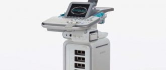
- Ultrasound. It is an informative diagnostic method. The study reveals the size, location, and contents of cysts.
- Biopsy . This examination is necessary to study the cellular composition of the formation and exclude a malignant process. A biopsy is performed with a needle under ultrasound guidance and material is removed. Punctures are made several times for more accurate results. The procedure takes just over one minute and is well tolerated. A sterile bandage is applied to the puncture site. Hematomas may appear at puncture sites . In 80% of cases, the histological response is a colloid node . If the obtained material does not contain a sufficient number of cells to establish a diagnosis, it is recommended to repeat the study.
- A fine-needle biopsy does not cause an increase in formation - if there was no such trend before the biopsy, then it will not exist after this study.
- Computed tomography with contrast. Gives more accurate ideas about education: qualitative assessment and structure. Lymph nodes and parathyroid glands are also studied.
Diagnostics
The tumor can be felt by the endocrinologist by palpation during the examination. After this, an ultrasound is performed to identify the structure of the formation and clarify the size. Next, a biopsy is needed, which will show the severity of the disease, identify atypical cells, if any, and purulent inclusions (if the tumor has not festered, then it is considered uncomplicated).
During puncture, treatment of a thyroid cyst can be performed simultaneously: the contents are completely removed from it and sclerotherapy is performed by introducing special drugs into the empty cavity. Thanks to this, the growth of the formation stops and the patient’s recovery is accelerated at an early stage.
To get a complete clinical picture, especially if there is a suspicion that the tumor is degenerating, additional studies are needed:
- CT (reveals the structure);
- laryngoscopy (if it is difficult to swallow and the voice has changed);
- angiography (if the doctor suspects that large blood vessels are involved in the process);
- bronchoscopy (for a large tumor that prevents the patient from breathing);
- tests to determine hormonal levels.
However, it is important to understand that it is impossible to predict the “behavior” of a tumor even after a thorough examination.
It often happens that a stable small tumor suddenly begins to grow. Causes may include stress or a viral infection. This is due to the fact that pathology mainly occurs due to hormonal changes, which are influenced by many external factors.
During pregnancy
Often the first ultrasound of the gland is performed during pregnancy and cyst-like formations are detected. If there are no abnormalities in the function of the gland, then there is no threat of miscarriage. The threat exists only in a hypothyroid state. There is also no data on the danger of benign formations for the development of a child if the function of the gland is within normal limits. A pregnant woman can undergo dynamic ultrasound of the gland and even a fine-needle biopsy if necessary, but scintigraphy with iodine isotopes is contraindicated.
If a nodular pathology is detected, the pregnant woman must be sent for examination to endocrinological institutes or dispensaries. Benign formations, including nodular goiter , cyst , adenoma , are not an indication for termination of pregnancy and are not subject to surgical intervention during pregnancy. If a diagnosis of “follicular tumor” is received, surgical treatment is carried out after childbirth.
For cysts with normal gland function, iodine is prescribed (200 mcg/day) and ultrasound monitoring is done every three months. The main goal of conservative treatment is to prevent the growth of the formation or reduce its size. In forms that occur with hypofunction, hormone replacement therapy ( Eutirox , L-thyroxine ) is prescribed. The dose is selected under the control of TSH ( thyroid-stimulating hormone ).
Considering that during pregnancy the need for hormones increases, therefore TSH is monitored once every 3 months and the dose of the drug is adjusted accordingly. A pregnant woman's diet should be complete and contain a sufficient amount of protein (cottage cheese, meat, eggs) and iodine (seafood, fish, tomatoes, beets, seaweed, kiwi, persimmon), given that the need for it during pregnancy is 150-200 mg per day. day.
Treatment
For a small cystic node (up to 1 cm), dynamic monitoring of the neoplasm is carried out: for this you need to undergo an ultrasound of the thyroid gland every few months. For cysts larger than 1 cm, the cystic node is punctured and its contents are pumped out. After this, the contents of the cyst are analyzed. If it is benign, then puncture can completely cure the cyst. But benign cysts often recur (fill with fluid again). If the cyst recurs, repeated aspiration can be performed, or a sclerosing procedure can be performed (by injecting 96% alcohol into the cyst). If the outcome of the procedure is favorable, the cyst does not recur, and a scar forms in its place. If the malignant nature of the neoplasm is detected, a surgical intervention is performed in which the entire thyroid gland is removed along with the malignant neoplasm. In case of a benign cyst, which is large in size or in case of a recurrent cyst, as well as when calcium deposits are found in the cyst, partial removal (resection) of the thyroid gland is performed.
Diet
Diet for thyroid disease
- Efficacy: therapeutic effect after a month
- Timing: constantly
- Cost of food: 1600-1700 rubles per week
Nutrition for thyroid pathology should be complete. If you live in an area with a natural iodine deficiency, then your diet must include seafood (mussels, squid, seaweed, scallops). It is advisable to use iodized salt. On the advice of a doctor, you can take iodine preparations ( Iodomarin ) orally. For any pathology of the gland, you need to pay attention to the content of vitamins and microelements in the diet. Useful:
- Selenium - bran, beans, eggs, whole grains, chickpeas.
- Zinc, which is rich in seafood, pumpkin seeds, peas, beans, peanut butter, sesame seeds.
- Magnesium is found in bran, nuts (cashews, almonds), soybeans, buckwheat, oatmeal, brown rice, and spinach.
- Vitamin D , which can be obtained by introducing cottage cheese, cheeses, vegetable oil, fish oil, cod and other fish liver, and seafood into the diet.
- Antioxidants, which are rich in vegetable oils, nuts, vegetables, fruits.
- Sea fish of fatty varieties contains vitamins A and D , omega-3 acids , B vitamins , and essential amino acids . Fish, seafood, olive and flaxseed oil should prevail in the diet of patients. In addition, sea fish are a source of calcium, phosphorus and magnesium.
Prevention
Prevention of thyroid diseases is:
- Proper nutrition with sufficient iodine content, especially in areas with a deficiency of this element.
- Daily walks.
- Periodic intake of vitamin and mineral complexes, including selenium, iodine, zinc, germanium.
- Annual ultrasound examination of the gland.
- Elimination of excessive insolation.
- Exclusion of physiotherapy procedures performed on the neck area.
- Clinical observation of patients with cysts that cannot be removed.
- If surgical treatment is performed, follow-up ultrasound examinations are required once a year after removal of the cyst.
You can do without surgery!
As noted above, among the many diseases of the thyroid gland, goiter is the leader, including those accompanied by the formation of cysts and nodules.
- In this case, a fine-angle aspiration puncture biopsy technique is performed to determine the nature of the formation - benign or malignant. Then the question of further treatment tactics is decided. If the formation is benign, then the patient is observed and periodically punctured twice a year. If the formation is malignant, it is subject to surgical treatment,” says Tatyana Dublic, an endocrinologist at the Diagnostic Medical Center.
Today there is a standard examination, it includes an ultrasound of the thyroid gland, examination of hormonal levels, fine-angle puncture biopsy if the nodule is more than 1 cm in size. By the way, in young people under 18 years of age, a puncture is performed even if the nodule has not reached these sizes.
— People over 40 years of age must have their thyroid gland examined. For healthy people, it is enough to visit an endocrinologist once every 5 years, if violations are detected - according to the doctor’s testimony, emphasizes Tatyana Dublic. — It is equally important to determine thyroid hormones, because from time to time problems with the thyroid gland occur under the guise of other diseases. And sometimes the hardware looks perfect at first glance, but at the same time it does not work properly. That is why abroad they first determine the hormonal levels and only then do an ultrasound.
Endocrine diseases of the thyroid gland are very often treated by the joint efforts of several specialists: an endocrinologist, a gynecologist and a mammologist. Since women suffer from thyroid diseases more often than men, they are required to undergo an examination (ultrasound of the thyroid gland, pelvic organs and mammary glands) once a year.
Very often, people do not even suspect that they have problems with the thyroid gland, and turn to specialists with completely atypical complaints. If a goiter, for example, occurs with dysfunction of the thyroid gland, then tachycardia and disturbances in heart rhythms are observed, then, most likely, the patient is referred to an endocrinologist by a cardiologist. Patients may be concerned about constipation, hair loss, hoarseness, muscle pain, swelling, menstrual irregularities in women, infertility and much more - such complaints can initially lead to a gastroenterologist, nephrologist, trichologist, gynecologist and other specialists. But after a more thorough examination, it turns out that in fact the problem lies precisely in the disruption of the thyroid gland.
Important!
Patients should not remain indifferent to their health and, even if nothing hurts, undergo regular preventive examinations, because the golden rule “prevention is better than cure” has not been canceled! Modern techniques make it possible to detect thyroid disease in the early stages and successfully treat it without surgery.
Author: Victoria Telepneva
You can make an appointment for a consultation with medical specialists by phone. 425-888 or via the feedback form.
Consequences of a cyst in the thyroid gland
Is a cyst on the thyroid gland dangerous? Let's consider the possible consequences:
- Gland dysfunction. It is possible to develop both hypothyroidism and hyperthyroidism .
- If the size is sufficient, compression of the esophagus (swallowing is impaired), trachea (breathing is difficult), and recurrent nerves (hoarseness and hoarseness appear). In severe cases, neck deformity develops.
- What else is the danger of a cyst on the thyroid gland? of cystic degeneration should be taken into account . The greatest danger is represented by isthmus cysts, since with this localization degeneration into malignant tumors is more often observed.
- The main thing about malignant tumors is their ability to grow infiltratively (the tumor grows not only into the gland, but also into the surrounding tissues).
- Suppuration of the cyst is also dangerous and in this case resection of the gland is indicated.
- Bleeding into the cyst is also possible.
Forecast
The prognosis for nodular formations depends on the histological form. With a benign form of the cyst, complete recovery is possible. If the gland is functioning fully, patients have no complaints, but it should be observed by an endocrinologist, since cysts can recur. Malignant tumors of the gland in the absence of metastases are cured in 70% of cases. Malignant tumors that give distant metastases and grow into other organs have an unfavorable prognosis.
List of sources
- Petrov V. G., Antonova E. V., Nelaeva A. A. et al. The use of laser-induced thermotherapy in the treatment of benign nodular pathology of the thyroid gland. Endocrine surgery. 2013. - No. 1. P.42-48.
- Aleksandrov Yu. K., Mogutov M. S., Patrunov Yu. N. et al. Minimally invasive surgery of the thyroid gland. M., 2005.
- Ushakov A.V. Benign diseases of the thyroid gland. Clinical classification. - M.: Clinic of Dr. A. V. Ushakov, 2013. - 384 p.
- Sleptsov I.V. Thyroid nodules. Modern principles of diagnosis and treatment. - M.: Publishing house "Elite", 2014. - 96 p.
- Valdina E.A. Thyroid diseases/Guide. 3rd ed., revised and expanded. - St. Petersburg: Peter, 2006. - 368 p.
Treatment of benign thyroid nodules
- hormone therapy,
- surgical treatment,
- minimally invasive procedures.
Minimally invasive procedures
- Ethanol sclerotherapy.
For cystic thyroid nodules (the nodule is filled with fluid), the surgeon, under ultrasound control, removes fluid from the node and gradually injects 95% ethyl alcohol (ethanol) into the cyst, which causes the death of the node cells. After 4-5 sessions, the cyst in the thyroid gland can decrease by 90%. - Laser-induced thermotherapy.
Under ultrasound control, a quartz light guide is inserted into the tissue of the thyroid nodule, through which laser radiation is supplied to the node. Under the influence of light energy transmitted by the laser, the node heats up and its cells die. The dead cells are subsequently replaced by scar tissue. - Radiofrequency thermal destruction.
It is used mainly to suppress the activity of large autonomously functioning nodes that cause thyrotoxicosis. Such formations are most often found in elderly patients with severe comorbidities and contraindications to surgical treatment. The technique is as follows. A needle is inserted into the thyroid node, from which conductors equipped with temperature sensors are extended into the tissue of the node. Using a radio frequency generator, a high-frequency electromagnetic field is created on the conductors, which heats the tissue of the node to 105 degrees, which leads to irreversible damage to the cells of the node.
Surgical treatment
As we said earlier, only after FNA of the thyroid gland and obtaining a histological report of the node tissue are the indications for surgery determined.
If histology reveals a “colloid node” (benign formation), and the patient has no complaints, surgery is not indicated for him. At the same time, there are situations in which a “colloid nodule” is also detected during FNA of the thyroid gland, but surgical treatment is recommended for these patients.
This happens in the following cases:
- When the thyroid nodule increases to a large size, and compression of the neck organs appears, accompanied by disturbances in the swallowing process or a feeling of suffocation.
- When the growth of a node leads to deformation of the anterior surface of the neck.
- If the growth of the node is accompanied by an increase in the amount of thyroid hormones and leads to the appearance of thyrotoxicosis.
The extent of surgical intervention depends on the degree of damage to the thyroid gland. This may be the removal of one lobe - hemithyroidectomy in the presence of a single node, or removal of the entire gland - thyroidectomy in the case of multiple nodes.
If FNA followed by histological findings definitely confirms the malignant nature of the nodule (papillary carcinoma, follicular carcinoma, medullary carcinoma, squamous cell carcinoma, anaplastic thyroid carcinoma), the only correct solution would be to remove the thyroid gland completely.