Adrenal hyperplasia is a very dangerous pathology. This is due to the peculiarities of the function of paired glands. They produce special hormones - glucocorticoids, androgens, aldosterone, adrenaline, norepinephrine - which, in turn, regulate the vital functions of the human body.
- Causes of adrenal hyperplasia
- Symptoms
- Nodular adrenal hyperplasia
- Diffuse adrenal hyperplasia
- Nodular adrenal hyperplasia
- Hyperplasia of the adrenal cortex
- Congenital adrenal hyperplasia
- Diagnostics
- Treatment
- Prevention
Hyperplasia is an excessive and rapid increase in cell tissue. If this happens to an organ, it retains its shape, but acquires a significantly larger volume. The adrenal glands consist of a cortex and medulla. The pathological process of hyperplasia most often occurs in the adrenal cortex. Various types of tumors are most often diagnosed in the medulla.
In most cases, the disease is congenital, hereditary, or can arise as a result of various unfavorable factors, both internal and external. Also, adrenal hyperplasia can be a concomitant pathology of certain diseases. It is detected in 40% of patients with Cushing's syndrome, which is diagnosed in old age or even in old age. If a nodular form of hyperplasia is present, then one or more neoplasms are identified, the size of which can reach several centimeters.
Causes of adrenal hyperplasia
The causes of the disease depend on the type of pathology. If during pregnancy the expectant mother experienced severe forms of functional disorders of the body, then the child may be diagnosed with congenital pathologies.
Adrenal hyperplasia can be of several types:
- Hypertensive.
- Virilnaya.
- Salt-wasting.
The virile type is caused by the activity of androgen secretion, which leads to a noticeable increase in the size of the external genitalia. In addition, hair growth and acne appear much earlier and in greater quantities, and muscles develop very actively. The hypertensive subtype is diagnosed if androgens and mineralocorticoids are produced too actively. This process negatively affects the vessels of the fundus and kidneys and leads to the development of hypertension syndrome.
Salt-wasting type of hyperplasia is caused by increased production of androgens with a lack of other hormones produced by the adrenal cortex. This type of change leads to hypoglycemia and hyperkalemia. They, in turn, are dangerous because they cause dehydration, weight loss and vomiting.
Adrenal hyperplasia can also be caused by the following factors:
- stress,
- nervous and psychological overload,
- unstable emotional state.
They can lead to an excess of cortisol, the most important hormone among glucocorticoids.
Etiology
Depending on which enzyme gene defect led to the development of the disease, several forms of the disease are distinguished, of which the most common is 21-hydroxylase deficiency (95% of cases). It is caused by mutations in the CYP21A2 gene, most often caused by recombination between the functionally active CYP21A2 gene and the CYP21A1P pseudogene, located close to each other on the short arm of the 6th chromosome. A pseudogene that carries multiple mutations is inactive, that is, it does not encode a protein.
Thus, there are three groups of mutations leading to the development of CAH. Mutations of the first group are associated with gene conversion, the transfer of fatal pseudogene mutations to the active gene, and occur in 70% of cases. Mutations of the second group, accounting for about 30%, are associated with the formation of chimeric genes from an active gene and a pseudogene due to large deletions. Mutations of the third group are rare and are associated with spontaneous mutations in an active gene.
In the classical form, both alleles of the CYP21A2 gene carry severe mutations: large deletions, nonsense mutations, or chimeric genes. The most common mutations are In2G, Q318X, I172N and some others. Patients with NF CAH are heterozygotes or, most often, compound heterozygotes, that is, they have different mutant alleles. In 1/2 - 2/3 of cases, a combination of a “severe” mutation on one allele of the gene and a point mutation on the other allele is observed, while the clinical picture is determined by the “milder” mutation. The most common mutations associated with the development of NF CAH are V281L, P30L, P453S.
Symptoms
When the disease occurs, there are disruptions in the metabolic processes in the body and symptoms caused by a deficiency or, conversely, an increase in one or another glucocorticoid are detected.
With non-classical types of hyperplasia, the following symptoms are revealed:
- Pubic and axillary hair grows early.
- Too tall for his age.
- Extremely high levels of androgens.
- Hirsutism is terminal body hair growth.
- The growth zones close too early.
- There are no periods.
- Infertility.
- Acne.
- Bald patches at the temples.
Manifestations of adrenal hyperplasia are very different and depend on what pathology is caused. Most often, doctors identify the following:
- Changes in blood pressure.
- Muscle atrophy and numbness.
- Diagnosis of diabetes.
- Weight gain, moon-shaped face.
- The appearance of stretch marks on the stomach and thighs.
- Detection of osteoporosis.
- Mental changes (memory deterioration, psychosis, etc.).
- Gastrointestinal diseases.
- Decreased immunity.
If a person often experiences thirst and the urge to urinate at night, this should be a reason to contact a doctor as soon as possible.
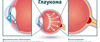
The adrenal glands are a “factory” of hormones
The adrenal glands are small paired organs located, as the name suggests, above the kidneys. Their main task is to produce hormones - substances that regulate all vital processes in the body. For example, glucocorticoid hormones are responsible for metabolism, mineralocorticoid hormones are involved in water and salt metabolism, androgens and estrogens are analogs of sex hormones, adrenaline and norepinephrine are stress hormones. The adrenal glands are surrounded by a dense connective tissue capsule and are embedded in adipose tissue. Bundles of the capsule (trabeculae) penetrate into the internal parts of the gland, forming partitions that divide the organ into layers and zones. The adrenal glands are, in addition, surrounded by the renal fascia, to which they are connected very firmly. The right adrenal gland has the shape of a truncated cone, flattened from front to back. From the front it resembles a triangle with rounded corners - “Napoleon's tricorne”. The apex of the left adrenal gland is flattened and is shaped like a crescent. Front view of isolated right and left adrenal glands
Nodular adrenal hyperplasia
With Cushing's syndrome, 40 out of a hundred patients are diagnosed with “bilateral adrenal hyperplasia.” The nodules become 2-4 cm in size. Sometimes these are single formations, sometimes multiple. In many cases the structure is characterized as lobular. The disease is found mainly in older people. Nodular hyperplasia of the adrenal gland causes the growth of an autonomous adenoma.
This type of hyperplasia is hereditary and is transmitted in an autosomal dominant manner. There are no characteristic symptoms. Presumably, the pathogenesis has an autoimmune basis. The older the patient, the more pronounced the clinical picture. Possible symptoms:
- symptoms of mucosal neurofibromatosis
- Carney syndrome (spotty pigments on the skin)
- dysfunction of the conduction and excitation of neurons in muscle structures, which is expressed by weakness in the body and cramps
- dizziness
- headache
- flickering "flies" before the eyes
- kidney dysfunction
Incidentally discovered tumors
With the introduction of ultrasound examination and high-resolution radiological methods of topical diagnostics, such as computed tomography and magnetic resonance imaging, into medical practice, formations in the adrenal glands began to be detected in patients examined for various reasons, which had not previously manifested themselves clinically. In most cases, we are talking about relatively small neoplasms ranging in size from 0.5 to 6 cm in diameter. These tumors became known as incidentalomas. The incidental discovery of an adrenal tumor should prompt the physician to more carefully examine the patient for symptoms of adrenocortical, adrenomedullary disease, or malignancy. Tactics regarding adrenal incidentaloma depend, firstly, on whether it is a source of excess production of any hormone, and, secondly, whether it is a malignant tumor. Hypersecretory formations of the adrenal glands require specific therapy and, most often, surgical intervention. In general, the approach to adrenal incidentalomas is such that hypersecretory and malignant formations require removal, as well as tumors of large sizes (more than 5 cm) and suspicious for malignancy, while formations that have been proven to be benign, such as a simple adrenal cyst, myelolipoma and Adrenal hematoma requires only regular (every six months) CT monitoring.
Diffuse adrenal hyperplasia
Diffuse hyperplasia is characterized by preservation of the shape of the adrenal glands and the formation of 1 or more nodules. Ultrasound is almost never used for diagnosis because it does not provide reliable, accurate results. And CT and MRI methods are relevant today for detecting pathology. Hypoechoic structures of a triangular shape and surrounded by fatty tissue are detected.
Adrenal hyperplasia can be of a mixed form: diffuse nodular. A person exhibits the following symptoms:
- panic attacks
- weakness in the body, even after a good rest
- arterial hypertension, periodically manifested
- excess weight
- excessive amount of body hair, etc.
"Crisis" tumor
Pheochromocytoma is a tumor of chromaffin cells that produces excess amounts of catecholamines (adrenaline, norepinephrine and dopamine). Pheochromocytoma may develop from chromaffin tissue of the adrenal medulla (90%) and may be non-adrenal in location. The disease occurs at any age. The main symptoms of the disease in the vast majority of patients are arterial hypertension, hypermetabolism and hyperglycemia. The classic clinical picture is considered to be periodic rises in blood pressure, accompanied by some vegetative symptoms, reminiscent of the effects of the administration of adrenaline or norepinephrine. In the initial period of the disease, these attacks, or crises, occur rarely - once every few months or even years. Over time, the frequency, duration, and severity of attacks usually increase. The onset of a crisis is quite often characterized by the appearance of unaccountable fear, sometimes a feeling of chilliness, paresthesia, marbling or pallor of the skin. Sometimes, on the contrary, there is pronounced redness of the facial skin, shiny eyes, dilated pupils, and frequent urge to urinate. A crisis can begin with paresthesia, convulsions, and vasoconstriction of the extremities. The crisis ends as suddenly and quickly as it began. Blood pressure returns to its original values, the pallor of the skin gives way to redness, and sometimes profuse sweating and excessive secretion of the salivary glands are observed. After an attack, general weakness and weakness persist for a long time. The diagnosis of pheochromocytoma is established when elevated amounts of catecholamines or their metabolites are detected in 24-hour urine. The most reliable is urine analysis collected within 3 hours after the attack. The accuracy of the method reaches 95%. It is recommended to conduct such studies several times. Tumor detection is usually carried out using ultrasound, CT and MRI. If the diameter of the tumor is more than 10 mm, then the sensitivity of these methods approaches 100%. Ultrasound is important in establishing the location of pheochromocytoma. However, its effectiveness largely depends on the experience of the specialist; Moreover, for tumors of extra-adrenal localization, the method is not very informative. Treatment of tumors that produce catecholamines is only surgical. However, surgical interventions for pheochromocytoma are classified as very complex, primarily due to the high degree of surgical risk associated with the likelihood of severe hemodynamic disturbances. In this regard, the cooperation of the surgeon and anesthesiologist and the choice of the most rational method of preoperative preparation and anesthesiological care are of particular importance. During preoperative preparation, the main attention should be paid to the prevention and relief of hypertensive crises. After surgery, hormone replacement therapy is often prescribed - a constant intake of adrenal hormones.
Nodular adrenal hyperplasia
Nodular adrenal hyperplasia is also known as nodular hyperplasia. Most patients are in childhood or adolescence. The disease is associated with Itsenko-Cushing syndrome and hypercortisolism. Causes: dysfunction of the adrenal glands or taking excessive amounts of corticosteroids.
Symptoms:
- muscle atrophy (mainly on the lower extremities)
- uneven obesity (face, chest, abdominal fat, neck)
- osteoporosis of the chest and lower back
- compression fractures with severe pain
- thin and very dry skin
- vascular pattern on the skin
- the formation of purple or crimson striae
- areas with hyperpigmentation
- depression and lethargy, but there may also be the opposite state: very strong joy and emotionality
- heartbeat disturbances
- absence of menstrual cycle
- a lot of hair on a woman's body
- diabetes
If the disease is detected very early and treated correctly, the prognosis will be favorable.
Therapeutic tactics for adrenal tumors
When identifying an adrenal tumor larger than 1 cm, it is first necessary to exclude or confirm the hormonal activity of the formation, which can manifest itself as hypercatecholaminemia, ACTH-independent hypercortisolism, and primary hyperaldosteronism.
At the second stage, to diagnose the malignant potential of a tumor, it is recommended to evaluate quantitative densitometric parameters using three-phase multislice CT:
- density of the tissue component before contrasting (native);
- density in the tissue phase of contrast (arterial and venous phases);
- density in the delayed (10 minutes after contrast administration) contrast phase (washout phase).
When obtaining high-density CT values in the native phase, contrast delays in the delayed phase - the malignant potential of the tumor should be assessed as high.
In the differential diagnosis of adrenal tumors, needle biopsy has no proven advantages and is associated with low sensitivity, specificity and a high likelihood of complications. The same thing seems to apply to such a diagnostic method as PET CT; it has been revealed that not all neuroendocrine malignant tumors of the adrenal gland accumulate a contrast agent, so this method is not specific and reliable, and can give a false-negative result.
Hyperplasia of the adrenal cortex
Along with hyperplasia of the cortex of this organ, there may also be a disorder in the production of hormones that affect the human genital area. Congenital pathology occurs due to abnormal changes in genes, which leads to disruption of the production of a hormone such as cortisol. Its level in the body decreases, and ACHT increases in the blood, and bilateral hyperplasia is detected.
Symptoms:
- pigmentation of the external genital area is higher than normal
- masculine traits prevail
- acne appears
- at an early age, hair begins to grow under the armpits and on the patient’s pubis
- Critical days begin for the first time at a late age
The disease resembles an adrenal tumor. Therefore, it is important to check the level of hormones in the body, for which the patient’s blood and urine are examined.
The influence of heterozygous mutations in the CYP21A2 gene on the development of CAH
An important problem in the study of CAH is the question of what effect carriers of mutations in the CYP21A2 gene have on patients, since the frequency of occurrence of heterozygotes ranges from 1:60 to 1:10 among various ethnic groups, among Ashkenazi Jews it reaches 1:3.
Some researchers report that patients who are heterozygous for mutations in the CYP21A2 gene have an increased risk of developing hirsutism, premature pubarche, and severe acne.
According to Vassos Neocleous et al., women who carry mutations in the CYP21A2 gene exhibit clinical signs of hyperandrogenemia, laboratory tests show increased levels of 17-hydroxyprogesterone and decreased concentrations of 11-deoxycorticosterone and aldosterone in the blood compared to normal. According to the results of other studies, single mutations in the CYP21A2 gene are often observed in girls with premature pubarche.
In a study by Admoni et al. all carriers were divided into 2 groups: heterozygous family members of patients with the classical form and symptomatic carriers who had clinical manifestations of androgen excess and also had a mutation in one of the alleles. The level of 17-hydroxyprogesterone in all heterozygotes was higher than in people with a normal genotype. It was noted that the V281L mutation occurs most often in heterozygotes. In carriers of this mutation, the level of 17-hydroxyprogesterone was higher than in carriers of other single mutations. In addition, this mutation was much more common among symptomatic heterozygotes (58%) than among family members (22%). The mechanism of formation of hyperandrogenemia associated with the V281L mutation in the presence of one normal allele has not been fully studied, however, Admoni et al. hypothesized that in this case the cause is a dominant-negative effect, as a result of which the mutant enzyme competes with the normal one, sharply reducing its activity. Other studies support this hypothesis. Thus, it was noted that patients with V281L have an increased risk of developing hirsutism, while among heterozygotes with other mutations it was equal to the population risk. Therefore, it can be concluded that clinical attention should be paid to V281L carriers, since the risk of developing complications of hyperandrogenemia in these patients is higher than in heterozygotes with more severe mutations. This is probably due to the fact that when one allele of a gene is affected by a severe mutation, the enzyme synthesized from it is completely devoid of activity, which makes a dominant-negative effect impossible. For timely administration of therapy, it is important to monitor symptomatic heterozygous patients before and after puberty, since there is a risk of developing additional clinical manifestations.
Congenital adrenal hyperplasia
The course of congenital hyperplasia can be classic or non-classical. The first type includes the lipoid form, diffuse, with pronounced loss of salt and with a deficiency of 21-hydroxylase. Cases of lipoid adrenal hyperplasia are recorded very rarely; the body lacks steroid hormones and 20.22 desmolase, which often ends in death. If the sick baby does not die, his sexual development is significantly inhibited, and severe organ failure is also recorded. With congenital pathology with salt loss, when the body has a deficiency of 3β-hydroxysteroid dehydrogenase, female infants may develop male external genitalia. And boys with this pathology are very similar to children of the opposite sex.
Diffuse congenital adrenal hyperplasia develops with 17α-hydroxylase deficiency; such cases are very rare. Children develop hypotension, as well as a lack of potassium in the body. In female patients, sexual development is delayed, and in male patients, manifestations of so-called pseudohermaphroditism are characteristic.
Congenital adrenal hyperplasia in most cases is detected after birth or within 12-24 months after birth. The pathology is mainly characteristic of girls. Not only the genital area suffers, but also other systems in the body. Only early diagnosis and professional therapy can prevent sad consequences.
Adrenal insufficiency
MMA named after I.M.
Sechenova N
Adrenal insufficiency (adrenal insufficiency, hypocorticism, NN) is a clinical syndrome caused by insufficient secretion of hormones from the adrenal cortex as a result of dysfunction of one or more parts of the hypothalamic-pituitary-adrenal system (HPA).
According to the initial localization of the pathological process, NN is divided into primary
(damage to the adrenal cortex itself, 1-NN) and
central forms: secondary
, resulting from a violation of the secretion of adrenocorticotropic hormone (ACTH) and
tertiary
, developing with a deficiency of corticotropin-releasing hormone (CRH). Secondary and tertiary NN are combined into central forms due to the complexity of their differential diagnosis in clinical practice. They are often designated “secondary NN” (2-NN).
Reasons 1-NN
can be:
1. Autoimmune destruction of the adrenal cortex (80–85% of all cases of 1-NN):
- isolated 1-HH of autoimmune origin;
- 1-NN in the framework of autoimmune polyglandular syndromes.
2. Tuberculosis of the adrenal glands (5–10%).
3. Adrenoleukodystrophy (about 6% of all cases of 1-NN).
4. Metastatic lesion of the adrenal cortex.
5. Damage to the adrenal glands due to disseminated fungal infections.
6. HIV-associated complex.
7. Iatrogenic 1-NN (bilateral adrenalectomy for Itsenko-Cushing disease, bilateral hemorrhage in the adrenal glands during anticoagulant therapy).
Central forms
adrenal insufficiency: hypothalamic-pituitary diseases (panhypopituitarism, pituitary tumors, surgical interventions on the pituitary gland, etc.).
1-NN is a relatively rare disease - from 40–60 to 100–110 new cases per 1 million adults per year. The true incidence of central forms of NN is unknown, but its most common cause is suppression of the HPA axis during chronic glucocorticoid therapy. Due to the fact that 1-HH is the most common type in clinical practice (more than 95%), consideration of various aspects of this type of hypocortisolism is given the main place in the article.
The clinical picture of the disease associated with the destruction of the adrenal glands by a pathological process was first fully described in 1855 by the English physician Thomas Addison (1793–1860). Since then, 1-HH of tuberculous and autoimmune etiology has been designated as Addison's disease
.
Etiology of primary hypocortisolism
Autoimmune damage to the adrenal cortex
Autoimmune destruction of the adrenal cortex (autoimmune adrenalitis) is currently the main cause of 1-NN. It accounts for up to 90% or more of 1-HH cases in developed countries. It should be noted that if in the second half of the 19th and early 20th centuries autoimmune destruction accounted for no more than 15-20% of all cases of Addison's disease, then throughout the 20th century the ratio of etiological factors of primary hypocorticism gradually changed towards the predominance of autoimmune destruction over tuberculosis. Thus, in the 40–50s, adrenal tuberculosis accounted for 48% of cases of 1-NN, while in the 80–90s this figure decreased to 15% (Fig. 1). In the near future, due to a significant increase in the incidence of tuberculosis, we can expect a slight increase in the frequency of 1-HH tuberculous etiology.
Rice. 1. Dynamics of the etiological structure of primary hypocortisolism (% of the total number of those examined)
The results of studies in the early 90s showed that specific immunological markers of autoimmune destruction of the adrenal cortex are antibodies to the adrenal steroidogenesis enzymes 21-hydroxylase (P450c21), 17a-hydroxylase (P450c17) and the side chain cleavage enzyme (P450scc). With isolated 1-HH, antibodies to 21-hydroxylase are of greatest importance. While the significance of these antibodies as a serological marker of 1-HH autoimmune genesis is beyond doubt, their pathogenetic role in the destruction of the adrenal cortex remains completely unknown.
Autoimmune polyglandular syndromes
A fundamental aspect when discussing the etiology of 1-NN is autoimmune polyglandular syndromes (APS). APS is a primary autoimmune lesion of two or more peripheral endocrine glands, usually leading to their failure, often combined with various organ-specific non-endocrine diseases of autoimmune origin. Currently, based on clinical and immunogenetic characteristics, APS types 1 and 2 (APS-1 and APS-2) are distinguished (Tables 1 and 2).
APS-2
is the most common, but less studied version of APS. In turn, the most common variant of APS-2 is Schmidt's syndrome, which is a combination of 1-NN and autoimmune thyropathies (autoimmune thyroiditis or diffuse toxic goiter). Less common is the combination of 1-NN with type I diabetes mellitus (Carpenter's syndrome).
Many of the diseases within APS-2 are associated with histocompatibility antigens - HLA-B8, HLA-DR3, HLA-DR4, HLA-DR5. However, at present, no significant immunogenetic differences have been identified between isolated autoimmune endocrinopathies and those within APS-2. In most cases, APS-2 occurs sporadically, but many cases of familial forms have been described, in which the disease is observed in different family members over several generations. APS-2 is approximately 8 times more common in women, manifests itself in middle age (between 20 and 50 years), and the interval between the clinical debut of its individual components can be more than 20 years. In 40–50% of patients with initially isolated chronic NN (1-CNH), another autoimmune endocrinopathy sooner or later develops.
APS-1
(APECED-Autoimmune polyendocrinopathy-candidiasis-ectodermal-dystrophy, MEDAC-Multiple Endocrine Deficiency Autoimmune Candidiasis, candido-polyendocrine syndrome) is a rare disease with an autosomal recessive type of inheritance or less frequently occurring sporadically, which is characterized by the
classic triad
described by Whiteker:
mucous- cutaneous candidiasis, hypoparathyroidism, 1-CNH
. APS-1 usually debuts in childhood. In the vast majority of cases, the first manifestation is mucocutaneous candidiasis, which develops in the first 10 years of life, more often at the age of about 2 years, with damage to the mucous membranes of the oral cavity, genitals, as well as skin, nail folds, nails, less common is gastrointestinal -intestinal tract (GIT), respiratory tract. Against the background of mucocutaneous candidiasis, 84% of patients develop hypoparathyroidism, which develops in 88% of patients in the first 10 years. The most significant discovery in recent years in the field of APS research is the discovery of a gene whose mutations lead to the development of APS-1. This gene is located on chromosome 21q22.3 and is named AIRE-1 (for autoimmune regulator). The discovery of the AIRE-1 gene is of general medical importance. The genetic component is assumed to be one of the principal factors in the development of most autoimmune diseases. However, from the standpoint of genetics, autoimmune diseases are polygenic or diseases with a hereditary predisposition.
With regard to APS-1, we are dealing with the only autoimmune disease known in human pathology (!) with a monogenic, i.e. Mendelian, the nature of inheritance, when the autoimmune process in most endocrine glands and many other organs is caused by a mutation of one single gene.
In our study, when dynamically assessing the ratio of the frequency of isolated 1-NN and 1-NN within the APS, it was revealed that if in the 30–50s of the twentieth century, 1-CN within the APS occurred in 13% of cases, then by 80– In the 90s, this figure increased to 34%, and therefore we can conclude that the next stage of the pathomorphosis of Addison’s disease is the gradual transition of this pathology into the category of APS, primarily APS-2.
Adrenal tuberculosis
As already indicated, destruction of the adrenal cortex by the tuberculosis process ranks second among the etiological factors of 1-NN. Adrenal tuberculosis develops due to hematogenous spread of mycobacteria. Usually, both the cortex and the medulla are involved in the process (the latter phenomenon appears to have virtually no clinical significance). As in the case of autoimmune damage, in the tuberculous process, NN clinically manifests itself only when 90% of the cortex of both adrenal glands is destroyed. With adrenal tuberculosis, in most cases, patients have traces of previous tuberculosis or an active process.
Adrenoleukodystrophy
Adrenoleukodystrophy (ALD, Siemerling-Creutzfeldt disease, melasma leukodystrophy) is the most common inherited peroxisomal disease with an X-linked recessive mode of inheritance, which is characterized by excessive accumulation of saturated long-chain fatty acids (LCFA), usually in myelin, manifested in predominantly white lesions. substances of the central nervous system, adrenal cortex and testicles. The disease is caused by a deletion of the ALD gene on the long arm of the X chromosome (Xq28), manifested by a deficiency of lignoceroyl-CoA synthetase. This, in turn, leads to disruption of the b-oxidation of saturated FA (having 24–32 carbon atoms) in peroxisomes and their subsequent accumulation together with cholesterol esters in the cells of the nervous system and adrenal cortex. The estimated prevalence of the disease is 1 in 100–150 thousand men.
There are at least 6 clinical phenotypes of ALD, which differ in form: from a severe childhood cerebral form to an asymptomatic course. In the same family, as a rule, different ALD phenotypes occur. The childhood cerebral form is phenotypically the most severe variant of the course of ALD. Patients are practically healthy until the age of 2–10 years, when adrenal insufficiency and severe progressive neurological dysfunction manifest. After manifestation, symptoms rapidly progress and death occurs within 2–4 years. Adolescent cerebral and adult cerebral forms occur in a similar way, but at a later age. The following 2 forms are of greatest interest to us. In adrenomyeloneuropathy (35% of cases of ALD), which, as a rule, manifests itself in the 3rd–4th decade of life, against the background of progressive neurological symptoms (spastic paraparesis of the legs, impaired vibration sensitivity, impaired sphincter activity), 2/3 of patients develop 1 -NN. Finally, in 10–20% of cases of ALD, the only manifestation of the disease is NN without any evidence of neurological dysfunction. More than half of women who are carriers of ALD have certain neurological manifestations of the disease and hypocortisolism of varying severity, which is associated with uneven and preferential inactivation of the X chromosome, which does not have a mutant gene.
Metastatic lesion of the adrenal glands
In itself, damage to the adrenal glands by tumor metastases is quite common. Thus, it was noted that metastases of breast cancer affect the adrenal glands in 58% of cases, bronchogenic lung cancer - in 36–40%, melanoma - in 33% of cases. At the same time, 1-HH develops very rarely, since, as indicated, this requires the destruction of 90% of the cortex of both adrenal glands. The most common tumor whose metastases cause the development of the clinical picture of hypocortisolism is non-Hodgkin large cell lymphoma; somewhat less frequently, the cause of the disease is metastases of bronchogenic lung cancer.
Damage to the adrenal glands in HIV infection
Subclinical 1-NN is detected in 8–12% of patients with HIV infection. Most often it develops as a result of damage to the adrenal tissue by an infiltrative process followed by its destruction (cytomegalovirus infection, fungal infections, Kaposi's sarcoma, lymphoma, etc.). The use of various medications that affect steroidogenesis (ketoconazole, rifampicin, phenytoin) can also cause 1-NN.
Pathogenesis and clinical manifestations of adrenal insufficiency
Primary adrenal insufficiency
1-HH is based on absolute corticosteroid deficiency
. Aldosterone deficiency leads to loss of sodium and water through the kidneys and gastrointestinal tract with the development of dehydration, hypovolemia, hypotension, and progressive hyperkalemia. Deficiency of cortisol, the main adaptogenic hormone of the human body, leads to a decrease in resistance to various endo- and exogenous stressors, against the background of which (most often against the background of infections) decompensation of NN occurs. Of fundamental importance are the loss of cortisol functions such as stimulation of gluconeogenesis and glycogenolysis in the liver, as well as the permissive effects of cortisol on thyroid hormones and catecholamines.
The clinical picture of 1-NN was described quite fully by Thomas Addison himself. In this regard, only minor additions have been made to this description over the past 150 years.
1-NN manifests itself in middle age (between 20 and 50 years). Currently, the disease is significantly more common in women, which is associated with the predominance of 1-HH of autoimmune origin. Addison's disease of tuberculous etiology affects men and women equally often.
Hyperpigmentation of the skin and mucous membranes
- the most famous and typical symptom of 1-NN, which is pathogenetically related to the fact that with 1-NN there is hypersecretion of not only ACTH, but its precursor - propiomelanocortin, from which, in addition to ACTH, melanocyte-stimulating hormone is formed in excess. Hyperpigmentation is most noticeable on exposed parts of the body (face, arms, neck), in places of friction (skin folds, places of friction with clothing), and places of natural accumulation of melanin. Hyperpigmentation of mucous membranes (oral cavity, gums, buccal mucosa at the level of teeth, friction points of dentures) is of fundamental importance.
Weight loss
– typical symptom of NN; a progressive increase in the patient's body weight practically excludes the diagnosis. Weight loss is usually significant, reaching 5–20 kg.
General and muscle weakness
– at the beginning of the disease it can be expressed moderately (decreased performance) and reaches significant degrees with decompensation of the disease (up to adynamia).
A characteristic symptom of 1-NN is mental depression
.
The cardinal symptom of 1-NN is arterial hypotension
. Severe systolic and diastolic hypertension in most cases allows us to exclude the diagnosis.
Dyspeptic disorders of varying severity
are almost always available. More often it is poor appetite and nausea, periodically occurring diffuse abdominal pain, less often - vomiting, upset stool. A characteristic symptom of 1-NN, pathogenetically associated with severe sodium loss, is an addiction to salty foods. In a number of cases, we observed patients using salt in its pure form, however, many patients did not present this complaint at all. Hypoglycemic attacks are a rare symptom of 1-NN; they more often occur with 2-NN due to the insignificant severity of other symptoms. In principle, it can be noted that none of the individual listed symptoms of 1-NN are specific to this disease. It is only the combination of these symptoms that matters.
In the clinical picture in patients with APS-2, manifestations of 1-NN prevail. Hyperpigmentation may be mildly expressed, especially with a combination of 1-NN and hypothyroidism. A typical mistake is to interpret a moderate increase in the level of thyroid-stimulating hormone (TSH) in the phase of 1-NN decompensation as primary hypothyroidism. This increase in TSH levels is associated with adenopituitary dysfunction due to hypocortisolism. The test must be repeated after achieving clinical and laboratory compensation of 1-NN, supplementing it with a study of the level of antithyroid antibodies and ultrasound of the thyroid gland. Typical signs of the development of 1-NN against the background of type 1 diabetes mellitus are a decrease in the dose of insulin required for the patient and a tendency to hypoglycemia, which, despite the seemingly milder course of diabetes, are combined with weight loss, dyspeptic disorders, and hypotension.
Secondary hypocortisolism
The most important pathogenetic difference between 2-NN is the absence of aldosterone deficiency. ACTH deficiency in this case leads to a deficiency of cortisol and androgens, but does not affect the production of aldosterone, which is practically independent of adenopituitary influences, the secretion of which is regulated by the renin-angiotensin-sodium-potassium system. In this regard, the symptoms of 2-NN will be quite poor. Symptoms such as arterial hypotension, dyspeptic disorders, and addiction to salty foods will not be expressed. The fundamental clinical difference of 2-NN is the absence of hyperpigmentation of the skin and mucous membranes. General weakness, weight loss, and, less commonly, hypoglycemic episodes come to the fore in the clinical picture. Diagnosis is facilitated by the presence of anamnestic or clinical data on pituitary pathology, operations on the pituitary gland, and long-term use of corticosteroids.
Acute hypocortisolism
The most common cause of acute hypocortisolism is decompensation or acute manifestation of chronic forms of HF, the etiology of which was discussed earlier. Thus, severe symptoms characteristic of chronic NN will almost always be identified. Less commonly, we talk about hemorrhagic adrenal infarction, the pathogenesis of which is based on DIC syndrome in septic conditions (Waterhouse-Friderichsen syndrome) and various coagulopathies. In the pathogenesis of acute hypocortisolism, circulatory insufficiency and dehydration are of fundamental importance. There are three main forms of acute NN.
1. Cardiovascular.
The phenomena of collapse and acute cardiovascular failure dominate.
2. Gastrointestinal.
Dyspeptic symptoms dominate: severe vomiting, diarrhea. This form must be differentiated from foodborne toxic infections.
3. Cerebral form
(meningoencephalitic). Patients are in prostration, often in a delirious state, with pronounced neurological symptoms.
As a rule, there is a combination of all three groups of symptoms of varying severity.
The diagnosis of acute HF is usually based solely on the clinical picture; Certain importance is given to identifying characteristic disturbances in electrolyte levels using express methods.
Diagnostics
Nonspecific laboratory changes
Chronic 1-NN is characterized by hyperkalemia and, less commonly, hyponatremia. In addition, normochromic or hypochromic anemia, moderate leukopenia, relative lymphocytosis and eosinophilia are often observed in the blood. These data are of limited value in clinical practice.
Hormone level studies
The first level test in the diagnosis of NN is the determination of the daily excretion of free cortisol in urine
. Studies such as determining the excretion of 17-hydroxycorticosteroids (17-OX), 11-hydroxycorticosteroids (11-OX) and 17-ketosteroids (17-KS) are considered uninformative and should not be used either for diagnosing NN or in clinical medicine in general. . (A typical diagnostic error is studying the level of 17-CS excretion to diagnose hyperandrogenemia in gynecological practice.)
Determining blood cortisol levels has limited diagnostic value, since in many patients with BN it is often at the lower limit of normal. However, a marked decrease in this indicator may be important - a cortisol level of less than 3 μg/dL (83 nmol/L) is absolutely diagnostic of BN.
With a detailed clinical picture of primary hypocortisolism, the detection of a significant decrease in the daily excretion of free cortisol in the urine makes it possible to make a diagnosis and begin treatment (Fig. 2).
Rice. 2. Scheme of laboratory diagnosis of primary hypocortisolism with a detailed clinical picture
With an erased clinical picture of 1-NN, as well as with borderline or questionable results of determining cortisol excretion, patients are advised to perform
a test with 1-24 ACTH
(the first 24 amino acids of the 39-amino acid ACTH molecule) (Fig. 3). A stimulation test with a study of the level of cortisol in the blood plasma 30 and 60 minutes after intravenous administration of 250 mg (25 units) of 1-24 ACTH per 5 ml of saline is a classic study in the diagnosis of primary hypocortisolism. An increase in cortisol levels greater than 20 mcg/dL (550 nmol/L) reliably excludes BN. For example, in a healthy person, maximum stimulation of the adrenal cortex is caused by the administration of only 1 mcg of 1-24 ACTH.
Rice. 3. Laboratory diagnosis of adrenal insufficiency with a blurred clinical picture
In clinical practice, it is extremely rare that there is a need to differentiate 1-NN and 2-NN using laboratory methods.
, determination of the level of ACTH
can be used , which will be increased with 1-NN (more than 100 pg/ml) and decreased with 2-NN. It should be noted that determining the ACTH level has no independent significance for diagnosing NN. In addition, 1-NN will be characterized by an increase in renin activity in the blood plasma (or an increase in renin levels), which is associated with aldosterone deficiency developing with 1-NN, which is not observed with 2-NN.
Approaches to laboratory diagnosis of secondary hypocortisolism are somewhat different (Fig. 4). In cases of recently developed 2-NN, for example immediately after surgery on the pituitary gland, administration of 1-24 ACTH will lead to an adequate release of cortisol, since the adrenal cortex has not yet had time to atrophy. The same applies to subclinical 2-NN, for example in large pituitary adenomas. In this situation, administration of such a large dose of ACTH as 250 mcg can cause an adequate response in the moderately hypotrophic adrenal cortex. Thus, a test with 1-24 ACTH will have diagnostic value only in cases of long-term and severe 2-NN. In general, tests with insulin hypoglycemia and metyrapone (metopyrone) are of greatest importance in the diagnosis of 2-NN.
Rice. 4. Laboratory diagnosis of secondary hypocortisolism
In
the insulin hypoglycemia test,
short-acting insulin is administered intravenously at a dose of 0.1–0.15 U/kg. The test will have diagnostic value if the patient experiences a decrease in glycemic levels of less than 2.2 mmol/l and develops hypoglycemic symptoms. If, against this background, the blood cortisol level exceeds 20 mcg/dL (550 nmol/l), we can talk about the normal functioning of the HPA axis and the absence of both 2-NN and 1-NN. The test is contraindicated in patients with severe cardiac and other pathologies, arrhythmias, and epilepsy.
Metyrapone (metopyrone) test
examines the presence of functional reserves of ACTH of the pituitary gland. Metyrapone blocks the adrenal enzyme 11b-hydroxylase, which converts 11-deoxycortisol into cortisol. Normally, when taking metyrapone, blockade of cortisol production will lead to stimulation of ACTH secretion, which in turn activates steroidogenesis proximal to the enzyme block and will lead to the accumulation of excess 11-deoxycortisol. The drug in a dose of 30 mg/kg is drunk by the patient simultaneously at night at 24.00. The next morning at 8.00 am, blood is drawn to determine the content of cortisol and 11-deoxycortisol. Normal HPA function is reflected by an 11-deoxycortisol level greater than 7 mcg/dL (200 nmol/L). BN is diagnosed when 11-deoxycortisol does not reach 7 mcg/dL and cortisol levels are low (less than 5 mcg/dL (138 nmol/L)). The latter indicates adequate blockade of 11b-hydroxylase. As in the test with insulin hypoglycemia, in the test with metyrapone, an increase in plasma ACTH levels of more than 150 pg/ml is normally observed.
Etiological diagnosis
After laboratory confirmation of the presence of NN in a patient, the next step is to determine its cause. In clinical practice, after establishing the diagnosis of 1-NN, it is most optimal to immediately exclude its tuberculous etiology. For this purpose, a chest x-ray and examination by a phthisiatrician are performed. In the absence of data on the tuberculosis process (and this is the most common situation), a diagnosis of presumably idiopathic (autoimmune) Addison's disease is made. A strong argument in favor of this diagnosis would be the presence of concomitant autoimmune endocrinopathies in the patient, i.e. actually APS. It should be noted that it is quite possible that the patient has a combination of old tuberculous changes in the lungs and 1-HH of autoimmune origin. Such cases have been repeatedly described in the literature and observed by us.
A test that could easily solve this diagnostic problem is the determination of circulating antibodies to 21-hydroxylase
(P450c21). These antibodies are a highly sensitive and specific marker of 1-HH autoimmune genesis. Thus, in our study, these antibodies were detected in 83–90% of cases with idiopathic 1-NN, which lasted up to 15 years, and were not detected in any patient with adrenal tuberculosis. Unfortunately, at present, this research has not received widespread clinical use.
Traditionally, in the etiological diagnosis of 1-NN , instrumental methods allowing visualization of the adrenal glands
(X-ray examination, computed tomography), a small role is assigned. However, a number of authors report that in most cases of adrenal tuberculosis there is organ enlargement according to computed tomography (CT) and magnetic resonance imaging (MRI), and in some relatively rare cases calcifications can be detected. These methods are of greatest importance in the etiological diagnosis of 2-NN.
A laboratory marker of ALD is a high level of FA.
This study is especially indicated when 1-NN is combined with various types of neurological symptoms (in particular, peripheral neuropathy). To establish the diagnosis of ALD, electromyography, as well as CT and MRI of the brain are of particular importance.
Treatment
Replacement therapy for acute hypocortisolism and severe decompensation of chronic hypocortisolism
1. Without waiting for the results of laboratory tests (if possible, preliminary blood sampling to determine the level of cortisol, ACTH, potassium, routine hematological and biochemical tests), as early as possible, begin intravenous administration of 2-3 liters of saline solution
(at a rate of 500 ml/hour; in a collaptoid state, in a stream), possibly in combination with a 5–10% glucose solution. During the 1st day, at least 4 liters of liquid are administered. The administration of potassium-containing and hypotonic solutions, as well as diuretics, is contraindicated.
2. 100 mg of hydrocortisone
intravenously
immediately, then every 6 hours for 1 day.
As an alternative (during delivery to the clinic) - 4 mg of dexamethasone intravenously
(or an equivalent dose of prednisolone - 40 mg), followed by transition to hydrocortisone therapy. In parallel - symptomatic therapy, most often antibiotic therapy for infectious processes that caused decompensation of the disease.
3. On days 2–3, with positive dynamics, the dose of hydrocortisone is reduced to 150-200 mg/day
(with stable hemodynamics, administered intramuscularly). Mineralocorticoids are not required until the daily dose of hydrocortisone is less than 100 mg/day.
With moderately severe decompensation, as well as newly diagnosed disease, therapy, as a rule, begins with intramuscular administration of 100–150 mg of hydrocortisone per day (for example, 75 mg in the morning, 50 mg at lunchtime and another 25 mg in the evening). After a few days, the dose of the drug is reduced and switched to maintenance replacement therapy with tableted corticosteroids. It should be noted that the pronounced positive effect of corticosteroid therapy has important diagnostic significance. In the presence of NN, patients literally notice a noticeable improvement in their well-being within days 1–3.
Replacement therapy for chronic hypocortisolism
1-NN replacement therapy involves the mandatory combination of gluco- and mineralocorticoids. A typical mistake is treating Addison's disease with glucocorticoid monotherapy. With 2-NN, there is no mineralocorticoid deficiency and there is no need to prescribe mineralocorticoids (9a-fluorocortisol), unless the patient has severe arterial hypotension.
Mineralocorticoid replacement therapy
Modern 1-NN replacement therapy with mineralocorticoids involves the use of only one drug – fludrocortisone
. The drug deoxycorticosterone acetate has insufficient mineralocorticoid activity and is inactivated upon first passage through the liver; It is currently not in use and has been discontinued.
Fludrocortisone is prescribed once a day at a dose of 0.05–0.1 mg (usually daily in the morning). In clinical practice, it is recommended to use the following criteria for the adequacy of fludrocortisone therapy:
- normal plasma potassium and sodium levels;
- normal or moderately increased level of plasma renin activity;
- normal (comfortable) blood pressure;
- absence of swelling, fluid retention (signs of drug overdose).
Glucocorticoid replacement therapy
The doctor has a large number of glucocorticoids in his arsenal, which can potentially be used for replacement therapy for NN.
The most used drug in the world for replacement therapy of NV is tableted hydrocortisone
, followed by the much less commonly used
cortisone acetate
. In Russia, tableted hydrocortisone has so far been practically not used and most patients receive synthetic glucocorticoids, less often cortisone acetate. A negative property of synthetic drugs is their relatively narrow therapeutic range. Particular caution should be exercised when prescribing long-acting drugs (dexamethasone), which are associated with a high incidence of overdose. The drug of choice in children and adolescents is hydrocortisone, since a number of studies have revealed growth retardation in children with NN during therapy with synthetic drugs. In this regard, when using synthetic glucocorticoids, a higher incidence of osteopenia syndrome can be expected than when using hydrocortisone therapy.
| Glucocorticoid replacement therapy regimens: 1. Using short-acting drugs: A. Twice-daily regimen: hydrocortisone - 20 mg in the morning and 10 mg in the afternoon (or cortisone acetate 25 and 12.5 mg, respectively). B. Three times a day regimen: hydrocortisone - 15-20 mg in the morning, 5-10 mg in the afternoon, 5 mg in the evening (or cortisone acetate 25, 12.5, 6.25 mg, respectively). 2. Using drugs of medium duration of action: prednisolone - 5 mg in the morning and 2.5 mg in the afternoon (much less often methylprednisolone - 4 and 2 mg, respectively). 3. Using long-acting drugs: dexamethasone - 0.5 mg per day once (late at night or in the morning). 4. All possible combinations of the listed glucocorticoids. |
On the other hand, when treating with hydrocortisone and cortisone acetate, certain difficulties are created by the relatively short period of action of the drugs.
With the classic two-time use of these drugs, patient complaints of weakness in the evening and early morning before taking hydrocortisone are quite typical. The following criteria for the adequacy of the glucocorticoid component of NN replacement therapy can be distinguished:
• minimally expressed complaints of weakness and low performance;
• absence of pronounced hyperpigmentation of the skin and its gradual regression;
• maintaining normal body weight, no complaints of constant hunger and no signs of overdose (obesity, Cushingoidization, osteopenia, osteoporosis).
Reliable objective (laboratory) criteria for the adequacy of glucocorticoid replacement therapy for NN are currently absent, and the selection of therapy is based almost exclusively on the clinical picture and the doctor’s experience.
Principles of treatment of APS and ALD
Treatment of APS consists of replacement therapy for insufficiency of the affected endocrine glands. It should be borne in mind that when hypothyroidism and NN are combined, L-thyroxine therapy is prescribed only after compensation for NN. When carrying out replacement therapy for a combination of hypoparathyroidism and 1-HH, it should be borne in mind that normally cortisol and vitamin D have opposite effects on the intestinal absorption of calcium. Thus, with cortisol deficiency, there is an increased risk of overdose with vitamin D preparations. On the other hand, the administration of large doses of corticosteroids, even with 1-H decompensation combined with hypoparathyroidism, can provoke severe hypocalcemia.
There are currently no proven effective treatment or prevention methods for ALD. Treatment options include diet, immunosuppressive therapy, bone marrow transplant, and gene therapy.
A promising direction for the treatment of NN is the addition of drugs of the main adrenal androgen dehydroepiandrosterone to the standard described therapy.
Literature:
1. Melnichenko G.A., Fadeev V.V. Laboratory diagnosis of adrenal insufficiency. // Problem endocrinol. 1997; 43 (5): 39-47.
2. Melnichenko G.A., Fadeev V.V., Buziashvili I.I. Etiological aspects of primary chronic adrenal insufficiency. // Problem endocrinol. 1998; 44 (4): 46-55.
3. Fadeev V.V., Buziashvili I.I., Dedov I.I. Etiological and clinical structure of primary chronic adrenal insufficiency: a retrospective analysis of 426 cases. // Problem endocrinol. 1998; 44 (6): 22-6.
4. Fadeev V.V., Shevchenko I.V., Melnichenko G.A. Autoimmune polyglandular syndromes. // Problem endocrinol. 1999; 45 (1): 47-54.
5. Czerwiec FS, Cutler GB Modern approach to corticosteroid replacement therapy. //Curr. Options in Endocrinol and Diabetes. 1996; 3: 239-46.
6. Oelkers W. Dose-response aspects in the clinical assessment of the hypothalamo-pituitary-adrenal axis, and the low-dose adrenocorticotropin test. //Eur. J. Endocrinol. 1996; 135: 27-33.
Diagnostics
The doctor must perform a clinical examination, prescribe laboratory tests, including hormones, as well as biochemistry. Functional indicators of the adrenal glands are determined using special methods, functional testing is possible. Laboratory methods include RIA and ELISA. When performing a radioimmunoassay, free cortisol is determined in the patient’s urine and blood. When performing ELISA, blood serum is examined for the amount of hormones present.
To diagnose adrenal hyperplasia, radiography is used, including aortography, angiography and tomography. Today, the shape and size of the adrenal gland can be determined using radionuclide scanning, as well as MRI. In some cases, doctors talk about the need for aspiration puncture, which is performed under ultrasound guidance.
Sources
- Fruzzetti F. et al: Influence of the ovaries on adrenal androgen secretion in polycystic ovary syndrome. Fertility and infertility;
- Rittmaster RS: Clinical relevance of testosterone and dihydrotestosterone metabolism in women, Am. J. Med.;
- Azziz R: Hyperandrogen-insulin-resistant acanthosis nigricans syndrome: therapeutic resonance. Fertility and infertility;
- Ibanez L, et al: Localization of the source of androgen excess in adolescent girls. Journal of Clinical Endocrinology and Metabolism;
- Barnes RB et al: Ovarian hyperandrogenism due to congenital disorders of adrenal virilization: evidence for perinatal masculinization of neuroendocrine function in women. Journal of Clinical Endocrinology and Metabolism;
- Buyalos R.P. et al.: Relationship between circulation, obesity and hyperinsulinemia on serum insulin-like growth factor binding protein-1 in polycystic ovary syndrome. Am. J. Obstet. Gynecol;
- Katz E, et al: Effect of systemic hyperandrogenism on the adrenal response to the hormone adrenocorticotropin. Fertility and infertility;
- Morris RS et al: Prorenin is elevated in polycystic ovary syndrome and may reflect hyperandrogenism, fertility, and infertility;
- Varenik-Shimankevich A. Androgenization syndromes in women. Clinical perinatology and gynecology;
- Yann R, et al: Ultrasound assessment of ovarian stromal hypertrophy in hyperandrogenism and ovulation disorders: visual analysis versus computerized quantification, Fertility and Sterility.
ONLINE REGISTRATION at the DIANA clinic
You can make an appointment by calling the toll-free phone number 8-800-707-15-60 or filling out the contact form. In this case, we will contact you ourselves.
If you find an error, please select a piece of text and press Ctrl+Enter
Prevention
If you have a history of congenital hyperplasia in your family, you need to contact a qualified geneticist. Prenatal diagnosis (diagnosis when the child is still in the womb) is important for prevention. A chorionic villus biopsy is performed in the first 3 months of pregnancy. In the second half, an analysis of the amniotic fluid is performed to determine hormone levels.
Screening testing of newborns is also important for the prevention of this pathology. To do this, blood is taken from the child's heel from a capillary. When the mother is still pregnant, it is necessary to exclude the effect of poisons in various forms and radiation on her body.