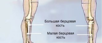
Radius bone of the hand
is a long tubular fixed paired bone in the forearm, the body of which has a triangular shape with three surfaces (anterior, posterior and lateral) and three edges (anterior, posterior and interosseous). It is located next to the ulna bone, so they are dependent on each other and interconnected. Below these bones connect to the bones of the wrist. This is how the wrist joint is formed. The radius is also responsible for the mobility of the forearm at the elbow, and the shoulder at the shoulder joint. But according to statistics, with almost the same structure and anatomy, the radius bone breaks much more often than the ulna.
What is a radius fracture?
A fracture of the radius is one of the most common household injuries; about 16% of all registered acute pathologies of the skeletal system are precisely such injuries. Humanity has encountered this type of fracture throughout its history; in burials more than 5 thousand years old, archaeologists find bones with traces of such injuries, and the first ancient, Egyptian, and Chinese treatises known to us already contain recommendations for the treatment of such victims. This pathology is so widespread, as a result of the mechanism of its occurrence, the victim receives an injury by falling on an outstretched arm, or by a strong blow with an outstretched arm against something quite hard.
This injury occurs more often in women after menopause; more than half of such injuries are suffered by them. This is due to the fact that during this period the calcium content in their bones decreases and they become more fragile, and even a small load can lead to injury. Next, we will take a closer look at how such damage occurs, what symptoms it has, how to treat it, and how dangerous a fracture of the radius can be.
Causes
The injury usually occurs when falling on an outstretched arm. If a person stumbles or falls into ice, then the probability of damage to the beam is very high. This injury often occurs in childhood or in people who play sports, which is associated with high mobility and a high incidence of falls. It may also occur in the event of an accident or at work.
In older people, the cause of the damage is osteoporosis. Even a minor impact causes a fracture.
Displaced radius fracture
A displaced fracture of the radius develops when parts of the broken bone move relative to each other. The types of such fractures are very different, and differ in the direction and type of movement of damaged bone fragments, their location, and the integrity of the skin.
There are several groups of such fractures:
- Closed - all fragments of a broken bone are under the skin, they are most favorable for the patient, the area of injury is sterile, the risk of possible complications is minimal among fractures of this type.
- Open - in which fragments of a broken bone tear the skin, and the area of injury is in contact with the external environment, such a wound is not sterile due to microorganisms entering it from the external environment, such injuries are dangerous due to possible infectious complications.
- Intra-articular - the fracture line is located completely or partially in the joint cavity, as a result, blood from the broken bone enters it, hemarthrosis develops, and there is a significant risk of disrupting the normal functioning of the damaged joint.
A change in the ratio of bones in the area of injury may be a consequence of the injury itself, for example, when the bone is crushed into fragments, or it may be a consequence of muscle work. This happens when they pull one end of the bone in their direction, and it becomes confused with the other part of the bone, to which this muscle is no longer attached. As a rule, with displaced fractures, both variants of the pathological process are observed simultaneously, which makes it difficult to ensure adequate restoration of limb function.
A characteristic external sign of a mixed fracture is a change in the externally visible shape of the limb, a characteristic deformation is observed, however, you need to understand that changes externally visible to the eye with such an injury occur only with severe destruction of bone tissue, and are relatively rare.
Transverse and longitudinal displacement of bone fragments is widespread. With this type of injury, a transverse or oblique fracture first occurs, which divides the radius into 2 parts. As a result, one of the parts of the bone moves to the side under the action of contracted muscles, in this case a transverse fracture with displacement is observed. If the fracture was longitudinal, then part of the bone fragments, under the influence of traumatic influence, moves up the arm, and they seem to slide relative to each other. In most cases, victims experience simultaneous transverse and longitudinal displacement of bone fragments.
Somewhat less common is a displaced fracture, called an impacted fracture. It looks like this: the patient falls on his arm, and one part of the radius bone seems to be driven into the other, the bone in this case is a little like a telescopic antenna, in which one part of the bone fits into the other.
Since the mid-20th century, the proportion of compression fractures among radial bone fractures has been increasing. This is directly related to the spread of motor transport and industrial equipment, and as a consequence to the increase in the number of victims in accidents related to equipment. The mechanism of injury in such situations differs from that typical for this pathology; bone damage occurs not as a result of a fall or a blow, but as a result of pinching a limb between two metal surfaces, as a result of which the bone is crushed, as if it were in a vice. Such injuries are characterized by extensive soft tissue damage and many small bone fragments at the site of injury.
The main diagnostic method for this type of fracture in modern medicine is x-ray examination. An x-ray taken in two projections allows the doctor to assess the position of the bones relative to each other and the severity of the injury.
Consequences and complications
The most common complication of a radius fracture is its malunion. At the same time, the severity of the consequences can vary widely depending on the location of the fracture/degree of bone deformation and can be represented by:
- Sudeck-Thurner syndrome (chronic pain syndrome/reflex sympathetic dystrophy). The pathology is progressive in nature and is accompanied by chronic pain syndrome, limb deformation with impaired function, trophic disorders, development of stiffness of adjacent joints/osteoporosis and often ending in disability.
- Instability of the wrist joint/impaired rotational function of the wrist joint.
- Shortening the forearm.
- Decreased grip strength in the hand.
- Slowing growth of the radius/development of radial clubhand.
- Deforming arthrosis of the wrist joint.
- Volkmann's contracture , developing against the background of long-term compartment syndrome.
Complications are observed mainly in the absence of timely/adequate treatment for complex radial fractures and non-compliance with recommendations for the duration of bone fixation and lifestyle correction for the healing period.
Non-displaced radius fracture
At least half of the cases of fractures of the radius occur without displacement, since the muscle mass of the forearm is much smaller than on the lower limb, or on the shoulder, then with incomplete fractures, muscle strength is not enough to displace the bone fragments relative to each other. In some cases, even a complete transverse fracture of the radius is not accompanied by displacement of bone fragments.
The most common type of non-displaced radius fracture is a fracture in the bone tissue. In traumatology, a fracture is usually called an incomplete fracture, when there is damage to only some part of the bone, but it does not extend to its entire thickness. As a rule, cracks are a consequence of household and sports injuries in relatively young people. Their bones are elastic and strong enough to withstand severe loads, and complete fractures from low falls or impacts are quite rare.
Externally, such a fracture manifests itself in the form of swelling and pain at the site of injury, unlike a displaced fracture and an open fracture of the radius, at the site of injury there will only be swelling and possibly a hematoma. On an x-ray with this type of pathology, a full fracture line may not be observed, but only damage to the periosteum and compaction of bone tissue at the site of damage.
Diagnostics
Most fractures are accompanied by severe symptoms - pain, swelling, displacement of a bone fragment, and the presence of a hematoma. However, X-rays are always ordered to confirm the type of injury. This diagnostic method is considered the most accessible and widespread.
In severe cases, traumatologists may refer the patient for a computed tomography or magnetic resonance imaging scan. These methods are mainly used to assess combined injuries, as well as before and after surgery.
Fracture of the radius in a typical location
A fracture of the radius in a typical location is the most common injury to the radius; destruction of bone tissue in this area occurs due to the anatomical features of the structure. In the area of the wrist joint, 3-4 cm from its articular surface, when falling on the hand, the maximum load occurs, and as a result, the bone cannot withstand it and is destroyed.
There are two main types of radius fractures in typical locations:
- Colles' fracture is a hyperextension of the wrist joint, in which the radius fractures in a typical location. With this type of injury, the distal (located further along the limb) bone fragment is mixed towards the dorsum of the forearm. Approximately two-thirds of radius fractures in a typical location are of this type. This type of fracture was first described in 1814 by Abraham Colles, a famous surgeon and anatomist who lived in Ireland.
- Smith's fracture is a flexion fracture of the radius, the victim in this case falls on his arm, the hand of which is bent towards the dorsum of the forearm. Thus, the distal bone fragment moves to the outer surface of the forearm. This type of typical radius injury was first described by Robert Smith in 1847. In fact, a radial fracture in a typical location is two types of fracture that mirror each other.
Currently, a significant proportion of victims with a radial fracture in a typical location are women over 45 years of age. This is due to the consequences of menopause, which negatively affects the strength of bone tissue, and as a result, the lack of resistance of bones to shock loads. An impact that would only lead to a bruise at the age of 20 can easily result in a fracture for a 50-year-old woman.
The peak of cases of such injuries in countries with cold climates occurs in spring and autumn, this is associated with ice, and an increased risk of falling, the number of people receiving bruises increases, and the number of fractures also increases.
X-ray anatomy of the wrist joint
The inclination of the articular surface of the radius in the direct projection is normally 15-25º. It is measured in relation to the perpendicular to the axis of the radius and a line along the articular surface. A change in the angle of inclination of the articular surface of the lower third of the radius is a sign of a fracture, both fresh and long-grown.
Palmar inclination is measured in the lateral projection in relation to the tangent line drawn along the palmar and dorsal eminences of the articular surface of the radius to the axial line of the radius. The normal angle is 10-15º. A clear change in angles is a sign of a fracture.
Complications after a fracture of the radius
Complications of radial bone fractures can be divided into two large groups:
- Immediate complications of injury are complications that arise due to the effect of damage resulting from a bone fracture on the normal functioning of the limb.
- Long-term consequences of injury are complications that arise as a result of incorrect treatment or disruption of normal healing after injury.
Immediate complications include:
- Ruptures and injuries to the nerves that provide sensation or mobility to the limb. Bone fragments can, with their sharp edges, damage or rupture large nerve trunks, depriving the area below the site of injury of signals from the brain. As a result, the ability to voluntarily move the affected area may partially or completely disappear, and sensitivity may be lost.
- Injuries to the finger flexor tendons, bone fragments moving towards the dorsal surface of the forearm can damage the bundle of tendons leading to the hand, and as a result, the victim completely or partially loses the ability to move the fingers.
- Tight swelling of Turner's hand, as a result of which reflex immobility of the fingers develops; the patient cannot make voluntary movements with them, but if he tries to move them, he experiences severe pain. Severe osteoporosis develops in the wrist bones and cysts.
- Trauma to large main vessels, followed by intracavitary hemorrhage, such damage can lead to the development of long-term complications.
- Complete or partial rupture of muscles, or separation of muscles from places of attachment to bone tissue, leads to the impossibility of subsequent voluntary movements of that part of the limb, the movement of which was carried out by the affected muscle.
- Acute infectious complications, with open fractures, infection can enter the wound, which in turn can lead to the formation of acute osteomyelitis. This pathological condition manifests itself in the form of purulent melting of bone tissue with high fever and intoxication.
Long-term consequences of injury include:
- Ischemic contracture is a violation of the mobility of the joints of the affected limb due to an incorrectly applied plaster cast, which compresses the soft tissues, disrupting the blood supply, and as a result, adhesions are formed that impair the mobility of the involved joints.
- Violations of the bone structure due to inadequate reposition, an incorrectly applied plaster cast may not hold bone fragments well enough, and during the time required for healing they will take an incorrect position, and in this position they will be fixed by the growing bone tissue.
- Long-term infectious complications, as a rule, manifest themselves in the form of chronic osteomyelitis. This chronic purulent-septic disease develops as a result of the penetration of an infectious agent into the bone tissue, which, in the process of its life activity, begins to gradually destroy the bone tissue, forming purulent cavities in the bone. The presence of these cavities causes intoxication, pain in the affected bone, and can lead to a pathological fracture, due to a decrease in the strength of the bone tissue in the affected area.
- Long-term consequences of hemarthrosis, if there is a fracture of the radius inside the articular, blood inevitably enters the joint cavity. Blood in the joint leads to the formation of a fibrin clot, and this protein aggregation adheres to each other the surfaces of the joint from the inside, and the person can no longer freely and fully bend the affected joint.
Swelling after a radius fracture
Swelling at the site of injury is a typical sign of a bone fracture, and injury to the radius is no exception. Let's look in more detail at how it can be dangerous with such a fracture, and what to do with it. In most cases, swelling does not pose a significant danger, but it should not be taken lightly.
If you do not take into account the magnitude of the increasing edema when applying a plaster cast, then its increase in the confined space of the plaster splint will lead to compression and ischemia of the tissue, which, in turn, can cause the formation of ischemic contracture.
An equally dangerous complication is tight Turner's edema, as a result of which the patient loses the ability to move the hand, and without timely medical care this can lead to long-term loss of mobility in the affected joints.
On the subject: How to relieve swelling at home?
You should carefully monitor the condition of the hand and tissues visible from under the plaster splints, since the presence of edema under the bandage is difficult to detect, and its long-term existence is dangerous not only due to ischemic, but also thromboembolic complications. That is, in the area of edema, due to the slowing of blood flow, blood clots can form, which can subsequently move through the vessels and lead to serious health problems.
Pathogenesis
The pathogenesis of a pathological fracture is based on quantitative/structural changes in bone tissue, which significantly reduce bone strength. These include a decrease in bone mass and, as a consequence, a decrease in the mechanical strength of the bone and structural changes - disturbances in the microarchitecture of the trabeculae, an increase in the porosity of the cortical bone and the accumulation of trabecular microfractures, which directly affects the strength of the bone, regardless of its mass. Thus, the pathogenesis of pathological fractures involves a significant decrease in bone mass per unit volume and a violation of the strength/structural characteristics of bone tissue.
Treatment of a radius fracture
Treatment of a radius fracture, like any other fracture, consists of the following stages:
- First aid can be provided by anyone, even without medical education. The goal of first aid is to reduce pain, ensure rest of the affected limb, and prevent damage to the soft tissue surrounding the fracture site. If the fracture is closed, then you need to fix the limb in a safe position; if the fracture is open, you need to stop the bleeding and apply a protective bandage to the injury site. After which measures should be taken to transport the patient to a medical facility.
- First medical aid is carried out by a doctor or other specialist with a medical education. It is performed directly at the site of injury or in the emergency room. The task of assistance at this stage is to assess the condition of the victim in order to determine the scope of further treatment and prevent the development of further complications. To do this, it is necessary to assess whether the patient actually has a fracture of the radius, to differentiate it from a dislocation and sprain. After confirming the fact of a fracture, the patient is immobilized. The purpose of this procedure is to prevent displacement of fragments of the injured bone. If the patient is in the emergency room, then a decision is made on the need for hospitalization or treatment at home.
- Qualified medical care is provided by a traumatologist; the task of this type of care is to restore the anatomical and functional integrity of the injured limb.
It is necessary not only to properly heal the bone, but also to ensure the mobility of all fingers and hands and maintain their sensitivity. This goal can be achieved in three ways:
- Therapeutic treatment of a fracture of the radius. This technique is one of the most ancient, but is still effective. We have received information that already about 5 thousand years ago people knew methods of conservative treatment of fractures, and they actively used them. This is also evidenced by archaeological finds, where on the bones of skeletons we see traces of skillfully restored fracture sites.
The essence of this method of treatment is as follows: the bone fragments are positioned by the hands of the traumatologist in such a way that their position coincides as much as possible with the structure of the bone before the injury. Then, the bones are fixed in this position with a plaster or polymer bandage, and the limb is kept in it until a callus has formed and the bones are once again a single unit.
The method is the safest, but in about 20% of cases, fractures heal unevenly and curvature of the broken bone is observed, often unnoticed by the patient, but in some cases leads to serious problems.
- Closed or open reduction followed by fixation with knitting needles. This method of treatment, compared to the therapeutic treatment of fractures, is relatively young, and began to be actively used only at the end of the 19th century. The essence of the method is as follows: through the bone fragments through the skin or by making an incision, passing needles, or applying a plate, and bolting the parts of the bone in the same position.
The advantage of the method is its highest reliability, bolted fixation, metal knitting needles, all this is done under the visual control of the surgeon, the bones are always fixed in the correct position. However, this technique is not without its drawbacks: firstly, it is still an operation, and it has all the risks typical for an operation, and secondly, metal structures are a foreign object, and sometimes they are rejected by the body, which leads to serious complications.
- Using an external fixation device. This method of treating fractures is the youngest; such a device was first patented in the USSR in 1952. At its core, the technique involves installing a percutaneous compression-distraction osteosynthesis device on the patient’s limb.
Simply put, needles are inserted through the patient’s skin into fragments of broken bones, then these needles are fixed on a special cylindrical frame into which the victim’s limb is placed, the fastenings of the needles on the frame are mixed so that the parts of the broken bone are in a position that repeats the structure of a healthy bone, then all this is fixed, and the formation of a bone callus is expected to connect the damaged areas.
This technique allows you to completely restore the bone structure even after the most complex comminuted fractures, but there is a risk of infectious complications that penetrate the patient’s body along the wires passing through the skin.
On topic: 12 folk methods for home treatment
Treatment (conservative, surgical)
There are two treatment options:
Conservative. It is used for simple fractures, without fragments or displacements. The patient is given a plaster cast, and the arm heals under it on its own. Sometimes conservative treatment is used to restore fragments and displacement. The procedure may fail and the limb may become deformed over time. The entire treatment takes from 4 to 6 weeks plus a rehabilitation period.
Operational. It is carried out under anesthesia, cutting the arm and restoring the anatomical structure of the bone. The fragments are fixed with special titanium plates and screws. Transosseous osteosynthesis according to Ilizarov is also performed, and a special fixation apparatus is used to restore the arm. At the Laditsen Medical Center, the operation is performed using Dr. Veklich’s improved design, without the use of traumatic needles. After the operation, you do not need to wear a cast, just an elbow bandage. Recovery occurs faster, and the patient returns to his usual lifestyle.
Rehabilitation after a fracture of the radius
Complete recovery after a fracture of the radius consists not only of restoration of the bone structure, but also of complete restoration of limb function, in particular mobility and sensitivity.
Even with completely adequate treatment, prolonged immobility in the joints and muscles of the upper limb leads to the fact that it is difficult for the patient to make movements in joints that were previously easily accessible to him. The process of recovery from injury takes a long time and requires the patient’s desire to work and patience. Let's take a closer look at what needs to be done for a complete recovery.
How to develop a fracture of the radius? Exercises
Developing joints and muscles in case of a fracture of the radius should begin as early as possible; the timing of the start of these activities greatly depends on the type of fracture you have, and what treatment method was used by the doctor to treat it. If the fracture is treated conservatively, then after 3-5 days, after the swelling subsides, you should begin to exercise your fingers.
Start the exercises with passive movements, take the finger on your broken hand with your healthy hand and gently begin to bend it in all joints, thus stretch all fingers except the thumb for 5-7 minutes 3 times a day. After a week of such training, you can move on to active movements; the patient can begin to bend his fingers independently, without the help of a second hand. It is very important to distribute the load correctly; if pain appears during the exercise, or swelling begins to return, the exercises should be stopped.
If after a week your swelling does not subside, and exercises with your fingers cause pain, then you should consult a doctor; such a problem is a sure sign that the plaster cast applied to you does not provide reliable fixation of bone fragments.
Simultaneously with the beginning of passive movements in the fingers, you need to begin active movements in the elbow and shoulder joints, raise and lower your arm, bend it at the elbow, do these exercises for 3-5 minutes at least 2 times a day. Gradually increase the load.
After 3-4 weeks, if active movements of the fingers do not cause pain, begin to increase the load on these joints, take a lump of plasticine and start kneading it in your fist, do this as often as possible, for a week. After the plaster is removed, you can proceed to exercises with a wrist expander; exercise with it at least 3 times a day, for 5-7 minutes.
It is very important to do exercises on fine motor skills; by the end of the 4th week, start drawing or writing with the affected hand; if you couldn’t do this with it before, then try picking up rice or buckwheat grains one grain at a time, this will allow you to maintain not only the strength and mobility of your joints , but also coordination of finger movements. You can type texts on a computer keyboard as a coordination exercise.
If you perform all these exercises while you have a plaster splint installed, then after it is removed, the rehabilitation period will be significantly reduced.
Diet
Diet for fractures
- Efficacy: therapeutic effect after a month
- Timeframe: 2 months
- Cost of food: 1600-1800 rubles per week
Throughout bone fusion, dietary nutrition is indicated, the main task of which is to accelerate the fusion process. The diet for fractures should contain animal protein in an amount of about 100 g/day, which includes amino acids involved in the formation of bone tissue ( arginine , lysine , glutamine , proline , glycine , cystine ). The main products containing animal protein are dietary red meats, chicken, fish, chicken eggs, cottage cheese and dairy products.
Calcium, magnesium, phosphorus and zinc play a vital role in the formation of bone tissue, so the diet should include foods containing these elements. Sources of calcium are dairy products (cottage cheese/cheese), milk, fermented milk products, hazelnuts, lettuce, sesame seeds, spinach. It should be taken into account that for effective absorption of calcium, the diet must contain foods rich in vitamin D (fish oil/fatty sea fish).
The main sources of organic phosphorus compounds are meat, fish, milk, beef liver, beans, yolk, sturgeon caviar, walnuts, buckwheat/oatmeal, and dairy products. The required amount of magnesium in the body will be ensured by the inclusion in the diet of such products as wholemeal products, legumes, wheat, oatmeal and buckwheat (kernels), hazelnuts, milk powder, bananas, coffee beans, almonds.
And zinc is rich in bran, yeast, legumes, seafood, cereal grains, beef, dairy products, mushrooms, cocoa, sesame seeds, pumpkin seeds, peanuts, sunflower seeds, potatoes, onions.
B vitamins in the diet ; D ; WITH ; A , E , which are catalysts for the reactions necessary for the healing of fractures. Thus, vitamin D from fish oil, chicken yolk, fatty sea fish (sprats); vitamin C is found in rose hips, sea buckthorn, fruits/berries; group vitamins in offal (kidneys, pork/beef liver), cereals, walnuts, sweet peppers, hazelnuts, milk, yeast, garlic; vitamin E - in cold-pressed vegetable oils. A fracture of the radius heals on average in 27-35 days, and throughout this entire period, as well as for another 1-2 months, you must follow a diet.