One of the most serious complications of hip joint pathologies, accompanied by poor circulation in the head of the femur, is its aseptic necrosis. Its development is one of the most common reasons for the need for hip replacement. Otherwise, it will lead to disability, but the pathology most often occurs in people of working age. Therefore, it is important not only to promptly detect aseptic necrosis of the femoral head, but also to begin its treatment immediately. At the same time, you need to be prepared for the fact that the most effective method of combating the disease is surgical intervention.
What is aseptic necrosis of the femoral head
Aseptic necrosis of the femoral head (AFH) is a severe chronic disease that, over time, leads to destruction of the bone tissue of the femoral head and limited mobility of the hip joint. The term “aseptic necrosis” indicates the destruction and death of bone tissue in the absence of infection by pathogenic microorganisms.
According to medical statistics, ANFH occurs in 1.2-4.7% of patients with diseases of the musculoskeletal system, and it is 8 times more common in young and middle-aged men.
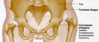
The head of the femur itself is the spherical end of the longest and largest tubular bone in the body - the femur. It connects to it through a thin neck (the most vulnerable place of the femur). Its surface is covered with thin and normally perfectly smooth hyaline cartilage. With it, it adjoins the hole in the pelvic bone, called the acetabulum, which is also covered with cartilage tissue.
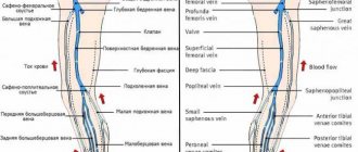
Thus, the head of the femur and the acetabulum form the hip joint. At the point of contact between the surfaces covered with hyaline cartilage, a small gap remains, called the articular. And the entire joint is surrounded by soft tissues that form the joint capsule. Its inner surface is called the synovial membrane. It produces a special lubricant (synovial fluid), which ensures smooth sliding of the femoral head in the acetabulum. The head of the femur is rather poorly supplied with blood, which makes it highly susceptible to degenerative-dystrophic changes.
The mechanism of development of the pathology is not yet completely clear, although there are 2 theories. According to one of them, ANFH is a consequence of a violation of the integrity of the bone when it is injured. The second theory suggests that the gradual destruction of bone tissue occurs against the background of impaired microcirculation and the development of ischemia. Therefore, there is a suppression of the activity of cells that form bone tissue, while maintaining the former rate of natural destruction.
It is often possible to trace the effect of both of these factors: trauma and ischemia. In any case, the death of the femoral head occurs, although it can be affected either completely or partially.
ANFH can be unilateral or bilateral.
Symptoms and differences from coxarthrosis
The symptoms of ANFH are determined by the stages of the pathology. The onset is marked by asymptomatic symptoms or minor pain for no apparent reason. At the same time, the normal range of motion in the joint is maintained, pain is noted when the hip rotates inward.
The progression of the disease is expressed by paroxysmal, severe pain. Such a symptom may indicate the presence of collapse or fracture of the femoral head, inherent in the final stage of degenerative changes. They lead to a decrease in range of motion and the presence of constant pain, crepitus and instability of the femoral head.
The clinical symptoms of ANFH are similar to the classic picture of coxarthrosis:
- pain in the groin that runs along the anterolateral surface of the thigh and radiates to the knee joint. The load only intensifies these sensations, with the pain moving to the lumbar region. They always bother a person, even at night;
- the range of motion in the affected joint is noticeably limited. These people need help with self-care;
- severe lameness on the affected leg when walking;
- rapidly progressive hypotrophy of the muscular frame of the thigh on the affected side;
- shortening of the thigh.
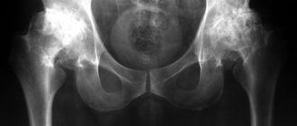
Necrosis of the right hip joint
Reasons for development
There are many factors that can lead to the development of ANFH. These include:
- dislocations of the hip joint, fractures of the femoral neck and the head itself, as well as surgical interventions;
- long-term use of corticosteroids, especially those intended for systemic use (tablets);
- alcohol abuse, which leads to metabolic disorders;
- long-term use of painkillers and NSAIDs;
- autoimmune diseases, including scleroderma, hemorrhagic vasculitis, systemic lupus erythematosus;
- decompression sickness, characteristic of divers, miners and representatives of other professions, which require them to work in conditions of rapid decrease in the pressure of the inhaled gas mixture;
- radiation sickness or radiation therapy performed to suppress a malignant tumor;
- cauda equina syndrome, caused by entrapment of the spinal roots at the level of the lumbosacral vertebrae.
However, in approximately 30% of patients, it is not possible to determine exactly what exactly caused the development of ANFH.
Causes of bone necrosis
The causes of necrosis of the knee joint are considered to be:
- vasculitis, inflammation of blood vessels leads to impaired blood supply;
- obesity, hyperlipidemia impede blood flow;
- Excessive physical overload;
- Professional sports;
- Joint injuries and their consequences;
- Alcoholism
- Osteochondropathies of the bones that form the knee joint can also provoke Osgood-Schlatter, Koenig and Larsen-Johansson diseases.
- Hereditary diseases and genetic predisposition
Stages of aseptic necrosis of the femoral head
There are 5 stages during the disease. Each of them is characterized by certain changes in the condition of the femoral head, which leads to the occurrence of typical manifestations. Each stage has a certain average duration, but the transition between them is quite conditional. And sometimes the disease develops so rapidly that very little time passes between the appearance of the first symptoms and the almost complete loss of the ability to move independently.
Therefore, the duration of each stage is individual and it is extremely difficult to predict the course of the disease in advance. It depends on the timeliness of the start of treatment, the patient’s lifestyle, the nature of existing concomitant diseases and the action of external factors.
Stage 1
Pathological changes are just beginning to occur in the bone tissue of the femoral head, so its shape remains normal. This is accompanied by pain, which initially appears only after physical exertion or changes in weather conditions, but gradually becomes constant. But sometimes acute pain, depriving a person of the ability to sit and walk normally for several days, appears suddenly. Over time, the pain decreases, but may worsen again after physical activity.
At stage 1 of the disease, pain is usually present directly in the hip joint, although it can radiate to the groin, lower back, buttocks and knees. In this case, patients do not notice mobility impairments.
In isolated cases, aseptic necrosis of the femoral head begins with the appearance of lower back pain, which can be mistakenly perceived as a sign of an intervertebral hernia or other spinal disease.
The duration of stage 1 of ANFH is about 6 months.
Stage 2
The head of the femur is one of the spongy bones. Therefore, with the gradual destruction of the bone tissue that forms it, it begins to deform and crumple. As a result, the pain becomes constant and quite severe. However, it does not go away completely even when a person is in a calm state and resting. This leads to problems with sleep and problems with movement, since physical activity continues to provoke increased pain.
With the development of a compressed or impression fracture of the femoral head, which is the determining factor in the transition of the disease to the 2nd stage, the muscles begin to gradually atrophy. This is especially noticeable with a unilateral process, as the thigh and buttock decrease in size.
There are also restrictions when performing circular movements with the leg. Attempts to move it to the side or make a rotational movement leads to a sharp increase in pain.
The duration of stage 2 is also about six months.
Stage 3
In the area where the femoral head is affected, the bone tissue begins to gradually dissolve. The resulting defect is filled with connective tissue, but the head itself becomes flatter. This is accompanied by thickening and shortening of the femoral neck, which leads to a decrease in the length of the affected leg and changes in gait and lameness. You can notice the shortening of the leg when the patient lies on his back or stomach with his heels brought together.
In 10% of cases, on the contrary, an increase in leg length is observed, which worsens the prognosis of the disease.
The 3rd stage of ANFH is characterized by constant pain, which tends to become stronger after any, even minor, physical exertion. But, unlike stage 2, they decrease when a person gives rest to the sore joint.
Due to changes and pain, it is difficult for the patient to walk. He can hardly pull his leg up to his chest. In such situations, in addition to the muscles of the buttock and thigh, the muscles of the lower leg also begin to atrophy. Therefore, without a cane or additional support, patients are practically unable to move.
Stage 3 lasts 1.5-2.5 years.
Stage 4
The connective tissue that has grown into the femoral head begins to transform into bone tissue, but this does not ensure the restoration of the spongy structure of the bone. As a result, cystic cavities and other changes in the structure of the femoral head are formed. But it remains flattened. Deformation of the acetabulum is also observed. It gradually flattens, which leads to a sharp limitation of the mobility of the hip joint.
This is accompanied by severe constant pain, which is present not only in the hip joint, but also in the lower back. Moreover, they do not disappear even at rest. There is also a pronounced decrease in the muscles of the lower limb. Patients cannot make rotational movements of the leg, and the amplitude of movement forward and backward is sharply reduced. As a result, they lose the ability to move independently.
The duration of stage 4 is from 6 months. It ends with the development of severe secondary arthrosis of the hip joint (coxarthrosis), which is called the 5th stage of aseptic necrosis of the femoral head.
Thus, the disease greatly reduces the quality of life and can ultimately put a person in a wheelchair and deprive him of the ability not only to work, but also to self-care. Therefore, it is important to diagnose it as early as possible and take measures to stop pathological changes.
Content
- Causes and risk factors
- Diagnostic methods
- Treatment methods
- Treatment programs
- Who carries out the treatment?
- Why do they contact us?
- Reviews from our patients
- Sign up for treatment
The mechanism of development of the disease is associated with deterioration of the blood supply to the femoral head for various reasons. As a rule, the disease is polyetiological in nature, which means that the disease can be caused by several causes at once. Long-term malnutrition leads to the destruction of bone architecture. As a result, deformation of the head develops, the bone becomes brittle, and microfractures occur.
Treatment begins with conservative therapy. Doctors do everything possible to help every patient. Surgical assistance is required in desperate cases, for example, when other methods of therapy have not brought the desired result. Let us consider in more detail the symptoms and treatment of necrosis of the hip joint.
Diagnostics
If you experience pain in the hip joint, it is important to contact an orthopedic traumatologist as soon as possible. This symptom may indicate the development of various joint pathologies, only one of which is ANKB. Therefore, the doctor must prescribe instrumental research methods:
- X-ray of the hip joint in 3 projections. The method makes it possible to establish not only the presence of ANKB, but also the stage of development of the disease. But it must be taken into account that at stage 1 there are no radiological signs of necrosis of the femoral head. This may cause a diagnostic error. Therefore, it is important to repeat the X-ray of the hip joint after six months.
- CT. It is prescribed, if necessary, to obtain layer-by-layer images of the hip joint and examine the characteristics of blood flow.
- MRI. The method allows you to detect the slightest changes in the condition of the hip joint, especially hyaline cartilage and other soft tissue structures.
- Densitometry. This diagnostic method makes it possible to measure bone density and detect signs of osteoporosis in the hip joint.
Additionally, biochemical blood and urine tests may be prescribed. The greatest attention is paid to the level of calcium, phosphorus, magnesium, DPID, pyridonine, osteocalcin.
ANGKB stages according to ARCO
However, in Russia the classification into five stages is more common:
- There are no radiological signs. The histological specimen shows signs of necrosis of the spongy substance of the head and bone marrow structures. Clinically expressed by aching pain and stiffness in the joint, increasing muscle weakness.
- multiple impression fractures. Against the background of necrosis, many microscopic fractures occur. The radiograph shows a homogeneous darkening of the femur, its height is reduced, the surfaces of the head in places are in the form of compacted facets, the joint space is widened. MRI data indicate a necrotic defect in the head.
- sequestration. The articular head is flattened and looks like structureless isolated fragments with different shapes and sizes. The neck of the bone shortens and thickens, and the joint space widens even more.
- reparative. The spongy substance of the femoral head is restored. On X-ray, sequestration-like zones are not noticeable; the shadow of the head is outlined, but with rounded cyst-like clearings.
- secondary deforming arthrosis. The bone structure of the femur begins to be traced, significantly changed, the congruence of the articular surfaces is disrupted.
Disease in dynamics.
Important: collapse of the femoral head occurs in an incredibly short period of time - 5 months.
Treatment of aseptic necrosis of the femoral head
The main treatment for ANKB is surgery. But if the disease is diagnosed at an early stage of development, there is a chance of delaying the operation or avoiding it. In such situations, complex conservative therapy is prescribed, which includes:
- drug therapy;
- orthopedic mode;
- exercise therapy;
- physiotherapy.
But for each patient, the orthopedist individually develops treatment tactics in accordance with the condition of the hip joint, the patient’s age, and the nature of concomitant diseases. Also, when the cause of the development of ANKB is established, treatment is prescribed to eliminate it.
Sometimes, to improve the well-being of patients, decompression tunneling is performed, which is a surgical procedure in which “tunnels” are made in the head and femur. Due to this, intraosseous pressure is reduced and pain is reduced.
Drug therapy
For each patient, a set of medications is selected individually. As a rule, for aseptic necrosis of the femoral head the following are prescribed:
- Medicines that improve blood circulation require long-term use for 2-3 months or more, followed by repetition of the course.
- Bisphosphonates are drugs that improve bone repair and prevent calcium loss. They are also prescribed for long-term hens, the duration of which can be from 8 months.
- Calcium supplements – help replenish the lack of calcium in the body. The course of treatment is 3 months. They need to be carried out 2-3 times a year.
- Vitamin D and its precursors help increase calcium absorption. Reception requires regular monitoring of calcium and phosphorus levels in the blood.
- Chondroprotectors are drugs that prevent deformation of hyaline cartilage. They can be administered directly into the joint, intramuscularly, or used in the form of capsules and powders. Treatment is long-term – at least 3 months.
- B vitamins – help increase the rate of bone tissue recovery and improve nerve conduction, which is especially important for cauda equina syndrome. They are used in courses of 20-30 days.
- NSAIDs are medications that help reduce pain and have an anti-inflammatory effect. Used upon request.
- Muscle relaxants are drugs that eliminate muscle spasm, which has a positive effect on microcirculation. Muscle spasms can occur reflexively in response to severe pain.
- Antiplatelet agents are medications that thin the blood and reduce the risk of blood clots.
A complex of drugs that affect the cause of the development of aseptic necrosis of the femoral head may also be prescribed.
At stages 3-4 of ANKB, plasma lifting or PRP therapy may be prescribed. The method involves injections of platelet-rich plasma from the patient's own blood. This direction has only recently entered medical practice, but it gives good results. Platelets help accelerate healing processes and enhance the synthesis of cartilage and bone tissue.
Orthopedic mode
With ANKBK, it is very important to strictly follow the recommendations received from the attending physician regarding the regimen and lifestyle. First of all, you need to give up bad habits (smoking, drinking alcohol). It is also necessary to avoid hypothermia and not to overload the hip joint, i.e., avoid lifting and carrying heavy objects, power loads, running, etc.
Patients who are overweight are advised to make dietary adjustments to achieve weight loss.
Exercise therapy
The most controversial and subtle issue in the treatment of aseptic necrosis of the femoral head is exercise therapy. At the initial stages of therapy, the maximum benefit will come from unloading the hip joint through bed rest and the use of mobility aids. But prolonged unloading of the hip joint can negatively affect the condition of the muscles and lead to persistent pain. Therefore, with the help of your attending physician, you need to find that fine line when you need to move from a gentle regime to increasing the load on the joint.
From this point on, it is recommended to walk, but still not give up the cane. In the future, they add climbing stairs and performing special exercises. The doctor also selects them individually.
Physiotherapy
In order to increase the effectiveness of other prescribed treatment methods and further enhance blood circulation in the affected area, as well as increase the speed of regeneration processes, courses of physiotherapeutic procedures are prescribed. Most often they resort to help:
- Hyperbaric oxygenation. The procedures are carried out in a special pressure chamber and involve specific exposure to oxygen-enriched air under conditions of high atmospheric pressure. This helps improve the quality of oxygen supply to the affected area.
- Shock wave therapy. The essence of the method is the effect of sound waves on the hip joint. This helps improve blood circulation, as well as eliminate calcium crystals that impede the restoration of the femoral head.
- Myostimulation. The procedures involve applying weak electrical discharges to the soft tissues of the thigh, which prevents muscle atrophy. It also helps eliminate muscle spasms.
Surgery for aseptic necrosis of the hip joint
Upon reaching stage 3 or 4 of ACHF, i.e., when there are signs of destruction of the femoral head, surgery is indicated. Previously, surgical interventions were carried out using different methods, but before the advent of joint replacement, they all gave either a short-term effect or led to complete immobilization. Therefore, today the gold standard for the treatment of grade 3-4 avascular necrosis of the hip joint is endoprosthetics.
The method involves replacing the head of the femur, and in case of damage to the acetabulum, replacing it with artificial prostheses. They completely replicate the shape of the natural components of the joint. Therefore, endoprostheses can completely eliminate pain and restore normal range of motion in the hip joint. Moreover, they are made from absolutely biocompatible and high-strength materials that do not cause allergic reactions.
The operation is performed under epidural anesthesia or general anesthesia. Moreover, after it you can get up and sit a little the next day. Exercise therapy is immediately prescribed - an obligatory part of rehabilitation. In general, recovery after endoprosthetics takes 3 months, after which the patient returns to a normal, fulfilling life.
Thus, ANFH is a formidable disease that can be difficult to detect in the early stages of development. But it is precisely at this time that it is possible to cope with the pathology using non-surgical methods. If the moment is missed or conservative treatment does not bring results, do not despair. Modern medicine has methods that make it possible to maintain normal mobility of the hip joint and avoid disability.
Pathogenesis
Currently, experts have two points of view regarding the pathogenesis of aseptic necrosis: traumatic and vascular.
This pathology can be provoked by various reasons: embolism , violation of the integrity of the arteries, arterial spasm, etc. The disease also develops as a consequence of joint injury or surgery.
Speaking about the non-traumatic development of aseptic necrosis of the head of the hip joint, it should be noted that the so-called “overfatigue” occurs in bone tissue as a result of overloads, microtraumas, as well as other unfavorable factors. Impulses go from the lesion to the cerebral cortex, causing feedback signals leading to vasospasm, stagnation of lymph and blood, and disruption of metabolic processes. As a result, decay products accumulate in the bone tissue, the properties of the bone change, as a result of which bone beams slowly collapse, local blood flow worsens, and the pathological process progresses.
The vascular theory suggests that necrosis of the femoral head is of ischemic origin and develops as a consequence of arterial embolism . This theory suggests that the disease is a consequence of changes in local blood circulation. Due to impaired blood flow, intraosseous blood pressure increases, which activates ischemic disorders.
Treatment with folk remedies
Folk remedies can be used as auxiliaries. However, they should not replace the main treatment under any circumstances.
- Pine buds . Mix fresh buds with sugar and place in layers in a jar. Store the container for a week in a dark place, after which drink the resulting syrup 2 times a day, 3 tbsp. l. It is also recommended to rub the syrup into the affected area. The course should last 2 months.
- Cabbage leaves . Large sheets of fresh cabbage should be greased with honey and applied to the affected area with the greased side. Wrap with plastic wrap and wrap with a warm cloth. The sheets should be kept for several hours, and it is advisable to carry out the procedure daily for a month.
- Turpentine bath . To prepare such a bath, add pine branches, 200 g of sea salt, and half a teaspoon of turpentine to hot water. After the salt has dissolved, you need to lie in the water for 10 minutes. Next, a mesh of iodine is applied to the lesion site.
- Animal fat . It is necessary to mix pork fat and nutria fat in equal proportions. Melt and rub this ointment into the affected area before going to bed, carrying out the procedure for a month.
- Mint and eucalyptus ointment . It is necessary to mix equal parts (50 g each) of eucalyptus, mint and aloe. Finely chop everything and boil until the mass becomes homogeneous. Apply the cooled mixture to the joint before going to bed.
Diet
Diet for sore joints
- Efficacy: therapeutic effect after 2-3 months
- Terms: 2-6 months
- Cost of products: 1700-1800 rubles. in Week
Nutrition should be complete. It is important to introduce foods into your diet that provide the body with vitamins, minerals, and collagen .
- There should be more dishes with gelatin in the diet.
- It is recommended to limit the amount of salt you consume.
- It is better not to eat fried, smoked, fatty foods; sweets should also be kept to a minimum.
- It is advisable to eat fractionally - more often and in small portions.
- Protein foods should dominate the diet.
Useful products for aseptic necrosis are:
- Dishes with gelatin.
- Berries, vegetables, fruits.
- Dried fruits.
- Porridge.
- Honey.
- Low-fat dairy products.
- Offal.
- Fish and seafood.