Fractures of the bones of the hand are injuries in which the integrity of the bones of the wrist, metacarpus or phalanges of the fingers is disrupted. These are common injuries, accounting for about a third of all bone fractures. This trend is associated both with the relative fragility of the hand, adapted for performing fine manipulations, and with its high activity. A fracture can occur as a result of a fall on a bent hand, a blow from a fist, the edge of the palm or fingers, or a direct blow to the hand.
The bone apparatus of the hand consists of 27 bones of three sections - the wrist, metacarpus and fingers. The wrist consists of 8 spongy bones arranged in two rows - proximal, closer to the forearm, and distal, closer to the metacarpus. The first, proximal row, starting from the thumb, are the scaphoid, lunate and triquetral bones, which form an articulation with the radius bone of the forearm - the wrist joint. The fourth bone of the proximal row, the pisiform, does not participate in the formation of the wrist joint.
The second, distal row - the polygonal, trapezoid, capitate and hamate bones - connect to the five tubular bones of the metacarpus, radiating from the wrist. The distal ends of the metacarpal bones form five metacarpophalangeal joints - the connections of the metacarpus with the fingers. The first finger of the hand consists of two phalanges, the rest - of three. The phalanges of the fingers - short tubular bones - are connected to each other by interphalangeal joints.
The bones most susceptible to fractures are the phalanges and metacarpal bones. Wrist bones are broken quite rarely. The vast majority of injuries to the bones of the wrist occur as a result of a fracture of the scaphoid bone; the lunate and pisiform bones are less commonly affected. Fractures of the hamate and distal carpal bones are practically never encountered in clinical practice.
Fractures of the bones of the hand are accompanied by sharp pain and swelling in the area of damage; when bone fragments are displaced, deformation of the hand is possible. A hematoma may appear at the site of swelling. With some fractures, you can feel the displaced bone fragments under the skin or hear their crepitus. The diagnosis can be established by a traumatologist, who will clarify the complaints, ask in detail about the mechanism of injury, examine and palpate the fracture area, and check the preservation of movements in the joints. The diagnosis is confirmed by an X-ray examination of the hand, which can visualize the fracture line, assess the degree of displacement of fragments and, as a result, determine treatment tactics.
Treatment of hand fractures includes mandatory immobilization with a plaster cast for a period of 3 to 8 weeks. In case of displacement of fragments, closed reduction is performed; if it is ineffective, skeletal traction or osteosynthesis is performed. Careful comparison of fragments and consolidation of the fracture are important to preserve not only the aesthetics of the hand, but also its full function.
Clinically Relevant Anatomy
Hand bones
The metacarpal bones are long, thin bones that are located between the wrist bones and the phalanges of the fingers.
- Each bone consists of a base, body and head.
- The proximal bases of the metacarpal bones articulate with the carpal bones.
- The distal heads of the metacarpal bones articulate with the proximal phalanges and form the knuckles.
- The 1st metacarpal bone is the thickest and shortest of these bones.
- The 3rd metacarpal bone is distinguished by a styloid process on the lateral side at its base.
- Soft tissues commonly injured in fractures include cartilage, joint capsule, ligaments, and fascia.
- In severe polytrauma, tendons and nerves adjacent to the fracture may also be damaged.
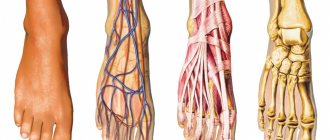
Vessels and nerves
The bones, joints, muscles and ligaments of the hands are abundantly supplied with blood . Blood saturates the tissues of the hand with oxygen, ensures high mobility and rapid tissue regeneration.
The ulnar and radial arteries approach from the forearm to the hand , then descend through the wrist joint to the palm and back of the hand, forming deep and superficial arches. On the back side, the vein diverges into four metacarpal arteries, and then each divides into two more digital arteries, which pass through the fingers to the nails. Networks of small capillaries supply blood to the fingers. Abundant branching of blood vessels protects the fingers from excessive blood loss when the hand is injured.
The innervation of the hand occurs thanks to the ulnar, median and radial nerves , which through their mutual action provide motor functions, tactile and pain sensitivity. Many nerve receptors run through the entire hand to the fingertips, contracting and relaxing muscles.
Reference! The nerve endings on the fingers are so sensitive that when the surface layer of the skin is cut with a piece of paper, the receptors react sharply to the entry of air and the person experiences pain more severe than from a cut with a knife.
Damage to the median nerve makes it difficult to flex and extend the hand , and simultaneous injury to the ligaments leads to complete loss of motor function. Compression or injury to the ulnar nerve causes loss of abduction and adduction of the fingers, particularly in the lower palm and little finger. The radial nerve is responsible for sensation in the dorsum of the hand and abduction of the thumb. If the radial nerve is damaged, it is impossible to clench your palm into a fist and unclench your hand.
Etiology
Metacarpal fractures (MCFs) typically occur secondary to a direct blow or a fall directly onto the hand.
- These fractures usually occur during sporting activities, especially contact sports. Almost a quarter of cases occur during sporting competitions.
- Sports injuries are a common cause among younger patients.
- Injuries related to physical work are a common cause in middle-aged patients.
- Falls are a common cause of fractures in older people.
- Bone fractures often occur after punching a wall or other hard object (hence the name “boxer's fracture”).
Preventive actions
To avoid injury, experts recommend following simple rules
- Observe safety precautions at work and at home.
- Be extremely careful when transporting heavy objects, playing sports, as well as under other circumstances associated with an increased risk of injury.
- Constantly monitor the condition of the musculoskeletal system, perform physical exercises that help strengthen bone tissue.
- Take vitamin complexes and minerals containing rich amounts of calcium.
Never perform any physical activities while intoxicated.
Metacarpal fractures
- Typically occurs in patients aged 10-40 years.
- Men are more often affected than women.
- Young people get PPK bones when punching a wall or getting hit in the arm.
- Older women suffer these injuries secondary to low-energy falls.
- The incidence of fractures of each metacarpal increases from the radial to the ulnar side.
- The incidence of 2nd metacarpal fractures is lower than the incidence of 5th metacarpal fractures.
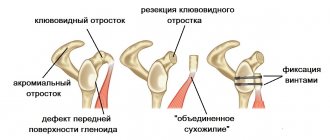
- Bennett's fracture is the most common fracture of the base of the thumb. This fracture is an intra-articular fracture and involves separation of the base of the 1st metacarpal bone in such a way that the part of the bone located closer to the ulna remains in its original position, and the other part is displaced outward.
PPK can be divided into three categories:
Types of Metacarpal Fractures
- A body fracture occurs when a person hits another person or object. In most cases, surgery is not necessary to treat this condition.
- Fractures of the metacarpal body often result from longitudinal compression, torsion, or direct impact. They are described by the appearance of the associated fractures and can be classified as transverse, oblique, spiral and comminuted.
- Fractures of the base of the metacarpal bone are rare and have minimal consequences because there is little movement of the joint. More common are fractures of the base of the fifth toe, which are the result of a longitudinally directed force.
Cervical injury
Damage to the metacarpal bones in the neck area occurs as a result of a sharp push. As a result, the head of the bone fits directly into its body without back cushioning. In this case, the bone itself does not move, but remains in place. Sometimes this condition may be considered normal, however, with a very strong blow, the bone can go deep into the cavity, which will noticeably shorten the finger.
If the fracture angle is slight, it can be treated without surgery, otherwise urgent surgery will be required. Leaving an injury untreated is not recommended, even if it is so small that it seems trivial. Over time, it will make itself felt: the consequences may be different, but restrictions on motor function and pain when trying to restore them are guaranteed.
Characteristics/Clinical picture
5th metacarpal fracture
Patients with metacarpal fractures typically have:
- Pain.
- Edema.
- Hematoma.
- Limitation of mobility.
- Deformation. Joint asymmetry may be observed; it may also appear that the joint is missing.
- Displacement of the fingers may also be noted.
- A fracture of the metacarpal head is associated with axial compression of the extended finger, which causes severe discomfort.
- When the base of the metacarpal bone is fractured, wrist movements or longitudinal compression increase pain.
- Any angulation of the ACC can lead to pseudoclaw deformity.
First aid to the victim
Some cases require first aid. Such injuries include open injuries with bleeding. The first step is to take measures to stop the bleeding and call an ambulance. Further hospitalization of the victim is carried out under the supervision of a traumatologist or other specialist.
With closed lesions, the injured arm must be immobilized to ensure that its mobility is limited. This is done in order not to accidentally touch the sore spot, and to avoid post-traumatic shock or loss of consciousness when damaged joints are displaced. The arm can be tied with any fabric available at hand. The main thing is that the fingers of the broken hand are bent.
Grade
The assessment includes:
- Standard radiographs of the hand (antero-posterior, lateral and oblique). In the vast majority of cases, this will be enough to confirm the diagnosis and formulate a treatment plan. Confirmation of more subtle lesions can be obtained using specialist views such as Brewerton (metacarpal heads), Roberts and Betts (thumb).
- A CT scan is sometimes necessary for base metacarpal fractures to rule out/confirm the presence of intra-articular displacement and determine whether surgery is necessary.
Diagnosis of injuries
To diagnose the condition of the injury, its characteristics and degree of complexity, specialists use the following methods.
- First of all, it is necessary to interview the patient, and then conduct a visual examination, take the necessary tests and determine the cause of the injury.
- Taking an x-ray. Photographs of the hand from both sides and from the side, depending on the location of the fracture.
- For multiple fractures with complications, computed tomography is necessary.
Typically, for experienced traumatologists, diagnosis is limited to a simple examination, and an x-ray is sufficient to confirm it.
Treatment
The goal of treatment is to restore the anatomy and function of the hand.
- Antibiotics and tetanus prophylaxis are treatment options for open fractures according to standard guidelines.
- Treatment will vary depending on the integrity of the skin (open or closed fracture), number of finger/metacarpal fractures, degree of bone fragmentation, displacement, rotation, etc.
- The general practitioner/specialist, after assessing the patient's condition, should perform gentle tests and imaging to determine whether surgery is required.
- If surgery is not required, a physical therapist will create a special splint that will keep the joint in the correct position while it heals.
Possible consequences
Hollow bones can be very fragile due to a lack of nutrients that help strengthen them. Therefore, it is worth paying attention to this and taking appropriate measures. If necessary, you can consult a doctor.
In case of a fracture, you must immediately contact a specialist and, if possible, provide first aid to the victim if he needs it. If you take care of your health, you can avoid numerous problems and complications.
Incorrect treatment or poorly performed surgery can lead to disability. Therefore, you should not self-medicate, ignoring the doctor’s recommendations. After minor injuries, motor functions can be restored or a repeat operation can be performed. But in severe cases, the rehabilitation course may take several months, and the result may be negative.
With open wounds, complications often arise that begin with improper healing and pathological changes. In this case, there is a risk of infection, which often leads to rotting of bone tissue.
Difficulties during treatment may arise if the recommendations of the attending physician are not followed or clinical conditions are completely ignored.
Physical therapy
The goal of rehabilitation is to restore strength and full range of motion.
- For this purpose, hand exercises with light resistance are prescribed. This is where resistance bands and a ball can be helpful, especially if there is scarring and limited flexor motion develops.
- Soft tissue repair can be more challenging (compared to bone repair).
- Rest and elevation are important, as is splinting (poor splinting can lead to stiffness, pressure sores, or even compartment syndrome).
Friends, on July 17 in Moscow, as part of the #RehabTeam project, Anna Ovsyannikova’s seminar “Rehabilitation of the hand after a fracture of the distal radius (fracture of the “radius in a typical place”)” will take place.” Find out more... In addition, on July 18, she will conduct a seminar “Rehabilitation of the hand after fractures of the metacarpal bones (Boxer fracture).” Find out more...
Physical therapists use a range of techniques to restore movement in the hand, wrist and fingers. These include:
- Eliminate swelling with massage and compression clothing.
- Soft tissue massage helps relieve muscle tension and pain.
- Developing a home exercise program for patients with specific recommendations for movement and strengthening exercises.
Here are the steps to follow for a stable fracture (you can use this or another of the stabilization methods below):
- A non-operative treatment involves strapping the injured finger to another finger. This can be done with or without a splint.
- Splinting for a fracture should be as follows: wrist extension 20 degrees; flexion of the metacarpophalangeal joint by 60-70 degrees and extension of the interphalangeal joint.
- If we are dealing with a stable fracture, it makes sense to start movements earlier.
- As a rule, active exercises to increase the range of motion without resistance can begin 2-3 weeks after surgical treatment (on intact or adjacent joints).
- Active movement. If the fracture is fixed, an active increase in range of motion may begin earlier. Most fractures are treated with immobilization, but active motion can begin after three weeks of therapy, starting with joints not affected during the initial immobilization. This phase usually lasts 3-6 weeks.
- Active movements involve sliding of tendons.
- Tendon gliding is important to prevent adhesions, increase circulation, and reduce swelling and compression at the fracture site.
Tendon gliding exercises
Exercises for tendon gliding
- The claw hand exercise can be used to improve the glide of the extensor digitorum tendon over the metacarpal bones.
- An exercise to block the deep flexor digitorum to improve the sliding of its tendon along the phalanges.
- A hook fist position that promotes selective gliding of the flexor digitorum profundus tendon.
- An exercise to block the superficial flexor digitorum to improve the sliding of its tendons along the middle phalanges.
- A straight fist position that promotes selective gliding of the superficial digital flexor tendons.
Passive movements
- Passive movements can be started after sufficient clinical healing after approximately 5-6 weeks of therapy.
- The timing of the onset of joint mobilization depends on the structures involved in the injury. If structures resisting force are not involved in the injury, joint mobilization can be initiated at the same time as active movement. Compression from a fracture can result in shortening, angulation, or rotation of the bone.
- Traditional passive range of motion exercises are designed to help joint cartilage heal and reduce swelling and stiffness.
- Resistive movement. Four weeks after injury, light resistance can be performed (this is true for most PKCs that are treated with immobilization). Active movements should only be continued if healing has not yet begun.
- Resistance exercises should also be postponed while the fracture is being fixed with pins (until the pins are removed). Light resistance exercises help in scar remodeling and improve movement. There are several types of resistance exercises, such as weight training. This type of exercise strengthens the superficial and deep flexors of the fingers.
- Functional exercises and work simulations should be incorporated into resistance exercises as soon as possible.
How to identify an injury
The origin of fractures is very diverse, however, the signs indicating injury have similar properties. General signs of injury are expressed as follows:
- manifestation of acute pain in the affected area;
- the presence of a bluish or purple tint to the skin;
- significant swelling;
- with some injuries, internal bleeding occurs, forming a pronounced hematoma;
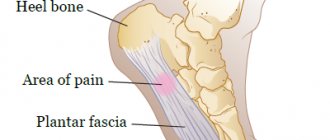
- if the bones of the foot are damaged, pain is felt when walking, which makes movement painful;
- There are cases when, with a strong blow, the bone undergoes severe deformation, and its head is difficult to feel.
Joints of the hand
The structure of the hand presupposes the presence of three main articular sections: the shoulder, elbow, and wrist. The hand includes many small articular elements , of which the following are distinguished:
- midcarpal (articulatio mediocarpalis), which has a separate capsule;
- carpometacarpal (carpometacarpea pollicis), having a saddle shape;
- metacarpophalangeal (articulationes metacarpophalangeae), responsible for attaching the fingers to bone structures, allowing them to bend, straighten, and rotate;
- interphalangeal (artt. interphalangeae manus), available on any finger in the amount of 2 pieces.
The joint element of the first (thumb) finger provides flexion-extension function.
Common pathologies
Human hands are susceptible to various inflammatory and degenerative processes . At the same time, a person can encounter diseases in the hand area even at a young age.
Tenosynovitis
Inflammatory process in the tendons , accompanied by pain, limited hand movements, and the inability to perform simple everyday tasks. Develops due to excessive muscle overload, injuries to the wrist, fingers, and shoulder. It can also occur against the background of chronic diseases (diabetes mellitus, sexually transmitted infections, rheumatoid arthritis), sore throat, flu, hormonal imbalance.
Tunnel (carpal) syndrome
Characterized by compression of the median nerve in the carpal tunnel . Occurs against the background of compression or traumatic injury to the hand. The pathology can be provoked by arthritis, arthrosis, tenosynovitis, tumor formations in burrow tissues, and diabetes mellitus. Accompanied by numbness, tingling, burning of fingers, pain in the hands, impaired grasping function, muscle atrophy, and discoloration of the skin.
Osteoarthritis
A pathological process occurring in the cartilage tissues of the joints, leading to degenerative changes and their destruction . The risk of developing the disease increases with age. However, even in young people, the health of the joints of the hands can deteriorate due to obesity, poor nutrition, hormonal imbalance, frequent hypothermia, injuries, and chronic illnesses. The patient experiences pain in the fingers and wrists during exercise, cannot perform full movements, and feels stiffness in the hands in the morning.
Gouty arthritis
inflammatory disease that affects the joints of the hands and feet. Accompanied by pain in the fingers, swelling, redness of the skin, and local fever. It can develop due to poor nutrition, the presence of concomitant pathologies, and the use of certain medications.
Raynaud's syndrome
A disease characterized by impaired blood supply to the blood vessels of the hands. In most cases, it is a symptom of other pathologies, such as rheumatoid arthritis. Causes occasional pain, pallor, cyanosis of the skin of the hands, a feeling of numbness and tingling in the fingers.
Coordinated work of all elements of the hand is possible if all its main parts function normally. Only then does a person remain able to work and can perform even the most complex tasks, for example, those associated with embroidery, typing, writing, driving, etc.
Story
Etymology
Greek physician Galen used to refer to the metacarpus
as μετακάρπιον.[4][5]
The Latin form metacarp
[4][6][7][8] more accurately resembles[4] its ancient Greek predecessor μετακάρπιον than metacarpus.[9][10]Meta- translated from Greek means beyond, and carpal - from the ancient Greek
καρπός
( karpós, "wrist").
In Anatomical Latin, adjectives such as metacarpium
,[11]
metacarpic
,[12]
metacarpius
,[13]
metacarpeus
,[14]
metacarpal
[15] and
metacarpalis
[10] can be found.
The form metacarp
[7][11] is more faithful to the later Greek form μετακάρπιος.[11]
Metacarpalis
, as in
ossa metacarpalia
in the current official Latin nomenclature,
Terminologia Anatomica
[10] is a compound consisting of Latin and Greek parts.[12] The use of such hybrids in anatomical Latin is frowned upon by some.[7][12]