Quick Transition Treatment of Acute Rheumatic Fever
In some cases, consultation with a cardiologist or neurologist is required.
Acute rheumatic fever (ARF) is a non-purulent complication that occurs 2-4 weeks after streptococcal tonsillopharyngitis.
Streptococcal tonsillopharyngitis (hereinafter referred to as GABHS-pharyngitis) is an acute infectious disease affecting the lymphoid apparatus and the pharyngeal mucosa.
Of particular danger are complications of GABHS pharyngitis, which are divided into:
- early (purulent), developing on the 4-6th day from the onset of the disease - otitis media, sinusitis, peritonsillar abscess, cervical lymphadenitis, pneumonia, etc.;
- late (non-purulent) - acute rheumatic fever, post-streptococcal glomerulonephritis, toxic shock.
Epidemiology of ARF
Acute rheumatic fever and rheumatic heart disease are called diseases of poverty and economic disadvantage. Complications caused by GABHS are the leading cause of death from cardiovascular disease in people under 50 years of age living in developing countries. ARF can occur at any age.
Worldwide, there are approximately 470,000 new cases of ARF and 275,000 deaths associated with rheumatic heart disease each year.
Most often, ARF occurs in children aged 5 to 15 years. ARF develops in 0.5-3% of cases if GABHS pharyngitis is not treated.
Reasons for the development of pathology
The immediate cause of rheumatism is an autoimmune process.
It occurs due to the entry of various pathogens - bacteria or viruses - into the body. In most cases, this is group A beta-hemolytic streptococcus. Human connective tissue antigens are similar to those of this bacterium. As a result, the antibodies of the immune cells begin to attack their body. All this is accompanied by increased activity of the immune system. Typically, rheumatism develops after infectious diseases such as tonsillitis or scarlet fever, especially if their treatment was inadequate. Hereditary predisposition also plays a significant role in the development of the disease. Most people are carriers of beta-hemolytic streptococcus, but only a small percentage of them develop rheumatic fever.
Diagnosis of ARF
To diagnose ARF, the major and minor Jones Criteria developed by the American Heart Association are used.
Five big criteria:
1. Carditis and valvulitis - develops in 50-70% of cases.
Damage to the layers and valves of the heart. Occurs within 3 weeks after GABHS pharyngitis. As a rule, it begins with endocarditis followed by the development of pancarditis; the mitral and aortic valves are most often affected. Disease progression can last for years after ARF and lead to heart failure.
2. Arthritis (migratory polyarthritis) - develops in 35-66% of cases.
Joint inflammation is the earliest manifestation of ARF; it occurs within 21 days after GABHS pharyngitis, lasts 4 weeks and goes away without a trace. The knee, ankle, elbow, and wrist joints are most often affected. Arthritis is migratory in nature - several joints are successively affected.
3. Damage to the central nervous system (rheumatic chorea, Sydenham chorea, “St. Vitus’s dance”) - develops in 10-30% of cases.
Sharp, irregular, involuntary movements of the limbs, muscle weakness, emotional disorders. Occurs 1-8 months after acute GABHS pharyngitis. Facial muscles are more often affected, and speech disturbances may occur. Emotional changes include outbursts of inappropriate behavior, including crying and restlessness. In 17-35% of patients it may develop into obsessive-compulsive disorder.
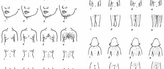
4. Rheumatic erythema - develops in less than 6% of cases.
A pink or pale red, non-itchy, ring-shaped rash. Localized on the body or limbs, but not on the face. The rash may appear, disappear, or appear again.
5. Subcutaneous nodules - develop in less than 10% of cases.
Dense, painless formations from a few millimeters to 2 cm. They persist for no more than a month. Localization of nodules is often above the bone, on the extensor surfaces, symmetrically. The skin over the nodule is not inflamed and mobile.
Minor criteria:
- Arthralgia is pain in the joints.
- Fever (above 38.5 °C).
- Increased erythrocyte sedimentation rate (ESR) - above 60 mm/h, C-reactive protein (CRP) - above 30 mg/l.
- Prolongation of the PR interval on the ECG.
The diagnosis of ARF is established on the basis of:
- the fact of having suffered GABHS-pharyngitis - confirmed by a positive rapid test, bacteriological examination at the time of acute infection or an increase in the titer of antistreptolysin-O (ASLO) already during the occurrence of complications;
- Jones criteria: 2 major criteria, 1 major and 2 minor criteria, or 3 minor criteria if the patient has previously suffered ARF.
All patients with suspected ARF must undergo an ECG and EchoCG (ultrasound of the heart) to identify morphological changes in the heart valves and signs of pathological regurgitation (backflow of blood). Laboratory tests according to indications (since they are nonspecific) - ESR and CRP. If signs of chorea are present, a neurological examination is necessary.
Publications in the media
Acute rheumatic fever (ARF) is a systemic inflammatory disease of connective tissue involving the heart and joints in the pathological process, initiated by group A β-hemolytic streptococcus, occurring in genetically predisposed people. The term rheumatism, widely used in practice, is currently used to designate a pathological condition that combines acute rheumatic fever and rheumatic heart disease.
Statistical data . Incidence: 2.1 per 100,000 population in 2001. The incidence of rheumatic heart disease in Russia is 0.17%. The predominant age is 8–15 years. Etiology. -Hemolytic streptococcus of group A, “rheumatogenic” serotypes M3, M5, M18, M24. The M protein on the streptococcal membrane has antigenic determinants similar to components of the heart muscle, brain and synovial membranes.
Genetic aspects . Ag D8/17 B-lymphocytes are detected in 75% of patients with ARF. Pathogenesis • Antistreptococcal antibodies are responsible for selective damage to the heart valves and myocardium with the development of immune aseptic inflammation, cross-reacting with heart tissue (molecular mimicry) • M-protein has the properties of a “superantigen” that causes activation of T- and B-lymphocytes without preliminary processing of Ag- presenting cells and interactions with class II molecules of the major histocompatibility complex.
Clinical picture • Onset of the disease. In more than half of the cases, 2–4 weeks after a streptococcal nasopharyngeal infection, fever, asymmetric joint pain, pain in the heart, shortness of breath, and palpitations occur. In other patients, the onset occurs as a monosyndrome (carditis, arthritis or chorea) • A repeated attack occurs with symptoms of carditis.
• Arthralgia and rheumatic polyarthritis (80% of patients) - typical reactive synovitis with fluid effusion into the joint cavity, swelling and redness of the periarticular tissues, sometimes with severe pain, tenderness and limitation of active and passive movements. Characteristic features:
•• damage to large joints (knee [most often], ankle, elbow, shoulder and much less often - wrist) •• symmetry of the lesion •• migrating, volatile nature of arthritis •• complete reversibility of the articular syndrome, no changes on radiographs, restoration of joint function • • in children, signs of arthritis, as a rule, completely disappear, and in adults a persistent course can be observed, leading to the development of Jaccoud's syndrome (painless deformation of the hands with ulnar deviation without inflammation in the joints and without dysfunction of the joint) •• with rheumatism, more often with repeated attacks, polyarthralgia rather than arthritis often occurs.
• Fever (90%). • Subcutaneous nodules (10% of patients), grain to pea-sized, localized in the periarticular tissues may appear during acute rheumatic fever. These nodules do not bother patients, they are painless, and the skin over them is not changed. Involution of the nodules occurs over a period of several days to several weeks. Observed only in children.
• Ring-shaped erythema - pale pinkish-red spots up to 5-7 cm in diameter with clear, not always smooth edges. Characterized by localization on the skin of the chest, abdomen, back and limbs, spontaneous disappearance and (rarely) recurrence. Occurs in less than 5% of patients. Erythema nodosum is not typical for rheumatism. • Chorea occurs in 10–15% of patients, more often in girls, 1–2 months after a streptococcal infection. It is chaotic involuntary twitching of the limbs and facial muscles. Characteristic is the complete disappearance of symptoms during sleep. • Rheumatic carditis (rheumatic carditis) can be primary (first attack) and recurrent (repeated attacks), with or without the formation of valve disease. Clinical symptoms of rheumatic carditis: •• Cardialgia . Characterized by prolonged stabbing, aching pain in the heart area, usually without irradiation. With pericarditis, pain is associated with breathing and intensifies in a horizontal position. •• In most cases an enlarged heart , which is not always detected physically. A chest x-ray is recommended. •• Arrhythmias as a manifestation of myocarditis ••• Sinus tachycardia (100 or more per minute), recorded at rest ••• Atrial fibrillation occurs, as a rule, when signs of mitral valve stenosis appear ••• Ventricular (rare) or supraventricular extrasystoles •• • Atrioventricular block with prolongation of the P-Q interval over 0.2 s or the appearance of Wenckebach (Samoilov-Wenckebach) periods. •• Decrease in sonority of the first tone at the apex of the heart, appearance of the third tone and murmurs. A gentle blowing systolic murmur with a tendency to increase in intensity and conducted into the axillary region is a symptom of mitral valvulitis. Protodiastolic murmur along the left sternal border is a sign of aortic valvulitis. •• Signs of congestive heart failure are rarely observed with primary rheumatic carditis; much more often they accompany recurrent rheumatic carditis. • Acute rheumatic fever lasts 6–12 weeks. There is no chronic or continuously relapsing course.
Laboratory data • Increased ESR, increased titers of antistreptolysin O, AT to DNase in a titer of more than 1:250 • Bacteriological examination of a throat smear reveals group A -hemolytic streptococcus. Instrumental data • ECG • Chest X-ray • EchoCG. Differential diagnosis • Reactive arthritis • Lyme disease • Infective endocarditis • Viral (Coxsackie B) myocarditis • Mitral valve prolapse • Functional heart murmur. Kissel-Jones-Nesterov diagnostic criteria (revised 1992) • Major criteria: •• carditis •• polyarthritis •• chorea •• annular erythema •• subcutaneous nodules • Minor criteria: •• clinical symptoms (fever, joint pain) • • laboratory changes (increased ESR, appearance of CRP, prolongation of the P–Q interval •• Additional signs: positive cultures from the tonsils for group A β-hemolytic streptococcus, increased titers of antistreptolysin-O and/or other antistreptococcal antibodies. The presence of two large or one large and two minor criteria in combination with the mandatory presence of additional signs allows us to consider the diagnosis of acute rheumatic fever reliable.Despite the presence of time-tested criteria, the diagnosis of ARF continues to be a problem, since individual criteria (fever, ESR, etc.) are not specific, and subcutaneous nodules and Annular erythema is rarely observed.
TREATMENT Management tactics . For acute rheumatic fever, hospitalization, bed rest, and a diet low in salt and rich in vitamins and protein are indicated. The basis of drug treatment is antibacterial and anti-inflammatory therapy. Drug treatment • Antibacterial therapy •• Benzylpenicillin 1.5–4 million units/day for adolescents and 400–600,000 units/day for children for 10–14 days, followed by a transition to benzathine benzylpenicillin or benzathine benzylpenicillin + benzylpenicillin procaine •• When if you are allergic to penicillins, you should choose a macrolide: roxithromycin for children 5–8 mg/kg/day, adults 150 mg 2 times/day, clarithromycin for children 7.5 mg/kg/day, adults 250 mg 2 times/day. It should be remembered that streptococcal resistance to erythromycin is currently increasing. • NSAIDs for 3.5–4 months (during treatment it is necessary to periodically conduct blood tests, urine tests, and liver function tests) •• Diclofenac 50 mg 3 times a day. • The use of GC is most justified for pancarditis. One of the treatment regimens is prednisolone 20–30 mg/day until clinical effect, then gradually reducing the dose over 20–30 days.
Prevention • Primary prevention : rational treatment of streptococcal diseases of the oropharynx for 10 days •• Aminopenicillins ••• amoxicillin 750 mg/day for children, 1500 mg/day for adults •• Cephalosporins ••• cephalexin for children with body weight <40 kg - 25– 50 mg/kg/day, adults 250–500 mg 2–4 times/day (daily dose 1–2 g) ••• cefaclor children 20 mg/kg 3 times/day, adults 750 mg 3 times/day ••• cefuroxime for children 125–250 mg 2 times a day, adults 0.25–0.5 g 2 times a day •• Macrolides ••• roxithromycin for children 5–8 mg/kg/day, adults 150 mg 2 times a day days •• Penicillins with β-lactamase inhibitors ••• amoxicillin + clavulanic acid (adults 375 mg 3 times / day) for 10 days. • Secondary prevention is indicated for patients who have had acute rheumatic fever to prevent relapses. The best results are achieved by year-round bicillin prophylaxis (benzathine benzylpenicillin 600,000–1,200,000 units (children), 2,400,000 units (adults) once every 3 weeks. Benzathine benzylpenicillin + benzylpenicillin procaine 1,500,000 units once every 10–12 days. D duration of secondary prophylaxis •• at least 5 years after the last reliable rheumatic attack for patients who have had acute rheumatic fever without carditis •• more than 5 years - for patients who have had rheumatic carditis •• with relapses for life • Prevention of infective endocarditis against the background of formed rheumatic heart disease with carrying out any surgical interventions (tooth extraction, abortion, abdominal surgery) - see Infectious endocarditis.
Synonym . Sokolsky-Buyo disease. Abbreviations. ARF - acute rheumatic fever.
ICD-10 • I00 Rheumatic fever without mention of cardiac involvement • I01 Rheumatic fever with cardiac involvement
Treatment of ARF
Eradication of GABHS infection is necessary regardless of whether there are signs of pharyngitis. Antibacterial therapy is carried out similarly to therapy for acute tonsillopharyngitis.
Symptomatic therapy:
- arthritis - non-steroidal anti-inflammatory drugs to relieve pain and prevent the involvement of new joints;
- carditis - treatment is carried out only if heart failure develops;
- chorea - usually does not require treatment, but sometimes it may be necessary to prescribe antipsychotics and anticonvulsants;
- erythema and subcutaneous nodules - no treatment.
Treatment and rehabilitation of the patient
Drug therapy and exercise therapy are mainly used. If the defects are significant, surgery may be necessary.
The following groups of drugs are used:
- glucocorticosteroids (prednisolone, dexamethazorne) - the main group that has anti-inflammatory and immunosuppressive activity;
- NSAIDs (aspirin, indomethacin, ibuprofen);
- antibiotics (penicillins, sulfonamides and others) - to combat infectious complications.
Cardiotonic, antiarrhythmic, diuretic and other drugs are used as symptomatic therapy.
For the purpose of further rehabilitation, sanitary-resort treatment, moderate sports, and physiotherapy (hydrotherapy, balneotherapy) are indicated.
Prevention of ARF
Primary is timely diagnosis and treatment of GABHS pharyngitis.
Secondary - prevention of new episodes of GABHS infection, including eradication of GABHS even in asymptomatic carriers.
The duration of antibiotic prophylaxis is determined based on the characteristics of the existing pathological process. If we are talking about post-streptococcal arthritis, it can be limited to 1-2 years. In case of ARF without carditis, the duration of taking antibiotics is 5 years or until the patient is 21 years old (whichever is longer), ARF with carditis without consequences is 10 years or up to 21 years old (whichever is longer), ARF with damage to the heart valves is 10 years or up to 40 years old ( whichever is longer), and sometimes for life.
How is ARF treated at the Rassvet Clinic?
We provide timely diagnosis and adequate treatment of GABHS pharyngitis, which reduces the incidence of ARF by almost 70%. When choosing antibacterial therapy, we always give preference to penicillin antibiotics - as the most effective drugs that have been proven to reduce the incidence of ARF. We never reduce the course of antibiotic therapy when there is clinical improvement. If GABHS pharyngitis is detected, we do not prescribe local treatment (rinses, sprays) to the detriment of systemic antibacterial therapy.
We provide adequate eradication therapy for GABHS in ARF in order to prevent relapses and progression of rheumatic heart disease. We offer a full examination for diagnosed ARF - ECG, ECHO-CG, consultation with a cardiologist and neurologist with the selection of the necessary therapy.
Author:
Chekaldina Elena Vladimirovna otorhinolaryngologist, Ph.D.