The world of bacteria is surprisingly diverse and very rich. They are found everywhere: in the air, soil, on human skin, on his mucous membranes. Under certain circumstances, bacteria become dangerous to humans, causing serious illness. Some of them can be easily treated with antibiotics or even regular antiseptics, while others are much more difficult to get rid of. Therefore, when making a diagnosis, as well as when prescribing treatment, gram-positive and gram-negative bacteria are isolated. This method of dividing microorganisms was proposed back in the 19th century, but is still used today.
World of bacteria
The kingdom of microorganisms is so diverse and complex that even modern science has not yet fully studied it. There are bacteria that survive at high temperatures and do not die even after prolonged boiling, while others die at the slightest change in temperature or composition of the external environment, for example after adding regular sugar. Some microorganisms thrive in hot springs, in acid, or feed on methane or other chemicals.
Bacteria are the most ancient organisms and are very widespread in the world. They are found everywhere: on the ocean floor, in the air, in the soil - even at great depths, in the body of living beings. Moreover, science has proven that there are 10 times more bacterial cells inside a person than their own. Some microorganisms simply live next to other living beings, while others actively interact with them. They can be beneficial or cause various diseases. Moreover, there are tens of times more beneficial bacteria than pathogenic ones.
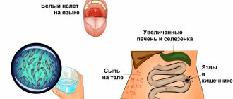
Many microorganisms are beneficial. For example, those that live in the human intestine are involved in digestion and protect it from infections. These are lactobacilli and bifidobacteria. About 40 million species of bacteria live in the human oral cavity, but only 5% of them are pathogenic. There are microorganisms that participate in the decomposition of waste. But, despite the fact that there are still more beneficial bacteria, their pathogenic types cause a lot of harm, as they cause dangerous diseases. Many people around the world still die from tuberculosis, cholera, tetanus, typhoid fever, botulism and other infections. Therefore, it is very important to learn how to interact correctly with the world of bacteria.
Features of microflora
A significant number of different microorganisms constantly live in the human body. Their presence ensures the normal functioning of all systems and organs.
Microorganisms, such as bacilli and cocci, are found in the mucous membranes of the mouth, nose, intestines, and in women also in the vagina.
In a normal state, when the immune system performs its functions in full, a balance is maintained between beneficial and pathogenic bacilli.
The cells of the mucous membrane contain glycogen, which serves as food for bacteria. Depending on the form, microorganisms are defined as rods and cocci, having a positive or negative gram reaction.
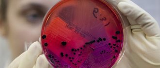
In modern gynecology, a smear is considered one of the methods for identifying the pathology of the female reproductive system. The microflora of the female genital organ includes mainly lactobacilli or gram bacilli.
All rods living in the vagina of women are divided into the following groups:
- obligate;
- transient;
- optional.
Obligate rods include those that are constantly, in the required quantity, present in the mucous membrane of the female genital organ. The sticks create a protective barrier that prevents the penetration of pathogenic microorganisms.
Transient cocci and bacilli include those that accidentally enter the vaginal mucosa. Among such representatives there may be beneficial, neutral, and pathogenic species.
Gram rods, which are examined after taking a smear, carry information about the properties of the microorganism.
Facultative microbes are not found in all women and this is a kind of norm. The qualitative composition of microflora depends on several factors.
Changes occur during menstruation, with age and with diseases of the genital area. There is a clear dependence on climate, intimate hygiene and the state of immunity.
The physiology of the female reproductive system is designed in such a way that lactobacilli and bifidobacteria mainly live and develop on the walls of the vagina.
In healthy women, these rods form an acidic environment in the vagina. In an acidified environment, the reproduction and development of other bacteria becomes impossible.
Thanks to this protective mechanism, a healthy environment is maintained in the vagina. An important feature of the rods is the ability to firmly attach to the mucous membrane, creating a reliable barrier to pathogenic bacteria.
A smear, when taken from women for preventive purposes, almost always shows a positive gram reaction.
READ Urogenital smear procedure - what is it?
When the normal state of microflora is disrupted, vaginal dysbiosis appears and develops.
Pathogenic bacilli and cocci are found in significant quantities. Accordingly, gram rods in the smear will have a negative sign.
This condition is usually caused by hormonal imbalance, long-term treatment with antibiotics, infectious infections and poor hygiene.
Dysbacteriosis often develops when the immune system is weakened. Treatment is carried out individually and only after the gynecologist takes a smear and makes the correct diagnosis.
Gram method
People have been looking for ways to treat infectious diseases for a long time. Once the existence of pathogenic bacteria has been discovered, scientists try to classify them in order to figure out how to fight them. The best method was proposed in 1884 by the physician Hans Christian Gram. It is quite simple, but informative and is still used today. This method distinguishes between gram-positive and gram-negative bacteria.
Dr. Gram used a purple dye to study microorganisms and noticed that some of them were stainable, while others were not. He found out that this is due to the characteristics of the cell walls of bacteria. Since these microorganisms consist of one, or less often two cells, it is very important for them to have a strong shell. Therefore, their cell walls have a complex structure. They protect the internal environment from the penetration of liquids. The structure of gram-negative bacteria is most complex. They are resistant to the penetration of saliva, gastric juice and other liquids.
The essence of the Gram method is that the medium under study is treated with aniline dye, fixed with iodine, and then washed off with alcohol. In this case, gram-negative bacteria become discolored, and gram-positive bacteria acquire a blue color. After repeated treatment with red dye, negative species may turn pink, and dead microorganisms are brighter in color.
Imbalance
Gram-positive and gram-negative bacteria live in almost all parts of the human body, on the skin, intestines, mouth and nose. Sometimes they cause disease, but in most cases microorganisms live in harmony with their hosts, providing vital functions necessary for human survival.
Reasons why the balance is disturbed:
- Poor diet and poor food choices.
- Excessive use of antibiotics.
- Lack of sleep.
- Emotional stress.
Overpopulation of one type of bacteria leads to infections and other health problems. With a decrease in good microorganisms in the intestines, the protective barrier is lowered, which allows harmful bacteria, toxins and large protein molecules to enter the body.
What happens when bad bacteria over-colonize:
- Intestinal permeability increases.
- Bad bacteria spreads into the urinary system, causing a urinary tract infection.
- Fatigue increases due to toxin overload.
- The activity of the immune system decreases, which contributes to the development of allergies and diseases associated with an autoimmune reaction, such as Crohn's disease, ulcerative colitis, rheumatoid arthritis, skin infections and vaginal tract infections.
- Diarrhea develops due to an intestinal reaction to harmful bacteria.
Bacteria are also present in healthy lungs. When the delicate balance of bacterial populations in the lungs is disrupted, it can lead to the development of diseases such as asthma and chronic obstructive pulmonary disease.
The main attention should be paid to the consequences of an imbalance in the microbial balance in the digestive system. It can have negative consequences not only for the gastrointestinal tract, but also for the nervous, metabolic and immunological systems.
Therefore, maintaining balance in the eternal battle between “good” and “bad” bacteria, both gram-positive and gram-negative microbes, is the path to health and wellness.
Application of the method in medicine
Gram's method of dividing microorganisms into gram-positive and gram-negative bacteria contributed to the improvement of microbiological research. It helps identify drug resistance of pathogenic species and develop new antibiotics to combat them. After all, the strong cell wall of gram-negative bacteria makes them insensitive to conventional antibacterial drugs. And the shell of gram-positive microorganisms, although very thick, is permeable to liquids and antibiotics.
The Gram method made it possible to divide all microorganisms into two large groups. Their features and characteristics help to choose more appropriate treatment for infectious diseases. Gram-positive bacteria, which quickly turn blue with aniline dye, form spores, exotoxins, and are therefore quite dangerous to health. But their shell is permeable to antibacterial drugs.
Like gram-positive, gram-negative bacteria are causative agents of serious diseases. They do not form spores, and in many cases are opportunistic. But under certain conditions they begin to release endotoxins and cause severe inflammation and intoxication. Due to the complex structure of the cell wall, they are almost insensitive to antibiotics.
The human body contains both types of these microorganisms. The correct ratio of gram-positive and gram-negative bacteria maintains the normal microflora of the vagina, intestines, and oral cavity. This helps protect the body from infections.
Why are they needed?
There are many types of bacteria that benefit humans:
- Microbes in the gut break down many of the proteins, lipids and carbohydrates into nutrients that you then absorb. Moreover, the microbes produce beneficial compounds, such as vitamins and anti-inflammatories, that the human genome cannot produce. Millions of bacteria inhabit the human digestive system, which help digest food and produce vital vitamins such as biotin and vitamin K.
- Bacteria in the gut work with the immune system to protect the body from disease.
- Microorganisms found inside the stomach help maintain the pH and acidity levels in the stomach.
- Protect the skin. The many bacteria on the skin (almost 200 different species in a healthy person) control the skin's environment and resources, preventing other harmful microbes from gaining a foothold.
- Beneficial bacteria that can produce oxygen are used to create antibiotics.
- Bacteria are used in food production to make yoghurts and fermented foods.
Gram-positive flora
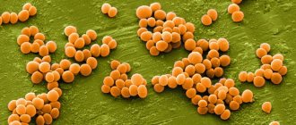
Most bacteria that can be stained with a purple dye, that is, have a permeable cell wall, are dangerous to humans. These include streptococci, staphylococci, listeria, bacilli, clostridia, mycobacteria, and actinomycetes. Particularly dangerous is Staphylococcus aureus, which affects a weakened body and, without treatment, quickly leads to the death of the patient. But they also include beneficial lactic acid bacteria.
Gram-positive microorganisms affect the respiratory tract, heart muscle, brain, and skin. They provoke purulent infection in wounds and blood poisoning.
Diseases they cause
It is gram-positive bacteria that cause such common infectious diseases as:
- tonsillitis, pharyngitis;
- sinusitis, otitis;
- rheumatism;
- blood poisoning;
- pneumonia;
- brain inflammation;
- anthrax;
- food poisoning;
- botulism;
- diphtheria;
- tetanus;
- gas gangrene.
Gram-negative bacteria
The list of them is quite large, but among them there are many that do not bring any harm to humans. These include mainly opportunistic microorganisms. Under normal conditions, they live in the human body without causing harm. The most common are the following gram-negative bacteria. Their types are varied:
- proteobacteria;
- pseudomonas;
- chlamydia;
- meningococci;
- Brucella;
- spirochetes;
- gonococci;
- Helicobacter.
Microorganisms that do not stain purple are also resistant to any antibodies and antibacterial drugs. Therefore, the diseases caused by them are very difficult to treat.
Pathogenicity
Most microorganisms pathogenic to humans are Gram+. Six genera of Gram+ organisms are typical human pathogens. Two of them, streptococci and staphylococci, are cocci (spherical bacteria. Bacteria are united in the kingdom Eubacteria or Bacteria. The kingdom is divided into several types: Gr...). The rest are rod-shaped and divide further to form spores.
Non-spore-forming: Corynebacterium and Listeria; spore-formers: Bacillus and Clostridia. Spore-formers can be divided into facultative anaerobesAnaerobes are microorganisms, including bacteria, energy process... Bacilli and obligate anaerobesAnaerobes are microorganisms, including bacteria, energy process... Clostridia.
What diseases cause
Under certain conditions, gram-negative bacteria can cause serious illness. This is due to the fact that the complex shell of these microorganisms, when destroyed, releases many toxins, which, spreading through the human bloodstream, cause severe intoxication. It turns out that it is not the bacteria themselves that are pathogenic, but the features of their cell membrane - the lipopolysaccharide layer, which causes the body's immune response. They lead to the development of inflammation. But if a person’s immunity is in order, he can easily cope with such microorganisms, and he is not afraid of infection.
Gram-negative bacteria include microorganisms that cause gonorrhea, syphilis, meningitis and respiratory diseases. These bacteria are especially common and cause damage to the respiratory and urinary tract and gastrointestinal tract. Gram-negative include such well-known infectious agents as Proteus, Escherichia, Enterobacteriaceae, and Salmonella. They cause salmonellosis, meningitis, typhoid fever, and dysentery. In addition, it is these resistant microorganisms that cause severe nosocomial infections. After all, they can survive even after serious disinfection.
Recommendations
- Fundamentals of Biology (March 18, 2021). "Bacteria".
- ^ a b
Madigan, Michael T.;
Martinko, John M. (2006). Broca's Biology of Microorganisms
(11th ed.). Pearson Prentice Hall. ISBN 978-0131443297. - Gibbons, N. E.; Murray, R. G. E. (1978). "Proposals for higher taxa of bacteria". International Journal of Systematic and Evolutionary Microbiology
.
28
(1): 1–6. Doi:10.1099/00207713-28-1-1. - ^ a b
Woese, C. R. (1987).
"Bacterial evolution". Microbiology Reviews
.
51
(2):221–271. Doi:10.1128/MMBR.51.2.221-271.1987. PMC 373105. PMID 2439888. - ^ a b c d f f gram h Gupta
, R. S. (1998).
"Protein phylogeny and signature sequences: reassessing the evolutionary relationships among archaebacteria, eubacteria, and eukaryotes." Microbiology and Molecular Biology Reviews
.
62
(4):1435–1491. Doi:10.1128/MMBR.62.4.1435-1491.1998. PMC 98952. PMID 9841678. - ^ a b c
Gupta, R. S. (2000).
"Natural evolutionary relationships among prokaryotes" (PDF). Critical Reviews in Microbiology
.
26
(2): 111–131. CiteSeerX 10.1.1.496.1356. Doi:10.1080/10408410091154219. PMID 10890353. S2CID 30541897. - ^ a b
Desvaux, M.;
Hébraud, M.; Talon, R.; Henderson, I. R. (2009). "Secretion and subcellular localization of bacterial proteins: a problem of semantic awareness". Trends in Microbiology
.
17
(4): 139–145. doi:10.1016/j.tim.2009.01.004. PMID 19299134. - ^ a b
Sutcliffe, I. K. (2010).
"A phylum-level perspective on bacterial cell envelope architecture". Trends in Microbiology
.
18
(10): 464–470. doi:10.1016/j.tim.2010.06.005. PMID 20637628. - ^ a b c d e
Gupta, R. S. (1998).
“What are archaebacteria: the third domain of life or monodermal prokaryotes associated with gram-positive bacteria? New proposal for the classification of prokaryotic organisms." Molecular Microbiology
.
29
(3):695–707. Doi:10.1046/j.1365-2958.1998.00978.x. PMID 9723910. S2CID 41206658. - ^ a b c d f f gram
Gupta, R. S. (2011).
"The origin of didermal (Gram-negative) bacteria: Antibiotic selection pressure rather than endosymbiosis likely led to the evolution of dual-membrane bacterial cells." Anthony van Leeuwenhoek
.
100
(2): 171–182. Doi:10.1007/s10482-011-9616-8. PMC 3133647. PMID 21717204. - ^ a b c
Marchandin, H.;
Teyssier, C.; Campos, J.; Jean-Pierre, H.; Roger, F.; Gay, B.; Carlier, J.-P.; Yumas-Bilak, E. (2009). “Negativicoccus succinicivorans gen. Nov., sp. Nov., isolated from human clinical specimens, the description of the family Veillonellaceae and the description of Negativicutes classis nov., Selenomonadales ord. Nov. and Acidaminococcaceae fam. Nov. In the bacterial phylum Firmicutes". International Journal of Systematic and Evolutionary Microbiology
.
60
(6):1271–1279. doi:10.1099/ijs.0.013102-0. PMID 19667386. - Yabe, S.; Aiba, Y.; Sakai, Y.; Hazaka, M.; Yokota, A. (2010). “ Thermohemmatispora onicobensis
gene.
nov., sp. Nov AND Thermogemmatispora foliorum
sp.
nov., isolated from leaf litter on geothermal soils, and description of Thermogemmatisporaceae fam. November and Thermogemmatisporales ord. November In the class Ktedonobacteria." International Journal of Systematic and Evolutionary Microbiology
.
61
(4):903–910. doi:10.1099/ijs.0.024877-0. PMID 20495028. - Sutcliffe, I. C. (2011). "Cell wall architecture in Chloroflexi: the shifting front line in a phylogenetic war for territory". Environmental Microbiology
.
13
(2): 279–282. doi:10.1111/j.1462-2920.2010.02339.x. PMID 20860732. - Hugenholtz, P.; Tyson, G. W.; Webb, R.I.; Wagner, A. M.; Blackall, L. L. (2001). "Study of a candidate subdivision of TM7, a recently recognized major lineage of domain bacteria with no known pure culture representatives." Applied and Environmental Microbiology
.
67
(1):411–419. Doi:10.1128/AEM.67.1.411-419.2001. PMC 92593. PMID 11133473. - Cavaletti, L.; Monciardini, P.; Bamonte, R.; Schumann, P.; Rohde, M.; Sosio, M.; Donadio, S. (2006). "A new lineage of filamentous, spore-forming, Gram-positive bacteria from soil." Applied and Environmental Microbiology
.
72
(6):4360–4369. Doi:10.1128/AEM.00132-06. PMC 1489649. PMID 16751552. - Gladwin, Mark; Trattler, Bill (2007). Clinical microbiology has become ridiculously easy
. Miami, FL: MedMaster. pp. 4–5. ISBN 978-0-940780-81-1. - Sahebnasagh, R.; Saderi, H.; Oulya, P. (September 4–7, 2011). Detection of methicillin-resistant
Staphylococcus aureus
strains from clinical samples in Tehran by detection of
MEKA
and
nuclear
genes
. First Iranian International Congress on Medical Bacteriology. Tabriz, Iran. - McDonald, Mhairi (2015). Avery's Neonatology: Pathophysiology and Treatment of the Newborn
. Philadelphia: Wolters Kluwer. ISBN 9781451192681. Access provided by the University of Pittsburgh. - Lau, S. K. P.; McNabb, A.; Wu, G. K. S.; Hoang, L.; Fung, A. M. Y.; Chung, L. M. W.; Woo, P. C. Y.; Yuen, K.-Y. (November 22, 2006). “Catabacter hongkongensis gen. Nov., Sp. Nov., Isolated from blood cultures of patients from Hong Kong and Canada." Journal of Clinical Microbiology
.
45
(2):395–401. Doi:10.1128/jcm.01831-06. ISSN 0095-1137. - ^ a b c
Johnston, C.;
Martin, B.; Fichant, G.; Polard, P; Claveris, J. P. (2014). "Bacterial transformation: distribution, common mechanisms and divergent control". Nature Reviews.
Microbiology .
12
(3): 181–96. Doi:10.1038/nrmicro3199. PMID 24509783. S2CID 23559881. - Michod, R.E.; Bernstein, H.; Nedelcu, A. M. (2008). "The adaptive significance of sex in microbial pathogens". Infection, genetics and evolution
.
8
(3): 267–85. doi:10.1016/j.meegid.2008.01.002. PMID 18295550. - " Emerging Infectious Diseases
Journal Style Guide."
CDC.gov
. Centers for Disease Control and Prevention.
Use of this knowledge in the treatment of diseases
When diagnosing a disease to determine more effective treatment, the Gram method is necessarily used to determine which microorganisms caused the disease: gram-positive or gram-negative bacteria. Antibiotics are prescribed depending on this. After all, the wrong treatment can only worsen the situation.
To determine the pathogen, sputum, nasal or vaginal discharge, and analysis of stool, synovial or pleural fluid are examined. These samples are examined using the Gram method.
The most difficult diseases to treat are those caused by gram-negative bacteria. They are mainly affected by a combination of two antibiotics or new generation drugs. Ampicillin or Amoxicillin, Chloramphenicol, Streptomycin, as well as a group of cephalosporins can be effective against them. They can cope with the outer membrane of such bacteria.
Knowledge about the structure of the bacterial wall has improved the effectiveness of the treatment of infectious diseases.
Difference between gram-positive and gram-negative bacteria
The key difference between gram-positive and gram-negative bacteria is that gram-positive bacteria have a thick peptidoglycan layer, hence the purple color.
The key difference between Gram-positive and Gram-negative bacteria is that Gram-positive bacteria have a thick peptidoglycan layer, hence appearing purple in color, while Gram-negative bacteria have a thin peptidoglycan layer, hence appearing in pink color at the end of the Gram staining method.
Bacteria are ubiquitous prokaryotes, single-celled and microscopic. They belong to the Kingdom of Monera along with the Archaea. Moreover, they have a very simple cellular organization. Structurally, they have no internal compartments; nucleus and membrane-bound other organelles. Moreover, bacteria can be classified based on several characteristics such as shape, genetic composition, cell wall composition, number of flagella, nutrition, biochemical reactions, etc. Gram staining is one of the techniques that is commonly used in the identification and characterization of bacteria . According to Gram stain, there are two categories of bacteria such as gram-positive and gram-negative bacteria. These two groups of bacteria differ from each other in the composition of their cell wall. Therefore, when stained gram by gram, they are colored differently.
1. Overview and main differences
2. What are gram-positive bacteria
3. What are gram-negative bacteria?
4. Similarities between gram-positive and gram-negative bacteria
5. Side-by-side comparison - gram-positive and gram-negative bacteria in tabular form
6. Summary
Method for determining “gram-positive” and “gram-negative”
If there are signs of infection in the body, the doctor will prescribe a Gram stain. This is necessary to determine the type of infection: bacterial, viral, fungal or parasitic, since each type is treated differently.
A sample for research can be in the form:
- sputum coughed up from the lungs;
- urine collected in a special container;
- blood donated in the laboratory;
- saliva;
- vaginal smears;
- feces
Using certain techniques, the type of microorganisms in the pleural and peritoneal fluid is determined.
The laboratory technician uses a special staining technique to make the cellular organisms easier to see under the microscope. If the Gram stain results are negative, it means that no bacteria were found in the sample. If positive, microorganisms were present. The results will also include information about the shape of the bacteria, which can indicate the type of infection.
Although the results may not identify the exact type of bacteria, they help the doctor get closer to figuring out what causes the disease and how best to treat it.
Gram stain results may also show the presence of a fungal infection (yeast or mold).
When treating diseases caused by gram-positive organisms, the doctor needs the following information:
- type of bacteria;
- antimicrobial resistance;
- whether bacteria produce toxins.
What are gram-positive bacteria?
Gram-positive bacteria are a group of bacteria that stain purple using the gram staining technique. The reason for the purple color is that Gram-positive bacteria have a thick peptidoglycan layer in their cell walls. Typically, the thickness of the peptidoglycan layer of Gram-positive bacteria is 20-80 nm, and teichoic acids are present on it.
A thick layer of peptidoglycan preserves the primary stain, which is crystal violet; hence, under a microscope they appear purple or crystal violet in color. Although the bleach removes the primary stain, the stain does not completely disappear from the thick peptidoglycan layer. Staphylococcus, Streptococcus, Bacillus, Corynebacterium, Listeria and Clostridia species are some examples of Gram-positive bacteria.
What are gram-negative bacteria?
Gram-negative bacteria are a group of bacteria that have a thin peptidoglycan layer in their cell wall. Consequently, they are unable to retain the primary stain. Characteristically, Gram-negative bacteria have an additional membrane called an outer membrane, which is absent in Gram-positive bacteria. In addition, this outer membrane contains lipopolysaccharides. Additionally, although the outer membrane is present, it is destroyed after application of the bleaching agent. The peptidoglycan layer then becomes porous and the crystal violet color leaves the cell wall completely.
Consequently, gram-negative bacteria appear in a pink secondary color. Compared to gram-positive bacteria, gram-negative bacteria are more resistant to cell wall-targeted antibiotics. This is due to the presence of an outer membrane. Moreover, their cell walls have two layers and there is a periplasmic space in the cell wall. Moreover, the cell wall is uneven and less rigid compared to gram-positive bacteria. Escherichia coli, Pseudomonas, Neisseria, Chlamydia are some of the gram-negative bacteria.
Classification
Together with cell shape, Gram staining is a rapid method used to differentiate bacterial species. Such staining, together with tests for growth and antibiotic sensitivity, as well as other macroscopic and physiological tests, constitutes the complete basis for classifying and subdividing bacteria (for example, see Figure and pre-1990 versions of Bergey's Manual
).
Hierarchy of species identification in clinical settings
Historically, Kingdom Monera has been divided into four divisions based primarily on Gram staining: Firmicutes (stain positive), Gracilicutes (stain negative), Mollicutes (neutral stain), and Mendocutes (stain variable).[ 3] Based on the 16S ribosomal RNA phylogenetic studies of the late microbiologist Carl Woese and collaborators and colleagues at the University of Illinois, the monophyly of Gram-positive bacteria was inferred,[4] with great implications for the therapeutic and general study of these organisms. Based on molecular studies from 16S sequences, the university recognized twelve bacterial phyla. Two of them were gram-positive and were separated by the proportion of guanine and cytosine content in their DNA. The high G+C type consisted of Actinobacteria and the low G+C type contained Firmicutes.[4] Actinobacteria include Corynebacteria
,
Mycobacteria
,
Nocardia
and
Streptomyces
genera. Firmicutes (low G+C) have a GC content of 45–60%, but this is lower than Actinobacteria.[2]
What are the similarities between gram-positive and gram-negative bacteria?
- Gram-positive and gram-negative bacteria have similar cellular organizations.
- In addition, both are prokaryotic unicellular microorganisms that have capsules.
- And they both share the same chromosome and are ubiquitous.
- In addition, both may contain plasmids as extrachromosomal DNA.
- Additionally, both reproduce asexually through binary fission.
- Likewise, both undergo transformation, transduction and conjugation.
- Moreover, antibiotics suppress gram-positive and gram-negative bacteria.
- Their cell walls contain peptidoglycan.
- And also both types of bacteria have a surface layer (S-layer).
- They respond to the gram staining procedure.
- In addition, they cause diseases in humans, plants and animals.
Why are they dangerous?
Although there are many more good microorganisms than bad ones, some bacteria are harmful. When a person comes into contact with harmful organisms, they can enter the body, where they begin to rapidly multiply and release toxins that damage body tissues, causing illness.
Bad bacteria are called pathogenic because they cause diseases such as sore throat, staph infections, cholera, tuberculosis and food poisoning.
Some types of gram-positive bacteria can cause serious infectious diseases:
- Urinary tract infections.
- Tuberculosis.
- Diphtheria.
Infections caused by gram-negative bacteria:
- Pneumonia.
- Meningitis.
- Peritonitis.
- Blood infections.
What is the difference between gram-positive and gram-negative bacteria?
The main difference between gram-positive and gram-negative bacteria is that gram-positive bacteria have a thick peptidoglycan layer in their cell wall, while gram-negative bacteria have a thin peptidoglycan layer in their cell wall. In addition to the peptidoglycan layer, gram-negative bacteria have an outer membrane, while gram-positive bacteria do not have one. Therefore, this is also the difference between gram-positive and gram-negative bacteria. Additionally, Gram-negative bacteria have a periplasmic space and two layers in the cell wall, whereas Gram-positive bacteria do not have a periplasmic space and have a single layer of rigid and smooth cell wall.
The following infographic describes more facts about the difference between gram-positive and gram-negative bacteria.
Summary – Gram-positive and Gram-negative bacteria
Depending on the bacteria, the primary stain is absorbed and retained; crystal violet during Gram stain, there are two types of bacteria namely gram positive and gram negative. Gram-positive bacteria have a thick layer of peptidoglycan in their cell wall, while gram-negative bacteria have a thin layer of peptidoglycan. This is the main difference between gram-positive and gram-negative bacteria. Because of this difference in cell wall, Gram-positive bacteria stain purple and Gram-negative bacteria stain pink when stained by gram. Additionally, Gram-negative bacteria have an outer membrane, whereas Gram-positive bacteria do not. Due to the presence of an outer membrane, Gram-negative bacteria are resistant to antibiotics directed at the cell wall, while Gram-positive bacteria are susceptible to them. So this summarizes the difference between gram-positive and gram-negative bacteria.
The world of microbes is quite diverse and rich. Pathogenic microorganisms live in soil, air, skin and mucous membranes of humans. Under the influence of certain factors, bacteria become aggressive and provoke dangerous diseases in humans. Some types of microorganisms can be easily cured with antibiotic therapy. And some bacteria are resistant to medications and are very difficult to get rid of. For this reason, when making a diagnosis and choosing a treatment regimen, bacteria are divided into gram-positive and gram-negative. This technique was developed back in the 19th century and is used in modern medicine.
What are there
Single-celled microorganisms do not have a formed cell nucleus, and their genetic material floats freely in the cytoplasm. They differ in the nature of the cell walls, in shape and in the genetic mechanism of variability.
Forms
Bacteria exist in the following forms:
- commas - for example, Vibrio cholera;
- spherical (cocci) – staphylococcus and streptococcus;
- rod-shaped (bacillus) - E. coli and salmonella;
- spiral (Spirilla) – Treponema and Borellia;
- Flagellate – tetanus bacteria.
Gram-positive and gram-negative bacteria
Some organisms have more complex shapes, such as stalked, square, or star-shaped.
In relation to oxygen
- Aerobic, which requires oxygen gas to complete its own energy cycle. These microorganisms cannot grow without oxygen.
- Anaerobic, which do not need oxygen to carry out metabolic functions; moreover, it is toxic to most of them.
- Facultatively anaerobic are microorganisms that do not require the presence of oxygen to grow or produce energy; they can use it, but can grow at the expense of anaerobic organisms; such bacteria are characterized by a high degree of adaptation.
- Microaerophilic, which grow better in an atmosphere with low oxygen content.
A bacterial cell is surrounded by 2 protective membranes: an outer cell wall and an inner cell membrane. Some bacteria, such as mycoplasma, do not have a cell wall.
Other microorganisms may have a third, outer protective layer called a capsule. Tail-like projections often cover the surface of bacteria—long ones called flagella or short ones called pili—that help them move and attach.
Types
Gram-positive and gram-negative bacteria are determined by the Gram staining method. This is a common method used to differentiate microorganisms based on their different cell wall components.
| Types of Gram-positive bacteria | Properties |
| Actinomycetes | They are responsible for the occurrence of caries and filamentous respiratory diseases. |
| bacilli | Associated with food poisoning |
| Clostridium | They have different strains that cause food poisoning, botulism, gangrene and tetanus. |
| Corynebacteria | The main group is not pathogenic for humans, but one of them is the causative agent of diphtheria. |
| Enterococci | They cause diverticulitis and endocarditis, but are quite resistant to many antibiotics. |
| Gardnerella | They are associated with vaginitis. |
| Lactobacilli | This genus of bacteria contributes to the formation of vaginal microflora. |
| Listeria | One of this genus of bacteria is dangerous to humans, is a pathogen of the fetus, and is responsible for meningitis in newborns. |
| Mycoplasma | This genus of bacteria does not have a cell wall and causes walking pneumonia. |
| Mycobacteria | They can cause tuberculosis. |
| Streptococci | They are causative agents of pharyngitis, some of them affect complications during pregnancy. |
Gram-positive bacteria do not have an outer membrane, unlike gram-negative bacteria. It is for this reason that the latter microorganisms are more resistant to antibiotics. If the infection is caused by gram-negative organisms, a strong dose of antibiotics and strict adherence to treatment will be required to completely get rid of the harmful bacteria.
| Types of Gram Negative Bacteria | Properties |
| Bortadella | They are short rod-shaped organisms, the whooping cough bacillus. |
| Chlamydia | This small parasitic bacterium is similar to a virus and is the cause of diseases such as psittacosis, trachoma and nonspecific urethritis. |
| Enterobacteriaceae | It belongs to a genus of anaerobic, non-spore-forming bacteria and can cause urinary tract infections, soft tissue infections, septic arthritis, central nervous system infections, osteomyelitis and more. |
| Helicobacter | This type of bacteria is rod-shaped and causes peptic ulcers. |
| Klebsiella | The rod-shaped bacterium causes hemorrhagic pneumonia. |
| Neisseria | Microbes of this species colonize the mucous membranes, and their pathogenic forms are the causative agent of bacterial meningitis. |
| Proteus | This rod-shaped bacterium causes urinary tract and kidney infections. |
| Pseudomonas | This is a genus of aerobic bacteria with high metabolic activity. |
When bad microbes enter the human body, they begin to actively multiply, release toxins, damaging body tissues and causing illness. Harmful bacteria are called pathogenic bacteria and cause diseases such as sore throat, staph infections, cholera, tuberculosis and food poisoning.
What are bacteria?
The bacterial world is so abundant and diverse that to this day medicine has not been able to fully understand it. Bacteria are microscopic organisms. Microbes are very common and live everywhere: in the air and soil, at the bottom of reservoirs and in living organisms. Some microorganisms are able to survive at high temperatures, while others die with a slight change in temperature.
Bacteria living in the human body are conventionally divided into pathogenic and opportunistic. Some opportunistic bacteria are essential for the proper functioning of the body, such as bifidobacteria and lactobacilli. With weakened immunity, these microorganisms can provoke the development of inflammatory processes.
And pathogenic microorganisms can cause various life-threatening infectious diseases, such as:
To select a regimen for the treatment of infectious pathologies, scientists classify bacteria according to different criteria. The Danish doctor Gram, taking into account the size and structure of the shell, developed a method that divides all microbes into two groups: gram-positive and gram-negative. The latter have a thin protective layer, and gram-positive microorganisms are characterized by a large cell wall.
Chemical disinfection
The chemical treatment method is universal and most effective for various fields of activity. All products from the Septolite line from the Satellite company are suitable for killing bacteria. All products are certified and have high antimicrobial activity. It is suitable for processing surfaces and tools. The online store also offers products for express treatment and hand disinfection.
Various concentrations and exposure times in solution can provide both bacteriostatic and bactericidal effects on pathogenic strains of bacteria. On the website you can familiarize yourself with the products and detailed instructions written in accessible language. We care about the health of our clients. With Satellite products you can be 100% sure of safety.
Return to list of publications
What is the essence of the method?
According to the developed method, the microbe is exposed to dyes and bleaching agents. The complete staining process occurs according to the following scheme:
- The cleaned glass slide is covered with a thin layer of biological material collected for research. Then it dries naturally.
- Fixation is carried out, causing protein coagulation.
- The main dye is applied and left for a few minutes.
- Next, the pigment is removed and Lugol's iodide solution is dripped onto the glass.
- After a minute, Lugol's solution is removed, and the microbes are washed until bleached with ethanol.
- The glass is rinsed with distilled water for about a minute.
- Next, additional dye is applied to the glass and left for 4-5 minutes.
After the expiration date, rinsing and drying are carried out again.
Bacterial reaction to pigments
The main dye is methyl violet or another pigment. Gram-positive microorganisms are able to form a bond with this substance. Exposure to an iodide solution strengthens this connection. As a result, ethanol or acetone is not able to destroy it. With the additional use of fuchsin or safranin, the bacteria do not stain again.
Due to the peculiarities of the cell membrane, Gram-positive bacteria are perfectly painted purple or dark blue. The wall of such microorganisms does not have an additional outer membrane, thanks to which they withstand unfavorable conditions. For this reason, gram-positive microorganisms are exposed to medications.
Gram-negative bacteria do not form compounds with the basic dye. Alcohol easily destroys all formed bonds and discoloration of microorganisms occurs in the smear. For this reason, an additional dye is used to identify them. Under its influence, gram-negative microorganisms acquire a crimson or pink color. Dead bacteria are painted entirely in bright colors, while living bacteria are only partially and very pale. The shell of such microorganisms has an additional outer membrane, which firmly protects them from the influence of antibiotics.
Biomaterial for research
To obtain material for diagnosing infectious pathologies, the following studies are used:
- Stool analysis. This procedure helps detect toxins produced by Gram-positive microbes.
- Microscopic examination of sputum.
- Vaginal smear examination. Allows you to calculate the proportion of Gram-positive and negative bacteria in vaginitis.
In addition, in rare cases, the doctor may prescribe a puncture of pleural, perinatal or synovial fluid.
The importance of the Gram technique for medicine
The human body contains representatives of both types of bacteria. Their correct ratio supports healthy microflora. If the balance is disturbed, dangerous pathologies can develop.
Thanks to the Gram method, it is possible to determine the resistance of microorganisms to medications, as well as to develop new types of antibacterial medications to combat diseases. After all, gram-negative microorganisms have a strong cell wall that protects them from the negative effects of antibiotics. And gram-positive bacteria are characterized by a more permeable coating for medications. The test results allow the doctor to prescribe effective treatment for the infectious disease.
The essence of the method
In microbiology, this method is considered complex. Bacteria are affected by primary and additional dyes, as well as a bleaching agent. The entire coloring process can be visually represented by the following steps:
- The slide is cleaned of fat and covered with a thin layer of the collected smear, after which it is dried naturally.
- Fixed physically or chemically, causing coagulation of proteins.
- The first dye is applied through filter paper and left alone for a couple of minutes.
- After that, the pigment is removed, Lugol’s solution is added to the glass and left for one minute.
- The iodine solution is removed, and the bacteria are washed with ethyl alcohol until discolored.
- Rinse the glass with distilled water for about a minute.
- Apply a second dye to the glass and leave it for no more than 5 minutes.
- Rinse again and dry with filter paper.
Gram-positive microorganisms
Representatives of this bacterial world are stained according to the Gram method in purple or blue. Most of these bacteria are dangerous to the human body. They can affect the myocardium, skin, respiratory tract, brain, and also provoke the development of purulent infection and sepsis.
The main representatives of gram-positive microscopic organisms and the diseases they cause are indicated in the table.
| Name of the bacterium | Name of the disease |
| Coccal | |
| Staphylococcus | Blood sepsis Purulent rashes on the skin Fungal infections |
| Streptococcus | Rheumatism Tonsillitis and Pharyngitis Erysipelas Pneumonia Sepsis |
| Enterococci | Gastritis and Diverticulitis Meningitis Endocarditis and Pleurisy Dysbacteriosis Wound infections Prostatitis Cystitis and Urethritis Colpitis and Vulvitis Pyelonephritis Enteritis Peritonitis and Sepsis |
| Pneumococci | Otitis Generalized pneumonia Sinusitis |
| Rod-shaped | |
| Firmicutes | Toxic infections Anthrax |
| Clostridia | Botulism Gas gangrene Tetanus |
| Listeria | Encephalitis Bacillus Toxic infections Anthrax |
| Club-shaped | |
| Corynebacteria | Diphtheria |
| Actinobacteria | |
| Mycobacteria | Leprosy Tuberculosis |
| Actinomycetes | Actinomycosis |
Staphylococcus aureus is considered the most dangerous and aggressive. It penetrates the body and, if left untreated, can be fatal.
Gram-negative microorganisms
Representatives of this group do not turn dark blue or purple and are more difficult to treat. The list of these organisms is very large. It mainly includes opportunistic organisms that do not cause harm to the body.
The most common representatives and the diseases they cause are listed in the table.
| Name of the bacterium | Name of the disease |
| Chlamydia | Chlamydia Conjunctivitis |
| Spirochetes | Leptospirosis Syphilis Age-related typhus |
| Spirilla | Sodoku Gonococcus Gonorrhea |
| Salmonella | Salmonellosis Typhoid fever |
| Helicobacter | Stomach ulcer |
| Escherichia coli | Digestive disorders |
| Proteus | Acute intestinal infections |
| Klebsiella and Legionella | Breathing problems |
| Catharlisnia | Moraxella catharalis |
| Meningococci | Meningitis |
The body of a healthy person easily copes with the above bacteria.
Use of knowledge about microorganisms in medicine
To calculate an effective treatment regimen, the doctor prescribes diagnostics using the Gram method, which makes it possible to identify the causative agents of the disease. Antibiotic therapy is prescribed taking into account which of the two types of pathogenic microbes according to Gram provoked the pathology: positive or negative. Incorrectly selected therapy can aggravate the condition.
Gram-negative microorganisms are difficult to treat. In addition, they can be fatal. Usually, to cure this type of bacteria, two antibacterial drugs or modern new generation medications are prescribed. Chloramphenicol and Streptomycin, Ampicillin or Amoxicillin are considered effective in the treatment of such bacteria. In addition, drugs from the group of cephalosporins are used for treatment.
Knowledge about the structure of the cell wall has significantly improved the effectiveness of therapy for infectious diseases.
Loading…
Implications for medicine
The etiological test developed by Gram turned out to be in demand not only in microbiology. The information gained from him also advanced medicine to new levels, especially the treatment of patients infected with gram-negative bacteria. Despite the fact that doctors classify gram-negative bacteria (gram-negative rods) as opportunistic microflora, it is a mistake to consider them harmless. They cause a violent response from the body and have high resistance, which often leads to death.
Patients experience severe inflammation of the bronchopulmonary system. It is caused by LPS, or the lipopolysaccharide layer of bacteria, which gram-positive microorganisms lack. Microbiology defines this component of the cell membrane as an endotoxin for the human body, regardless of the form of the bacterium: spirilla or rod. Once inside, it activates the synthesis of cytokines and the immune response.
As a result, localized inflammation begins to develop. Over time, toxins enter the bloodstream and are further distributed by the transport system throughout the body. The patient begins to experience increasingly increasing signs of intoxication. Respiration rate and body temperature increase, blood pressure drops. Toxic shock occurs, which, if left untreated, ends in death.
Thanks to Gram's discovery, doctors were able to find an alternative to penicillin and lysozyme, which are ineffective in such cases. For example, combining the latter with ethylenediaminetetraacetic acid, or EDTA. Successful clinical trials of ampicillin, streptomycin, chloramphenicol and nalidixic acid in the fight against gram-negative bacteria also helped.
Yulia Pyatirubleva