Retinal dystrophy is a general diagnosis for a group of diseases of the inner light-sensitive membrane of the eye with photoreceptor cells. This disease often goes unnoticed because it is practically asymptomatic. It can be caused by myopia, trauma, genetic predisposition and age-related changes.
What causes retinal dystrophy and how to recognize it at an early stage of the disease? People with various vision pathologies, especially myopia, are at risk, so it is in their interests to be regularly examined by an ophthalmologist, choosing a full preventive examination using modern diagnostic equipment. Early diagnosis increases the chances of a favorable prognosis.
About 40% of cases of diagnosing retinal dystrophy occur in patients with myopia. Among the reasons are stretching of the eyeball and, as a consequence, the retina, trauma, and disruption of normal blood flow to the organs of vision.
In people with good vision, without pathologies or refractive errors, retinal dystrophy is rare, accounting for about 5% of cases. What should alert a person at risk? Most often, the disease occurs without symptoms, but there are a number of signs of the disease that should attract attention.
Retinal dystrophy - causes and symptoms of the disease
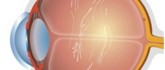
The retina is an important structure of the optical system of the eye; it has a complex structure that allows it to convert electromagnetic radiation into nerve impulses and perform primary processing. The retina interacts with the brain, receiving and transmitting information, so it is very important to keep it healthy and functional. Unfortunately, not everything depends on the person. Among the common causes of retinal degeneration and thinning are age-related changes caused by the inevitable aging process of the body, heredity, and the consequences of injuries.
Retinal dystrophy - symptoms and first signs of the disease:
- narrowing of the field of view;
- decreased ability to navigate in poorly lit rooms;
- deterioration of visual acuity, decreased visibility of peripheral fields;
- distortion, curvature of lines and objects;
- the appearance of “flies”, spots before the eyes, as well as optical effects in the form of lightning and flashes;
- the appearance of cloudy areas.
Sometimes the cause of retinal pathology can be diseases such as diabetes, hypertension, and atherosclerosis. They are successfully treated by retinal laser coagulation, but the decision on the advisability of this procedure is made by the doctor, taking into account the existing pathologies.
Symptoms
Peripheral retinal dystrophy is characterized by nonspecific symptoms that resemble most visual impairments:
- a gradual decrease in visual acuity in one or both eyes - there is a need for brighter lighting when reading, etc.;
- gradual narrowing of the visual field - a person ceases to clearly see objects located on the periphery;
- scotoma - the appearance in the field of vision of an impenetrable or transparent spot, a continuous curtain or the illusion of a foreign object between the eye and the object in question;
- distortion - the perceived picture becomes wave-like, as if an object is being looked at through thick water;
- decreased twilight vision acuity;
- color inversion - distortion of color perception, for example, red is perceived as blue, etc.;
- metamorphopsia - disturbances in the perception of shape and localization of objects in space;
- inability to determine the state of an object relative to space, that is, to recognize whether it is moving or at rest.
The listed symptomatic manifestations may bother the patient simultaneously or appear sporadically one at a time. As the size of pathological lesions increases, they become more pronounced, and some of them persist forever. This is typical for all forms of retinal dystrophy.
Diagnosis of retinal dystrophy, important symptoms of the disease
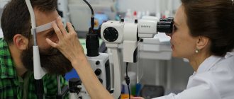
If you experience discomfort in the eyes in the evening, deterioration in visual acuity and the appearance of obvious distortions of visible objects, you should contact an ophthalmologist. Dystrophy is diagnosed during a complete examination of the retina, during an ophthalmological examination, which includes a set of important studies, including:
- determination of visual acuity;
- perimetry, assessing the condition of the retina in the periphery;
- fundus examination;
- measurement of intraocular pressure;
- ultrasound examination of the internal structures of the eye;
- checking the condition of the optic nerve, the viability of retinal nerve cells.
The most complete picture is provided by optical coherence tomography of the retina. This study allows you to visualize the structure of the retina, differentiate its layers, and determine the thickness of each layer. Tomography can reveal macular holes, which are characterized by detachment of nerve fibers while the remaining layers are intact.
Diagnostics
Peripheral retinal dystrophy can be detected before symptoms appear. The anomaly is clearly visualized during fundus examination using the following methods:
- ophthalmoscopy - an examination using a slit lamp, during which the doctor will see dystrophic changes on the retina, looking like light and dark spots of various shapes and configurations (the so-called cochlear mark, frost-like, in the form of radial stripes, lattice, etc.);
- perimetry - the method allows you to identify narrowing of the visual fields even before the patient begins to complain;
- visometry - measurement of visual acuity using special tables (it may correspond to the norm against the background of decreased peripheral vision);
- refractometry is an instrumental diagnostic designed to determine the degree of refractive error.
The most informative way to make a diagnosis is an ultrasound of the eye. This procedure allows you to identify the problem before the appearance of significant dystrophic processes, for example, at the stage of formation of traction. It also helps to establish the cause of the disease if it is changes in the size of the eye along the longitudinal axis, abnormalities of the vitreous body, vascular malformations, etc.
Causes of retinal degeneration and its types
Retinal dystrophy is divided into groups, depending on the cause that caused the disorder. The main cause of retinal degeneration is myopia. Due to severe myopia, the eyes become larger, the retina stretches, and pathological lesions form on it. In myopic people, peripheral retinal dystrophy is most common, caused, among other things, by impaired blood supply to the visual organs. In the treatment of peripheral dystrophies, laser coagulation is performed for prophylactic purposes, which reduces the risk of retinal detachment because it strengthens it. During surgery, the laser creates fusions between the retina and choroid of the eye.
Types of retinal dystrophy are acquired and hereditary:
- age-related disorders - age-related retinal dystrophy occurs in dry and wet forms (exudative), is an acquired disease, develops after 60 years, and is often accompanied by ailments such as cataracts;
- acquired pathologies - arise due to diseases, unhealthy lifestyles, injuries, disruption of receptors;
- congenital - Best's disease, cystic macular degeneration, achromatopsia (severe color vision impairment);
- juvenile, hereditary - cone dystrophy, juvenile retinoschisis, Stargardt disease (history of hereditary factor).
Changes in the central part of the retina occur against the background of excessive insolation, weakened immunity, after injuries, existing diseases of the cardiovascular and endocrine systems of the body.
Women with light eyes, skin, and hair are at risk. Dystrophy of the central part of the retina is often congenital.
What are PCRD and PVKHRD?
PCRD – peripheral chorioretinal dystrophy
This type of dystrophy directly affects the retina of the eye and its choroid (choroid). PCRD is considered a favorable type of dystrophy, since it does not carry any risks for the formation of a severe complication - retinal detachment. The most striking representative of the PCRD family is cobblestone dystrophy, which is harmless single or multiple foci of chorioretinal atrophy scattered along the periphery of the retina.
PVCRD – peripheral vitreochorioretinal dystrophy
In comparison with PCRD, PCRD is considered an unfavorable type of dystrophy, since in the pathological process, in addition to the retina and choroid, the vitreous body (lat. corpus vitreum), which is tightly fused to the area of chorioretinal dystrophy, is involved. It is believed that under conditions such as heavy lifting, sudden shaking of the body or head, childbirth, the vitreous body can provoke retinal rupture, thereby increasing the risk of retinal detachment. But, fortunately, in reality everything is not as scary as they used to think. Most PCVDs are harmless and simply require dynamic monitoring. There are types of PVCRD, the danger of which in most cases is greatly exaggerated. These include 2 types of dystrophies: lattice dystrophy (or “lattice”) and “snail track”. It is these two types of dystrophy that ophthalmologists constantly argue about.
"lattice" dystrophy
“snail track” dystrophy
How is Stargardt disease diagnosed - causes, symptoms and prevention
Stargardt disease begins in childhood and adolescence and often leads to disability. It manifests itself as dystrophic changes in the macular zone, significant deterioration of vision, impaired color perception (merging of red, yellow, green into a single color). Among the causes of the pathology is a mutation of the ABCR gene, a violation of protein production. Early diagnosis gives a high chance of successful treatment. This disease is largely genetically determined, but in the new generation it can be easier, without leading to disability. The pathology is transmitted according to an autosomal recessive type, that is, inheritance of the disease is not always transmitted to another generation and is not related to gender.
To obtain accurate diagnostic data, it is necessary to undergo an electrophysiological examination, visometry, perimetry, optical coherence tomography of the retina, fluorescein angiography (examines the condition of the capillaries and vessels of the retina), as well as a color vision test.
If there are cases of Stargardt disease in the family, it is also advisable to undergo molecular genetic analysis. Moreover, it allows you to detect the disease long before it manifests itself.
Progressive muscular dystrophies
Progressive muscular dystrophies (PMDs) are a heterogeneous group of inherited diseases characterized by progressive muscle weakness and skeletal muscle atrophy.
CLINICAL PICTURE
All PMD are characterized by muscle weakness of varying severity and muscle atrophy. The type of distribution of muscle weakness in PMD is one of the main diagnostic criteria. Each form of PMD is characterized by selective damage to certain muscles while sparing others nearby. In general, a typical myopathic symptom complex includes the following symptoms. • Symmetrical proximal muscle weakness of varying severity (muscle strength from 3-4 points in the early stages and up to 1-0 in the later stages of the disease), gradually developing muscle atrophy. • Govers' symptom: the patient, in order to rise from a squatting position, rests his hands on the floor, then rises, leaning his hands on his knees - “climbing on his own.” This early-onset symptom is caused by weakness of the muscles of the hips and pelvic girdle. • Difficulty walking up stairs - the patient helps himself with his hands. • “Duck” (waddling) gait associated with weakness of the pelvic girdle muscles. • Lumbar hyperlordosis, caused by weakness of the muscles of the pelvic girdle and back. • “Wing-shaped” scapula due to weakness of the serratus anterior muscle, as well as other muscles that fix the scapula. • Pseudohypertrophy of the calf muscles due to the development of connective tissue in them (muscle strength is reduced). • Walking on tiptoes due to Achilles tendon contractures. • Preservation of extraocular muscles, facial muscles. The myopathic symptom complex is most clearly identified in Duchenne and Becker PMD. • Duchenne PMD is characterized by an early onset of the disease (at 3-7 years), rapid progression, high CK levels, pronounced spontaneous activity according to needle EMG, and the absence of dystrophin in the muscles in immunohistochemical studies. As muscle weakness progresses, independent walking becomes difficult, and already at the age of 9-15, patients are forced to use a wheelchair, which provokes the development of kyphoscoliosis and osteoporosis. In the later stages, most patients develop dilated cardiomyopathy and weakness of the respiratory muscles. Intelligence is most often moderately reduced. • Clinical manifestations of Becker's PMD generally resemble those of the Duchenne form, but the course of the disease is milder: the onset occurs at a later age (from 2 to 21 years, on average 11 years), death occurs later (at 23-63 years ) . • Limb-girdle forms of PMD are also characterized by the development of a myopathic symptom complex. In terms of the age of onset of the disease, rate of progression and clinical manifestations, Erb's PMD resembles the Becker form, however, it is not characterized by cardiac pathology, in addition, the disease is noted in both boys and girls. In other limb-girdle forms, weakness of the facial muscles and cardiomyopathy are possible. • Landouzy-Dejerine PMD is characterized by severe weakness of the facial muscles (with the exception of a rare form without facial weakness), the symptom of “pterygoid” scapulae, weakness of the biceps and triceps brachii muscles with intact deltoid muscles, stepping. As a rule, the extraocular muscles (with the exception of one subtype) and the muscles of the tongue and pharynx, and the respiratory muscles remain intact. Some patients experience weakness of the pelvic girdle muscles (about 20% of patients are forced to use a wheelchair). Muscle atrophy is often asymmetrical. Many patients experience hearing loss, cardiomyopathy, or heart rhythm disturbances. • Emery-Dreyfus PMD is characterized by the presence of contractures (usually in the elbow, knee joints, and posterior neck muscles, due to which the head is slightly thrown back) and a scapulohumeral-peroneal distribution of muscle weakness and atrophy with preservation of the facial muscles. Cardiac arrhythmias and cardiomyopathy are common. The disease often debuts with contractures. • The main symptom of the ophthalmopharyngeal form is chronic progressive external ophthalmoplegia, followed by moderate bulbar syndrome. Subsequently, proximal muscle weakness develops in the arms and legs. • Distal myopathies are characterized by predominant weakness of the distal muscles. With Welander myopathy, the extensors of the hands are most affected; with Mioshi myopathy, the calf muscles are most affected: patients stand poorly on their toes and often stumble. With Govers' myopathy, tibial myopathy, the main symptom is stepping due to weakness of the peroneal muscle group, while Govers' myopathy is prone to further generalization: after 5-10 years, weakness of the hands and neck muscles is added, often about 1 toe and V on the arms . With tibial myopathy, which is common in Finland, isolated damage to the anterior tibial muscles is most often observed, and sometimes cardiomyopathy develops.
SYMPTOMS
With PMD Duchenne, Becker, limb-girdle forms, the most pronounced weakness is manifested in the lumbar-iliac muscles, muscles of the hips, deltoid, biceps and triceps muscles of the shoulder. Weakness in the distal muscles of the extremities is less pronounced. The facial muscles remain intact. Along with muscle weakness, hypotrophy of the affected muscles gradually develops, up to atrophy in the later stages. In this case, neighboring muscles can be completely clinically intact.
TREATMENT
Treatment should be carried out exclusively by a neurologist. Self-medication is unacceptable. There is currently no definitive treatment for PMD. The goal of treatment is to maintain muscle strength, prevent the development of contractures and joint deformities. Non-drug treatment Excessive physical activity, as well as insufficient physical activity, leads to an increase in muscle weakness. Daily exercise therapy helps maintain muscle tone and prevents the development of contractures. A physical therapy complex must include active and passive exercises, stretching exercises/contracture prevention exercises and breathing exercises. Active massage with muscle kneading can increase muscle weakness and fatigue, so a gentle massage is recommended. Patients tolerate physiotherapeutic treatment differently: some do not feel any improvement or even complain of increased muscle weakness. Surgical treatment In some cases, surgical treatment of contractures is possible, but it is necessary to remember the possibility of increasing muscle weakness during rehabilitation treatment (up to loss of the ability to walk). In some cases, implantation of a pacemaker is necessary.
Treatment and prevention of retinal dystrophy in Stargardt disease
Stargardt disease can affect the central cells of the retina, paracentral and centroperipheral. As for the treatment of pathology, at this stage of the development of science there is no treatment that would eliminate the cause of the disease and completely relieve its manifestations.
Patients are recommended stimulating conservative treatment of retinal degeneration:
- anabolic steroid;
- hardware treatment - laser and magnetic stimulation, electrical stimulation, electrophoresis with nicotinic acid and papaverine;
- revascularization operations;
- wearing sunglasses to prevent and reduce negative symptoms.
Most of the central dystrophies are age-related macular degenerations, in which the retina is not completely damaged and blindness does not occur, since peripheral vision remains. Treatment of central dystrophies depends on the characteristics of the course of the disease.
Types of pathology
Dystrophy can be congenital or acquired . The congenital form is not completely curable and constantly progresses, significantly worsening visual acuity. To slow down degeneration, it is necessary to strictly follow medical recommendations.
Acquired dystrophy is divided into:
- Central, or macular (macular degeneration) - affects the central part of the retina (macula), which is responsible for image accuracy and the ability to distinguish small details. As a result, central vision is impaired, but peripheral vision is not changed. In this case, problems arise when reading, writing, drawing, and driving a car.
- Peripheral - develops on the periphery of the retina. Does not affect visual acuity, but can lead to retinal rupture and detachment.
Treatment of age-related macular degeneration
The causes of age-related retinal degeneration are not known with certainty, but negative factors include lifestyle, nutrition, and chronic diseases. For the dry form of eye dystrophy, therapy is aimed at normalizing blood circulation and improving metabolic processes. Hardware stimulation and the use of low-energy laser radiation are quite effective (increase the antioxidant activity of the retina). Patients are prescribed vasodilators: Trental, Cavinton, Picamilon, Stugeron, vitamins with zinc and selenium, drugs with a high lutein content, antioxidants: Emoxipin, Tocopherol. It is also recommended that patients enrich their diet with vegetables, fruits, and leafy greens. In order to improve blood flow in the macular area, revascularization and vasoreconstructive surgeries may be offered if conservative treatment does not have the desired effect.
For the wet form of age-related macular degeneration of the retina, antioxidant drugs, diuretics to relieve swelling, hemostatic drugs and fibrinolytics (for hemorrhages), photodynamic therapy are prescribed to the patient: a special medicine is administered intravenously - a photosensitizer.
Timely initiation of conservative treatment of eye degeneration ensures high effectiveness of therapy, the main goal of which is to prevent the progression of the disease.
Treatment methods
Unfortunately, peripheral retinal degeneration cannot be completely and irreversibly eliminated. Modern medicine offers patients with this diagnosis complex treatment aimed at stopping the spread of dystrophic foci. In asymptomatic and early stages of the disease, conservative methods of therapy are used, consisting mainly of medications from several groups:
- Antiplatelet agents
are drugs that reduce blood viscosity, reducing the risk of thrombosis. The preferred form is tablets, sometimes injections are prescribed. Names of drugs - Clopidogrel, products based on acetylsalicylic acid. - Vasodilators
are drugs that relieve vasospasm and improve microcirculation. Along with tablets, injectable forms of medications are prescribed. Names - No-Shpa, Papaverine, Drotaverine. - Angioprotectors
are medications that protect and strengthen the walls of blood vessels. Prescribed in the form of tablets or intravenous injections. Names of drugs - Ascorutin, Complamin and their analogues. - Microcirculation stimulants
are agents that have the property of increasing vascular conductivity and reducing blood viscosity. These include Pentoxifylline in the form of tablets or injections. - Restoring and regenerating drugs
are medications and dietary supplements that enhance metabolic processes, restore microcirculation and regeneration. It is preferable to use local agents - drops and intraocular injections. Names of drugs - injection solution Retinalamin with polypeptides, eye drops Taufon, Emoxipin. - Vitamin complexes
are preparations enriched with B vitamins, retinol, keratin, lutein and other compounds beneficial to the eyes. Recommended in the form of capsules or tablets. Names of drugs - “Okyuvite-lutein”, “Blueberry-Forte”, etc.
If peripheral retinal dystrophy continues to progress after 2 courses of conservative therapy, physiotherapeutic procedures are used:
- electrophoresis;
- exposure to magnetic fields;
- photo-, electrical and laser stimulation;
- ILBI procedure (intravenous laser irradiation of blood).
If these measures do not lead to a noticeable slowdown in the progression of the disease, minimally invasive or full-fledged surgical intervention is performed. Laser coagulation of the retina is considered the most effective and least traumatic, during which the doctor “solders” the retina to the vascular base and at the same time eliminates scar formation.
If the disease is at an advanced stage, when the vitreous body has come out through a hole in the retina, vitrectomy is performed: the vitreous body is removed, the retina is fixed and the eyeball is filled with polymer.
Retinal dystrophy - alternative treatment
Traditional methods of treating retinal dystrophy can be effective at the initial stage of the disease. When the first symptoms appear, it is necessary to begin conservative treatment recommended by an ophthalmologist, and to enhance the therapeutic effect, you can use the experience of traditional medicine. First of all, you need to diversify your diet with foods rich in selenium, zinc, lutein, vitamins C, A, carotene, B1, B6, B9, B12 and E.
The menu should include dishes with beef, turkey, seafood, chicken eggs and other animal proteins. It is also useful to eat foods rich in Omega-3 unsaturated fatty acids: salmon, herring, shrimp, mackerel, flax seeds, almonds, pistachios, pine nuts.
By eating right and following your doctor’s instructions, you can avoid the unpleasant symptoms that accompany the disease.
Thus, by reducing the amount of salt consumed, a person with a diagnosed retinal pathology that has an edematous form will feel significant improvements in his condition. Quite effective are eye compresses with decoctions of caraway seeds and cornflower flowers, May herbs, pine needles, rose hips and onion peels in equal proportions, as well as with nettle. It must be poured with boiling water and left for six to seven hours.
It is also necessary to drink blueberry and rosehip tea throughout the entire period of treatment to alleviate the symptoms of the disease. Folk remedies will not help get rid of the disease, but they are effective in preventing its progression.
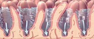
Structure of the cornea
The human cornea has five layers. The outer layer, consisting of 5-6 layers of cells, is the anterior epithelium, which performs a protective function. Beneath the anterior epithelium lies Bowman's membrane, a thin membrane of dense tissue that supports the outer layer.
Fig. 1 Structure of the human cornea
Under Bowman's membrane there is stroma (its volume accounts for up to 95% of the volume of the entire cornea). The stroma is made up of special cells - keratocytes, arranged in a strict order, which ensures the transparency of the cornea.
Recently, Duha's layer (discovered in 2013) has been distinguished in the cornea - a thin layer located between the stroma and Descemet's membrane.
The strongest tissue of the cornea adjacent to the stroma - Descemet's membrane - is a dense membrane that also performs a protective function.
The last layer is the endothelium (posterior epithelium of the cornea), which is a layer of one row of cells that perform the function of a “pump” - they control the flow of fluid into the cornea from the anterior chamber.
Retinal tears
Based on their type, retinal tears are divided into:
- perforated;
- valve;
- by type of dialysis.
Hole breaks
most often occur as a result of lattice and small cystic dystrophies, there is a hole in the retina.
A rupture is called a valve rupture when a section of the retina partially covers the rupture site. Valvular tears
result from vitreoretinal traction, which pulls and tears the retina.
When a tear forms, the area of vitreoretinal traction will be the valve apex. Dialysis
is a linear tear of the retina along the dentate line - the site of attachment of the retina to the choroid. In most cases, dialysis is associated with blunt trauma to the eye.