The eyeball consists of several membranes. Its innermost shell, lining the cavity of the eye from the inside, is called the retina, or retina. On the outside, the choroid, or choroid, is adjacent to it; on the inside, the retina borders on the vitreous body. The thinnest layer of retinal tissue contains light-sensitive cells (photoreceptors) that are responsible for image perception. The processes of these cells, folding into nerve fibers, carry information about the image to the corresponding parts of the cerebral cortex.
Retinal diseases can occur both due to direct damage to its structures and as a result of systemic diseases of the body. Changes in the retina are diagnosed in patients with diabetes mellitus, hypertension, pathologies of the kidneys and adrenal glands, after traumatic brain injuries, and with some infectious diseases. Major retinal pathologies include:
- Dystrophic and vascular lesions (angiopathy, retinopathy, maculopathy, hemorrhage)
- Retinal tears and detachments
- Inflammatory changes (retinitis)
- Retinal tumors
There are groups of patients whose risk of developing retinal diseases is increased. These include people with moderate and high myopia, elderly patients with diabetes and high blood pressure, and pregnant women.
At an early stage, many retinal diseases may not cause any discomfort. So if you or a loved one is at risk, don't skip your annual eye exam with a qualified ophthalmologist.
Retinal hemorrhages
Hemorrhages in the retinal area can occur as a result of injury to the eyeball, vascular pathologies or dystrophic diseases of the retina. Retinal hemorrhages are found in diabetes mellitus, thrombosis of the central retinal vein, defects of the cardiovascular system, blood diseases, and burns.
The blood in the cavity of the eyeball has a toxic effect on the internal structures of the eye, changes the structure of the vitreous body and reduces the transparency of the optical media of the eye, which is accompanied by a deterioration in the quality of vision. Severe complications of retinal hemorrhage are increased intraocular pressure and secondary glaucoma, retinal detachment, degenerative changes in the retina, etc.
To treat hemorrhages in the retina, conservative methods are used (injections of drugs that accelerate the resorption of hemorrhages), laser treatment (laser vitreolysis, laser coagulation), and surgical techniques (vitrectomy with replacement of the clouded vitreous).
Read more about the treatment of retinal hemorrhages >>>
Retinal edema: symptoms and treatment methods
Our retina is the thinnest inner layer of the eye. It is responsible for the perception of light waves and forms the image that is transmitted to our brain. Normally, the retina fits tightly to the fundus of the eye and connects to the optic nerve. Its other side is in contact with the vitreous body.
If for any reason retinal edema develops, a person’s vision noticeably deteriorates. Glasses don't help with this. With timely treatment, the process is reversible, vision can be restored or stabilized for a long time. In the absence of therapy, degenerative changes inevitably occur in the retinal tissue, and blindness occurs.
Symptoms of retinal edema
Symptoms vary depending on the location of the swelling and its extent.
If the swelling affects the center of the retina - the macula, then visual disturbances will be significant. The macula contains the largest number of light-sensitive receptors and is the area of greatest visual acuity. Any changes in its work immediately affect the quality of vision.
For macular edema:
- the image is blurred, especially in the central visual area;
- the eye focuses poorly both near and far (in some cases, vision is worse in the morning, then improves);
- straight lines and shapes of objects are distorted;
- when reading, individual letters fall out, lines become bent;
- color perception is impaired (pink shades dominate);
- excessive sensitivity to bright light appears, even to the point of pain.
If the edema has limited lesions and occurs on the periphery of the retina, the symptoms will be mild and will not bother the person too much. With such distribution they speak of focal retinal edema. It is discovered in most cases only at an appointment with an ophthalmologist, during a preventive examination.
Symptoms of retinal edema may be mild. It can only be detected at a doctor's appointment.
Swelling may develop in only one eye. In this case, the second eye partially compensates for visual disturbances, which is why a person does not immediately notice signs of swelling, which is why it is dangerous.
Retinal edema is a progressive pathology. Gradually, the swelling covers other zones (from the center to the periphery or vice versa) and becomes diffuse. Symptoms get worse. Under the influence of accumulated fluid, the retina peels off and scars, which threatens irreparable loss of vision.
After diagnosing swelling, it is necessary to carry out treatment as quickly as possible to avoid its further spread and pathological changes in tissues.
Reasons for the development of retinal edema
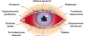
Retinal edema is not an independent disease. This is a complication of some other diseases or conditions, as a result of which vessels with abnormal permeability appear in the retina. Moisture “sweats” through them and accumulates in the surrounding tissues.
The cause of the development of retinal edema can be:
- Ophthalmic pathologies that lead to retinal dystrophy or degeneration, for example:
- angiopathy;
- glaucoma;
- retinopathy;
- various inflammatory processes;
- occlusion of the central retinal vein;
- high myopia.
- Systemic diseases that impair the functioning of blood vessels throughout the body. At risk are people suffering from diabetes, arterial hypertension, atherosclerosis, and obesity.
- Eye injuries that cause disruption of the blood supply to the eye.
- Age-related changes in eye tissues.
As they age, cell metabolism products accumulate in them, and thickenings form under the pigment layer of the retina. Which in turn causes inflammation and the growth of new pathological blood vessels in the eye - angiogenesis. Age-related macular degeneration usually develops after age 65. In approximately 90% of cases, it occurs in a “dry” form, that is, without swelling. But sometimes it begins to progress and turns into a “wet” form. The latter is very dangerous due to its rapid development. Irreversible changes in the retina occur within a few days or weeks.
Treatment methods for retinal edema
Ophthalmologists have various methods for treating retinal edema in their arsenal. Under certain conditions they apply together.
- At the earliest stages, conservative therapy is possible. Anti-inflammatory drugs and medications are prescribed to improve blood supply and metabolism in the tissues of the eye.
- With a focal form of edema, that is, when the focus of edema is small and not in the center, laser coagulation is successfully performed. It allows you to strengthen the retina in the periphery and avoid the progression of pathology. You can read more about this procedure in our article. However, laser coagulation has limited use. The macula is not affected.
- For macular edema of the retina, surgical treatment is recommended.
Previously, this diagnosis necessarily required complex interventions, for example, macular translocation. Now such operations are performed extremely rarely, only for certain dystrophic forms of the retina.
In most cases, for more than 10 years, ophthalmological surgeons have been doing without a scalpel and using intravitreal injection of an angiogenesis inhibitor (IVVIAG).
IVVIAG ophthalmology: what is it
This method is based on the use of monoclonal drugs. This is a modern medical approach known as “targeted therapy.” It is good because it allows you to purposefully influence the very cause of the development of pathology.
The retina can be affected by “targeted therapy”. IVVIAG is one of the methods of such therapy.
The growth of “irregular” vessels in the retina is provoked by a specific protein, VEGF, which appears as a result of a deterioration in its blood supply.
The therapeutic drug contains multiple cloned antibodies to the VEGF protein. The doctor uses an injection to inject them into the vitreous body of the eye. The VEGF protein molecules bind and it completely stops its activity. This suppresses retinal neovascularization and reduces vascular permeability.
Such drugs are inhibitors of angiogenesis (inhibition means “suppression”). To treat retinal edema, ophthalmologists use Lucentis or its analogue, Eylea. They are similar in their effectiveness, differing in the nuances of their mechanism of action.
Thanks to the action of angiogenesis inhibitors:
- slight swelling of the retina disappears completely;
- with diffuse edema, large areas are fragmented into small foci, which can then be corrected by laser coagulation;
- the structure of the retina improves;
- visual acuity improves.
In some cases, despite a decrease in edema, the visual acuity of patients does not change. This is explained by too large degenerative changes in the retinal tissue and the duration of the disease.
The sooner treatment for retinal edema begins, the more favorable its outcome and the shorter the therapeutic course.
The choice of inhibitor drug always remains with the attending physician. This is preceded by a thorough examination of the eye tissues and their interpretation based on the patient’s general medical history. The patient is informed about everything in detail.
Features of the IVVIAG operation
The procedure for administering the drug "Lucentis", despite its apparent simplicity, requires high professionalism of the doctor, since any intervention with retinal edema can provoke inflammation and worsen the situation. It is carried out only in the operating room, under conditions of strict asepsis.
Duration of the operation: 3–4 minutes. Anesthesia: local drip.
Do I need to stay in the hospital: no, the procedure is outpatient.
Brief description of the operation:
- The surgical field around the eye and eyelid is treated with an aseptic solution, and an eyelid dilator is installed in the eye.
- To eliminate involuntary movement of the eye and the risk of accidental damage, the eye is fixed with special tweezers.
- After administering the drug for half an hour, the doctor monitors whether the intraocular pressure in the operated eye changes, and only then allows the patient to go home.
Before and after the procedure, antimicrobial agents must be instilled for three days.
Restrictions after surgery: immediately after the injection, it is better to stop driving, even if there are no sensations. They may appear after the painkiller wears off.
How long does treatment last: the course of treatment usually includes several injections at intervals of a month. The duration of treatment depends on the severity of the swelling. Throughout the therapy, visual acuity and the condition of the retina are monitored. Laser tomography helps with this.
Retinal edema is a serious pathology that does not go away on its own. Her treatment is more effective the sooner it is started. Do not neglect preventive examinations by an ophthalmologist, and if any alarming symptoms occur, do not delay in contacting a specialist. Our ophthalmologists are always at your service!
Good vision to you!
Retinal inflammation
Inflammation of the retina of the eyeball (retinitis) can occur as a unilateral or bilateral process. The causes of retinitis are varied: infectious diseases of a bacterial and viral nature (including tuberculosis, syphilis, AIDS, herpes), toxic eye damage due to other pathologies, allergic diseases, chronic heart and kidney diseases.
Manifestations of retinitis can be different and depend on in which parts of the retina the pathological process is localized. Common symptoms for any form of retinitis include changes in visual fields (scotoma) and a decrease in its acuity. Scotomas are focal loss of visual fields; their size and location directly depend on the location and size of the inflammatory focus. If retinitis is untimely or incompletely treated, persistent vision deterioration occurs, which is especially noticeable in poor lighting conditions.
Treatment of retinitis should be aimed at identifying and eliminating the cause that caused the inflammation of the retina. The most effective is an integrated approach, including drug treatment, injections of anti-inflammatory and antibacterial drugs, and laser coagulation of retinal vessels.
Read more about the treatment of retinal inflammation >>>
Inflammatory disease of the retina - retinitis
Retinitis is an inflammatory disease of the retina, which can be either unilateral or bilateral. This inflammatory disease of the retina can be either infectious or toxic-allergic in nature. Retinitis can occur due to a number of infectious diseases. For example, such as: AIDS, syphilis, viral and purulent infections, etc.
Symptoms of retinitis depend on the location of the process on the retina.
But the main one is a decrease in visual acuity and a change in the field of vision. There are cases where retinal damage is initially limited to small areas, which then enlarge, leading to progressive loss of vision. Retinitis is treated with medication.
Retinal detachment (detachment)
It is a separation of the retinal layer and the underlying tissues of the choroid. In the event of a tear or rupture of the retina, intraocular fluid leaks between it and the choroid, leading to disruption of the normal nutrition of photoreceptor cells. If you suspect retinal detachment, you must immediately contact an ophthalmologist to provide qualified assistance. The ability to maintain the patient’s visual acuity will directly depend on how quickly and completely assistance is provided for retinal detachment. Delay in this case threatens irreversible loss of vision and blindness!
When retinal detachment is detected, surgical treatment methods are used: extrascleral filling, laser coagulation of tissue around the area of tissue detachment, vitrectomy (replacement of the vitreous body of the eye) to remove blood from the cavity of the eyeball and restore the transparency of the optical media of the eye.
Read more about the treatment of retinal detachment >>>
TsSHRD (TSSKHRP, TsSH)
Central serous chorioretinopathy (CSC) is one of the most common pathologies of the posterior segment of the eye. The cause of the disease is considered to be distress, a situation of “stress” that is extremely common in the modern world. Patients are young people, under 50 years old, mostly men. Complaints: the appearance of a translucent spot in front of the eye, a decrease in the size of objects, impaired color vision - everything seems a little yellowish-brown.
This disease of a non-inflammatory nature, in 50–70% of cases, resolves on its own, but requires dynamic observation and active intervention for a duration of more than 3–6 months.
The observation is based on regular optical coherence tomography of the macular zone in order to assess the dynamics of the height and extent of exudative retinal detachment in the macula.
To identify the point (or points) of filtration - sources of subretinal fluid, FA (fluorescein angiography) is used. The treatment method is laser coagulation of points of fluid leakage under the retina according to FA data. An outpatient painless procedure, after which fluid is reabsorbed under the retina.
Retinal reattachment after laser photocoagulation of leakage points.
Tumors of the retina of the eye
Retinal neoplasms can be benign or malignant, affect one or both eyes, and are detected in young (usually children) and adulthood. Gradually growing, the volumetric formation of the retina occupies most of the eye, and therefore the patient develops an asymmetric protrusion of the eyeball noticeable to the naked eye. Vision in the affected eye disappears, the eye loses mobility.
For timely detection and treatment of retinal diseases, contact the Retina Center. Our specialists will conduct a qualified eye examination using modern diagnostic equipment and will accurately determine whether you require eye treatment. If your doctor recommends surgery, do not delay the decision for long; this will help avoid progression of the disease and preserve your vision.
Treatment of retinal tumors is carried out in specialized medical institutions; delay in seeking qualified help can lead to irreversible consequences.
Main “risk groups”
- People with moderate and high degrees of myopia;
- pregnant women;
- elderly people with diabetes.
The initial stage of the disease may not be accompanied by any symptoms , so if you are at risk, be sure to undergo diagnostics using modern equipment. This examination will reliably determine whether you need treatment. If you are scheduled for surgery, do not put off surgery for too long. Before surgery, you need to protect your eye from possible damage in every possible way.
If dystrophic retinal diseases with thinning and rupture in the periphery are detected, the retina is strengthened using a laser. Otherwise, any sufficiently severe stress can cause detachment, requiring immediate surgical intervention. It is better to prevent such a situation. Moreover, detachment can occur when urgent provision of qualified ophthalmological care is impossible (at the dacha, on a trip, etc.)
Retinal dystrophies
The cause of dystrophic changes in the retina is usually disorders in the vascular system of the eye. Most often, dystrophies are found in elderly patients, as well as with moderate and high myopia. The myopic eye is usually extended in the anteroposterior direction, and the resulting overextension of the posterior parts of the eyeball is accompanied by the appearance of dystrophic changes in the fundus. Retinal dystrophies can also occur with various vascular and inflammatory diseases of the eye.
The main method of treating retinal dystrophies is photocoagulation of altered tissues using an argon laser to strengthen thinned areas of the retina and delineate the area of detachment, if one has been identified.
Read more about the treatment of retinal dystrophies>>>
Macular degeneration
This is damage to the central part of the retina, which leads to irreversible impairment of visual functions, primarily visual acuity. The main cause of macular degeneration is the gradual aging process of the body and the eye in particular. Less commonly, the cause may be previous trauma, infectious, inflammatory eye diseases, severe myopia, and less commonly, hereditary causes. The initial manifestations of macular degeneration are “fogging”, curvature of objects, letters (letters “break”), colors become less bright. Over time, a more or less transparent spot (central scotoma) appears in the central part of the visual field. On the one hand, macular degeneration leads to significant visual problems, but it does not lead to complete blindness, since paracentral and peripheral vision remains intact.
Treatment of macular degeneration
Depending on the type (dry or wet), dystrophy is subject to laser or surgical treatment. The final treatment method is chosen individually and only after a comprehensive diagnosis.
Age-related macular degeneration (AMD)
The disease is most often detected in people over 50 years of age and is the main cause of irreversible vision loss. The risk of the disease increases with a genetic predisposition, as well as in patients with diabetes mellitus, hypertension, and atherosclerosis. Symptoms of the disease are associated with damage to the macula area of the retina.
The initial manifestations of AMD do not have any clinical manifestations and can only be detected during an examination by an ophthalmologist. As the disease progresses, symptoms such as distorted outlines of objects, blurred vision, and blurred vision appear.
The dry form of AMD does not require special treatment. To treat the wet form of age-related macular degeneration, intravitreal injections (injections into the vitreous cavity) of drugs that block the growth of defective vessels under the retina (anti-VEGF drugs) are used. In addition, photodynamic therapy and laser photocoagulation of the retina are indicated.
Read more about AMD treatment >>>
Causes
There are three reasons for the formation of retinal diseases:
- Infectious. If the provocateur is an infection, then inflammation develops, affecting the visual apparatus.
- Acquired or congenital. Associated with changes in the structure of photoreceptors. The shell cracks, breaks, or becomes thinner.
- Vascular. As a rule, pathology is a consequence of existing vascular diseases (for example, atherosclerosis, hypertension).
Most often, vision impairment due to retina diseases occurs in patients suffering from myopia, since their retina is stretched.
Diabetic retinopathy
Eye changes in diabetes mellitus cause disruption of oxygen delivery to the retina and lead to the development of diabetic retinopathy. The disease does not develop immediately, but 5–10 years after the onset of diabetes.
Depending on the form of the disease (diabetes type 1 or 2), retinal lesions will differ. A patient with type 2 diabetes often exhibits signs of diabetic maculopathy, leading to decreased visual acuity. In the insulin-dependent form of diabetes, changes in the retina are much more pronounced and are manifested by proliferative changes: the growth of new defective vessels (retinal neovascularization), hemorrhages and scarring of the retina, macular edema.
To treat diabetic retinopathy, laser coagulation of the retina, intravitreal injections of anti-VEGF drugs, and in severe cases, surgical treatment (vitrectomy surgery) are used.
Read more about the treatment of diabetic retinopathy >>>
Diagnostics
To assess the condition of the retina, identify signs of its damage and make a diagnosis, the following is carried out:
- Assessment of visual acuity.
- Determination of visual fields.
- Examination of the fundus, retina and other structures of the eye.
Refractometry, perimetry, visometry, ultrasound of the eyeballs and other diagnostic methods are carried out, which allow a comprehensive assessment of the condition of the eye and visual impairment.
At the appointment, the ophthalmologist may recommend that the patient contact other clinic specialists and undergo examinations to find the causes of the disease. This could be diabetes, hypertension and other pathologies that negatively affect the eyes. This will make it possible to prescribe treatment not only for the consequences, but also for the root causes of diseases, and to avoid further progression and relapses.


