Date of article writing: 08/19/2020 Quantity
()
Causes of pathology Symptoms of xerophthalmia Diagnosis Treatment Drug treatment Surgery Hardware treatment Prevention and recommendations
Dry eye syndrome (DES) is a pathological condition in which the conjunctiva and cornea are poorly moisturized. 10-20% of the population of our planet suffers from a similar problem. It occurs much more often in women (70% of cases) than in men.
The pathology is also known under other names - dry keratitis, dry keratoconjunctivitis, xerophthalmia.
Causes of pathology
The main cause of dry eye syndrome is a decrease in the synthesis of tears or their rapid evaporation as a result of changes in properties, which does not allow the formation of a full tear film.
Various factors can provoke the development of pathological dryness:
- long periods of work on a computer that require eye strain;
- air dried by air conditioners and heating devices;
- constant use of contact lenses, which impairs the quality of tears;
- poor nutrition leading to vitamin deficiency (especially vitamin A);
- low-quality cosmetics;
- external influences (ultraviolet radiation, strong wind, smoke);
- a decrease in estrogen synthesis in women during menopause and pregnancy, as a result of which tears become more liquid and are not retained on the cornea;
- injury to the organs of vision;
- incorrectly performed laser vision correction;
- surgical interventions (conjunctivoplasty, keratotomy, photoablation, correction of ptosis) that destabilize tear synthesis;
- taking certain medications (tranquilizers, antidepressants, antihistamines, hormonal contraceptives) that affect hormonal levels (estrogens concentration);
- regular use of eye drops that contain preservatives;
- ophthalmological diseases (conjunctivitis, blepharitis, neuroparalytic keratitis, lagophthalmos, ptosis, stye);
- chronic diseases (diabetes mellitus, Sjogren's syndrome, Felty's syndrome, rheumatoid arthritis, infections, pathologies of the endocrine system);
- hereditary predisposition.
The risk of developing keratoconjunctivitis sicca increases with age - 88% of cases are diagnosed in people over 50 years of age.
What is the ocular surface?
The ocular surface is a complex biological structure, including the surface layer of epithelial cells of the cornea, conjunctiva, nasolacrimal outflow system, as well as a secretory apparatus consisting of a complex of multidisciplinary glands and secretory cells that produce components of the tear film, which is an integral part of the ocular surface.
The ocular surface includes the glands of Meibom, the glands of Moll and Zeiss, which produce lipid secretion for the tear film, as well as the secretory glands of Wolfring and Krause, which provide the basal (main) production of fluid for the formation of the aqueous layer of the tear film. In addition, the secretory system of the ocular surface includes Goblet cells, Manz cells, the crypt of Henle and Becher goblet cells, which produce mucin, forming the layer of the same name.
In addition to these structures, the ocular surface includes the hair follicles of the eyelashes and the edges of the eyelids.
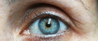
Symptoms of xerophthalmia
The following symptoms are noticeable with dry eye syndrome:
- constant dryness;
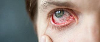
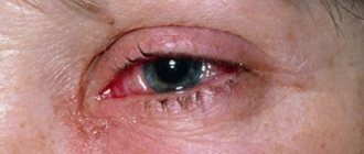
- swelling and redness of the conjunctiva;
- feeling the presence of sand;
- discomfort, burning and stinging;
- blurred vision;
- causeless lacrimation;
- mucous discharge;
- photophobia;
- desire to rub your eyes.
In 42-43% of people, vision deteriorates significantly and difficulties arise when reading.
If there is no treatment for dry eye syndrome for a long time, then complications gradually begin to develop, which worsen the quality of life and reduce performance. The cornea becomes thinner, becomes covered with scars, the eye tissue becomes infected, corneal erosions and ulcers form, perforation is possible, and diseases develop (filamentous keratitis, conjunctivochalasis).
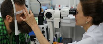
Diagnostics
At the first appointment, the ophthalmologist listens to the patient’s complaints, studies the medical history, and conducts an external examination.
Diagnosis of dry eye syndrome includes special tests:
- fluorescein instillation test - detects dry lesions and calculates the time of tear film rupture;
- Schirmer test (special pieces of paper that absorb tears are fixed under the eyelids for several minutes) – measures the exact total amount of tears produced;
- Norna test (fluorescein is instilled into the eye, the cornea is scanned, noting the time of the first rupture of the tear film) - determines the stability of the tear film, the speed and quality of its evaporation.
If necessary, additional examinations are carried out:
- biomicroscopy – determines the height of the tear menisci, assesses the condition of the conjunctiva, cornea, and tear film;
- thiascopy - examine the organs of vision in polarized light to determine the condition of the lipid layer.
Laboratory tests will also be required:
- crystallography - a drop of tear fluid is applied to a glass slide and then evaporated; the type of pathology is determined from the resulting crystals;
- osmometry – studies the osmolarity of tears, which makes it possible to assess the quality of the tear film and the severity of xerophthalmia;
- cytology of a smear taken from the conjunctiva.
Treatment
Before treating dry eye syndrome, it is necessary to eliminate the causes of the disease or at least minimize the adverse effects. If the pathological condition is caused by other diseases, the ophthalmologist will refer you to the appropriate specialist.
Correction of pathology is possible in several ways:
- medicinal;
- operational;
- hardware
Drug treatment
Most often, for xerophthalmia, drops with artificial tears are used, which are chosen depending on the severity of the condition. The drops activate the synthesis of tears, improve their properties, and help retain tears on the surface of the cornea.
Additionally, the doctor may prescribe retinol (vitamin A) and omega-3 fatty acids, steroid or hormonal drugs, and immunosuppressants. In case of infection, antibacterial ointments are used.
Surgery
If drug therapy does not have the desired effect, the ophthalmologist will recommend surgery:
- tarsorrhaphy - reduce the palpebral fissure by partially suturing the eyelids to inhibit the evaporation of tears;
- blocking the tear ducts - the ducts are cauterized or punctal plugs are inserted into them, thereby preventing the outflow of tears from the cornea;
- transplantation of salivary glands into the conjunctival cavity;
- keratoplasty - replaces a damaged area of the cornea.
Hardware treatment
The MAKDEL-02 device provides effective results in the treatment of dry eye syndrome:
- has analgesic and anti-inflammatory effects;
- normalizes the functioning of the lacrimal glands;
- relieves vasospasms;
- activates blood circulation;
- improves cell structure;
- accelerates regeneration processes;
- strengthens local immunity.
The device is used for medicinal purposes. If necessary, hardware treatment is combined with drug therapy and laser acupuncture.
Dry eye syndrome in ophthalmic practice
Dry eye syndrome (DES), or xerosis of the cornea and conjunctiva, is a complex disease that is widespread throughout the world and is one of the main problems of modern ophthalmological pathology. According to Russian researchers, this disease affects up to 12% of ophthalmological patients under the age of 40 years and over 67% of patients over 50 years of age [2]. The term “dry eye” itself appeared in Russian literature relatively recently. Previously, it was identified exclusively with Sjogren's disease - a severe systemic disease accompanied by a decrease or complete absence of secretion of all endocrine glands, especially the lacrimal and salivary glands. Currently, the concept of “dry eye syndrome” has been expanded and is defined as a complex of signs of damage to the corneal and conjunctival epithelium due to a decrease in the quality and/or quantity of tear fluid [1]. The latter forms a tear film (TF) on the surface of the eye, which performs a number of important functions, including trophic, protective and optical. Thus, a violation of the composition or production of SP can lead to quite serious damage to the anterior segment of the eye.
Xerosis of the cornea and conjunctiva occurs due to a number of pathologies. An important role in this process is played by pronounced anatomical disorders of the ocular localization, such as incomplete closure or excessive opening of the palpebral fissure due to cicatricial or paralytic lagophthalmos, endocrine ophthalmopathy, and buphthalmos. Corneal-conjunctival xerosis can also develop due to disruption of the trophism of the cornea or deformation of its surface, failure of the lacrimal gland, additional lacrimal glands after dacryoadenitis and inflammatory diseases of the conjunctiva. Also, a violation of the composition of SP is observed in the so-called menopausal syndrome [2]. A sharp decrease in tear production is observed in cases of disorders of the innervation of the lacrimal gland, such as facial paralysis and multiple sclerosis. Chronic meibomitis, in which the composition of the joint venture is disrupted, also leads to the development of a typical picture of dry eye syndrome. Recently, the so-called ocular office and ocular monitor syndromes, which occur in people of different ages as a result of systematic exposure of their eyes to conditioned air, electromagnetic radiation from office equipment and other similar sources, have become especially important [3]. One of the common causes of impaired stability of the joint, which has become increasingly important in recent years, is surgical interventions performed for refractive errors and cataracts [4]. It has been noted that dry eye syndrome can be caused by the use of certain medications, such as oral contraceptives, tricyclic antidepressants, antihypertensives, corticosteroids, as well as chronic instillation of beta-blockers used in the treatment of glaucoma. According to some data, the development of xerosis of the cornea and conjunctiva can be caused by taking cytostatics and anti-migraine drugs [2].
A typical initial symptom of dry eye syndrome is the sensation of a foreign body in the conjunctival cavity, which is combined with severe lacrimation, which is later replaced by a feeling of dryness. Patient complaints of burning and stinging in the eye are typical, especially when exposed to wind, smoke, air conditioning and other similar irritants, when using fan heaters. In addition to this, subjective signs of the disease are photophobia, deterioration of visual performance in the evening, and fluctuations in visual acuity during the working day. It is necessary to add pathognomonic signs to the above. In particular, a negative reaction of patients to instillation of even completely indifferent drops into the conjunctival cavity, for example, a solution of chloramphenicol 0.25% or a solution of dexamethasone 0.1%, is characteristic. In such cases, patients experience pain, burning or stinging in the eye [3].
The most common objective sign of the disease is a decrease or complete absence of tear menisci at the edges of the eyelids. Their place is usually filled by swollen and dull conjunctiva, “creeping” onto the free edge of the eyelid. Somewhat less often, in such patients one can detect the appearance of various “clogging” inclusions in the tear film. They are usually represented by tiny clumps of mucus, remnants of separated epithelial threads, air bubbles and other microparticles. They float in the thickness of the tear film, the tear meniscus and the lower conjunctival fornix, move along the corneal epithelium and are clearly visible in the light of a slit lamp. Another objective sign of dry eye syndrome is a characteristic discharge from the conjunctival cavity. When treating the eyelids, due to its high viscosity, it is drawn into thin mucous threads, which cause discomfort in patients. Based on the combination of the above symptoms, it is advisable to distinguish three degrees of severity of dry eye syndrome [1].
Grade I, mild, is characterized by:
- subjective signs - complaints of a feeling of “sand in the eye”, burning, photophobia, etc., arising from exposure to unfavorable factors;
- objective signs are increased tear production, hyperemia and swelling of the conjunctiva, the presence of inclusions in the tear film, the appearance of conjunctival discharge in the form of mucous threads.
II, average, degree has:
- subjective signs - a greater number of complaints and symptoms that persist long after the cessation of adverse factors;
- objective signs are a painful reaction to the instillation of indifferent eye drops, swelling of the bulbar conjunctiva with its creep onto the free edge of the lower eyelid, the absence of reflex lacrimation and the appearance of signs of deficiency of tear production.
III, severe, the degree is distinguished by special forms.
- Filamentous keratitis: multiple epithelial growths in the form of filaments, the free edges of which, moving towards the cornea, irritate the eye, which is accompanied by corneal syndrome. The conjunctiva is intact.
- Keratoconjunctivitis sicca: signs of filamentous keratitis are aggravated by degenerative changes in the conjunctival and corneal epithelium. The cornea loses its natural shine, gloss and becomes dull. Subepithelial opacities may be detected. Swelling and hyperemia of the conjunctiva at the edges of the eyelids are also observed.
- Recurrent microerosions of the cornea: periodic occurrence of superficial microdefects of the corneal epithelium that persist for a long time (up to 7 days). A pronounced corneal syndrome is characteristic; the disease recurs after 2–3 months.
Diagnosis of the disease
The diagnostic process for patients with dry eye syndrome is carried out in the traditional sequence. The initial ophthalmological examination of patients at the initial stage includes the following elements.
- Purposeful questioning of the patient, including clarification of the history of the disease and its possible connection with the professional activities of the subject.
- A standard examination of the organ of vision, but with “targeted” biomicroscopy of the cornea (Nidek, Paradigm), conjunctiva and free edges of the eyelids, including the use of sodium fluorescein 0.1%.
If signs of dry eye syndrome are detected, a clarifying examination is carried out, which includes three stages.
- Additional “targeted” biomicroscopy (Nidek, Paradigm) of the anterior segment of the eyeball using various vital dyes.
- Functional examination (determining the stability of the joint, studying the total and main tear production).
- Testing aimed at diagnosing pathological changes associated with dry eye syndrome.
The initial ophthalmological examination of patients is carried out according to generally accepted rules. More attention should be paid to complaints, which in some cases directly or indirectly indicate xerotic changes in the eye tissue. A targeted collection of anamnestic data regarding the general status, past illnesses, injuries and operations, treatment received, and professional activities of the subject is also necessary.
When biomicroscopy of the cornea and conjunctiva, it should be borne in mind that signs of dry eye disease are often masked by symptoms of other eye diseases, in particular those of a degenerative or inflammatory nature. To differentiate them, SCGTseng (1994) proposed a fairly simple rule: if changes suspicious for xerosis are localized in the so-called exposed zone of the surface of the eyeball, then they are associated with dry eye syndrome; when areas of pathology also involve the unexposed zone of the cornea and conjunctiva, their nature is most likely not xerotic.
Vital dyes significantly increase the capabilities of biomicroscopy: sodium fluorescein 0.1%, rose bengal 3% or lissamine green 1%, allowing one to obtain various complementary information.
The presence of initial, and even more obvious signs of dry eye syndrome is an indication for performing functional tests designed to assess the state of tear production and the strength of the precorneal joint.
The examination of a patient with suspected dry eye syndrome should begin with an assessment of the stability of the joint venture. Since the results of the test used for this according to Norn (1969) largely depend on the “invasiveness” of previous manipulations in the conjunctival cavity, they should be completely excluded. At the same time, studies by L. S. Beer et al. (2001) found that the most reliable results for assessing the stability of SP are obtained when using microvolumes (6–7 μl) of sodium fluorescein 0.1%. At the same time, their influence on the stability of the SP becomes minimal, in contrast to a whole drop (30–40 μl) of diagnosticum used in the Norn method.
The next stage of the functional study is to assess the state of total (main and reflex) tear production in each eye of the patient. Due to the fact that the deficiency of one component of tear secretion is often compensated by an excess of another (as a rule, a deficiency of the main tear production - reflex hypersecretion), the volume of total tear production may not decrease, and sometimes even increases. Due to these circumstances, it is necessary to distinguish the shares of each component of tear secretion, and not complete the study by limiting itself to measuring only total tear production, as is customary in the practice of most doctors. For these purposes, you should first measure the amount of total and then the main tear production, and then calculate the amount of reflex tear secretion. It should be noted that in patients with a mild form of dry eye syndrome, the clinical picture of which is dominated by microsigns of corneal-conjunctival xerosis against the background of hyperlacrimia, it is not advisable to conduct such studies. A generally accepted and now widespread clinical test characterizing the state of total tear production was proposed by Schirmer. In order to study the main tear production, you should refer to the Jones test (1966), which is similar to the Schirmer test, but includes preliminary instillation anesthesia.
Important additional information about the state of tear production can be obtained by studying the rate of tear secretion. The technique developed by V.V. Brzhesky and co-authors is based on determining the wetting time of a piece of hydrophilic (polyvinyl, cotton, etc.) thread placed at one end behind the lower eyelid of the subject. The use of local anesthetics or, conversely, irritating substances makes it possible to selectively assess the rate of basic or reflex tear production.
In general, the arsenal of diagnostic methods that allow obtaining comprehensive information about the pathogenesis, clinical course and features of functional disorders in patients with dry eye syndrome in each specific case is quite large. However, the rational choice of these methods in combination with the correct analysis of their results is impossible without the appropriate equipment.
Treatment of dry eye syndrome
Treatment of patients with dry eye syndrome is a very complex task and is still quite far from an optimal solution. It includes the use of both conservative and surgical methods. The most widely used are the so-called artificial tear preparations (natural tears, Vidisik, Korneregel, Lakrivit, Oftagel, Solcoseryl), which include hydrophilic polymers as a base. Artificial tears dripped into the conjunctival cavity form a fairly stable film on the surface of the eyeball, which also includes components of the patient’s tears, if its production is still preserved. In addition, the increased viscosity of the drugs prevents the rapid outflow of fluid from the conjunctival cavity, which is also a favorable factor.
Drugs used for instillation in the treatment of dry eye syndrome must meet the following characteristics:
- the physiological pH value should be close to 7.2–7.4;
- optimal viscosity;
- colorlessness and transparency.
When choosing a drug, you need to focus on the initial indicators of SP stability and the patient’s subjective feelings during trial quadruple instillations of the compared drugs. Subsequently, the optimal drug (or combination of drugs) for each specific patient is instilled with a frequency determined by the time of resumption of discomfort behind the eyelids. More detailed treatment regimens for drug therapy are presented in the table.
Currently, among the drugs approved for use in Russia, the most effective are Oftagel, natural tears, Vidisic and Korneregel [3].
Artificial tear drops have been used since ancient times. Among the large number of artificial tear eye drops registered in Russia, natural tears are the most widespread and recognized. The active substance of this drug is an original composition - Duasorb, a water-soluble polymer system, which, in combination with the natural tear fluid of the eye, improves the condition of the tear film. The application regimen is selected individually in each case. Natural tears are instilled 3 to 8 times a day. The patient may prefer a combination of eye drops, for example, natural tears (2-3 times) and some gel composition (2 times). A side effect of this drug is a decrease in the quality and quantity of the tear fluid, but only with long-term use.
Among the currently used pharmacological agents, preparations containing carbomer are of great interest. On the domestic market, such a product is the drug Oftagel. This drug is an ophthalmic gel containing carbomer 974P as the main component in an amount of 2.5 mg/g. Auxiliary components: benzalkonium chloride, sorbitol, lysine monohydrate, sodium acetate, polyvinyl alcohol and water. Carbomer, which is part of the drug, is a high-molecular compound that ensures a long-lasting and strong connection with the cornea, as well as an increase in tear viscosity, thickening of the mucin and aqueous layers of the tear film. Contact of the carbomer with the cornea lasts up to 45 minutes. The positive properties of the drug include its ability to prolong the absorption of other ophthalmic drugs when used simultaneously. It is not recommended to wear soft contact lenses during treatment. Rigid contact lenses should be applied no earlier than 15 minutes after instillation of Oftagel. It is well tolerated; side effects include mild blurred vision within 1–5 minutes after instillation [6].
Also among the most widely used artificial tear preparations with high viscosity is Vidisik, a hydrogel that can remain on the surface of the cornea and conjunctiva for a long time due to its high viscosity. The positive effect after instillation is ensured by the ability of the gel to change from a gel-like state to a liquid state due to the blinking of the eyelids. After a period of rest, the structure of the gel again acquires its original state (the so-called thixotropic property that Vidisik possesses). After instillation of the gel, the unpleasant sensations in the eye almost completely disappear; with keratopathy, epithelization of the cornea accelerates. It has been proven that Vidisik remains in the precorneal tear film 7 times longer than conventional tear substitutes and does not have allergenic properties. Prescribing Vidisik at night allows you to avoid using ointments to protect the cornea. But with long-term and constant use of the drug, a decrease in the production of natural tears may be observed [7].
One of the drugs of choice for dry keratoconjunctivitis and dystrophic changes in the cornea is Korneregel - a sterile gel with increased viscosity, which facilitates its long-term contact with the cornea and conjunctiva. The gel is well tolerated by patients and does not cause vision impairment. In addition to its tear-replacing effect, Korneregel also has a healing property, increasing the ability of the cornea to re-epithelialize. The high viscosity of Corneregel allows you to limit yourself to one, maximum two instillations per day. Also, the positive properties of this drug include cost-effectiveness, which is important for patients with a chronic form of the disease. Calculations carried out by S. Yu. Golubev and A. V. Kuroedov [8] showed that with long-term use of tear replacement fluids, Vidisik is more economical for the patient. Among the stimulators of corneal reparative processes, the use of solcoseryl and actovegin required the greatest expenses, while Korneregel turned out to be much more economical.
One of the new and very important directions in the treatment of patients with dry eye syndrome involves the creation of temporary or permanent conditions to reduce the outflow of tear fluid from the conjunctival cavity. This problem is now being solved using various means, including purely surgical ones. Polymer obturation of the lacrimal ducts has become most widespread. This procedure is indicated for patients with a marked decrease in the main tear production (Schirmer test result - less than 5 mm, Jones test - 2 mm and below) or with severe changes in the cornea (thinning or ulceration, filamentous keratitis). In the latter case, occlusion is necessary even with a slight decrease in the main secretion of tears (the result of the Jones test is 8 mm and below).
There are several models of long-term polymer obturators of the lacrimal ducts, among which two are most widely used: plugs-obturators of the lacrimal openings and obturators of the lacrimal canaliculi.
In order to assess the effectiveness of the planned long-term obturation of the lacrimal ducts, some experts recommend initially injecting collagen obturators into both lacrimal canaliculi, which dissolve on their own after 4-7 days. If a noticeable clinical effect is observed during this period, the same products are introduced into them, but made of non-absorbable silicone (first into the upper lacrimal canaliculus, and if the effect is insufficient, into the lower one).
Also very effective and relatively less traumatic is the operation of covering the lacrimal punctum with a free conjunctival flap (Murubu, 1996–2001). The latter is borrowed from the bulbar conjunctiva or separated from the ciliary edge of the eyelid. The results obtained indicate that the effect achieved is comparable to polymer occlusion of the lacrimal canaliculi [9].
In conclusion, it should be pointed out that, despite the apparent variety of methods for treating patients with dry eye syndrome, the problem under consideration is still not fully resolved. There is a need for further search for new, more effective therapeutic agents aimed at compensating for disorders of tear production and tear film stability.
Literature
- Brzhesky V.V., Somov E.E. Dry eye syndrome. - St. Petersburg: Apollo, 1998. - 96 p.
- Brzhesky V.V., Somov E.E. Corneal-conjunctival xerosis (diagnosis, clinical picture, treatment). - St. Petersburg: Saga, 2002. - 142 p.
- Brzhesky V.V., Somov E.E. Dry eye syndrome: modern aspects of diagnosis and treatment // Dry eye syndrome. - 2002. - No. 1. -S. 3–9.
- Kashnikova O. A. The state of tear fluid and methods of stabilizing the tear film in photorefractive surgery: Dis. ...cand. honey. Sci. - M., 2000.
- Somov E. E., Brzhesky V. V. Tear (physiology, research methods, clinic). - St. Petersburg: Nauka, 1994. - 156 p.
- Egorov A. E., Egorova G. B. A new long-acting artificial tear preparation Oftagel for the correction of dry eye syndrome // Clinical ophthalmology. - 2001. -No. 3 (2). — pp. 123–124.
- Moshetova L.K., Koretskaya Yu.M., Chernakova G.M. et al. The drug Vidisic in the treatment of dry eye syndrome // Dry eye syndrome: Special. publication of the Moscow Association of Ophthalmologists. - 2002. - No. 3. - P. 7–8.
- Golubev S. Yu., Kuroedov A. V. On the issue of choosing a cost-effective drug for the prevention and treatment of dry eye syndrome // Dry eye syndrome: Special. publication of the Moscow Association of Ophthalmologists. - 2002. - No. 3. - P. 12 - 14.
- Murube J., Murube E. Treatment of dry eye by blocking the lacrimal canaliculi //Surv. Ophthalmol. - 1996. - Vol. 40. - No. 6. - P. 463–480.
E. V. Polunina O. A. Rumyantseva , Doctor of Medical Sciences, Associate Professor A. A. Kozhukhov , Candidate of Medical Sciences, Russian State Medical University, International Center for Ophthalmic Surgery and Laser Vision Correction, Moscow