01 Aug 2021 at 10:37 MRI of the heart in Tushino 5052
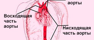
“Aneurysm of the aorta of the heart - what is it?” Many patients in whom a specialist has just discovered this disease ask a similar question. An aortic aneurysm is a widespread or limited dilation of the largest vessel in the human body. With this disease, the diameter of the aortic lumen exceeds the norm by two or more times. Aortic aneurysm is one of the most dangerous heart diseases.
Definition:
A cardiac aneurysm is a pathological condition that is accompanied by protrusion of the wall of the heart muscle. It is one of the complications after a myocardial infarction and can develop within several months after its onset. An aneurysm not only negatively affects the functioning of the heart and its contractile functions, but also poses a serious danger to life due to the possibility of sudden rupture.
Treatment of aortic aneurysm
A cardiac aortic aneurysm, the diameter of which exceeds 5 cm, is subject to surgical treatment due to the fairly high risk of its rupture. Surgery is performed under conditions of hypothermia and artificial circulation. In addition, surgical intervention is performed if the specialist observes too rapid growth of the aneurysm. The surgical mortality rate of this disease is about 15%. Contraindications to surgery are present if the patient suffers from severe heart disease.
Rehabilitation after treatment
During rehabilitation, it is important to follow a diet (eat as little fatty foods as possible, include apples, garlic, strawberries, and legumes in the diet). In addition to proper nutrition, it is very important to avoid stressful situations, control blood pressure, and monitor your weight (it should not exceed the norm). After the surgical intervention, an individual daily routine is drawn up for the patient. It is necessary to set aside time for rest. In this case, the patient undergoes regular examination by the attending physician.
Cause
Usually the cause of the formation of a cardiac aneurysm is a heart attack, most often of the left ventricle of the heart. When they talk about the development of myocardial infarction, the damaged parts of the heart are weakened, and under the influence of blood pressure they can bulge. Blood trapped in them forms a blood clot. Aneurysms occur in patients with atherosclerotic cardiosclerosis and cardiosclerosis of other origins. Much less common are aneurysms that arise due to other reasons: infections, injuries, and congenital defects.
Causes of heart attack
The direct cause of a heart attack is the sudden occurrence of an obstruction to blood flow in the branches of the coronary artery. In 95–97% of cases, such an obstacle is a blood clot formed as a result of atherosclerotic lesions of the arteries. In other cases, a heart attack occurs without any manifestations of atherosclerosis, and the main cause is a pronounced prolonged spasm of the coronary artery.
In extremely rare cases, myocardial infarction develops as a complication of other diseases (arteritis, developmental anomalies and injuries of the coronary artery, infective endocarditis, etc.). The likelihood of symptoms of an acute heart attack increases several times if the following risk factors are present:
- hyperlipoproteinemia (abnormal increase in the level of lipids and/or lipoproteins in the blood);
- old age (55 years and older);
- male gender;
- physical inactivity (lack of physical activity);
- obesity;
- smoking;
- diabetes;
- hypertonic disease;
- hereditary predisposition (presence of IHD in blood relatives).
Classification
Depending on the time period after myocardial infarction the aneurysm formed, the following forms are distinguished:
- Acute and subacute - develop in the early stages after a heart attack, often complicated by incomplete rupture.
- Chronic - formed after acute due to the replacement of a dead section of the heart muscle with connective tissue. They can be muscular, musculofibrous or fibrous.
Diagnostics
It is impossible to diagnose an aneurysm without an instrumental examination. Therefore, after collecting anamnesis and examining the patient, the doctor issues a referral to:
- ECG;
- Echo-CG;
- MRI;
- MSCT of the heart (multispiral computed tomography);
- Heart PET (positron emission tomography);
- myocardial scintigraphy (isotope scan) - intravenous administration of radioactive isotopes (radionuclides) to assess absorption by the heart muscle;
- EPI (electrophysiological study) - stimulation of various parts of the heart to detect life-threatening arrhythmias;
- coronary angiography and cardiac probing - X-ray contrast examination (catheterization) - insertion of a special catheter through the artery.
Symptoms
Symptoms of acute cardiac aneurysm are weakness, shortness of breath, fever, pulmonary edema, sweating, heart rhythm disturbances, and swelling of the veins in the neck. During the transition to the subacute form, symptoms of coronary insufficiency quickly develop.
With chronic cardiac aneurysm, signs of cardiovascular failure are observed: edema, angina attacks, rhythm disturbances. Complications of the disease can be thromboembolic syndrome with damage to peripheral vessels and arteries of the brain. If you experience such signs, immediately contact the cardiology center.
When an acute aneurysm ruptures, death occurs almost instantly. In this case, the patient experiences severe pallor, hoarse, difficult breathing, cold sticky sweat, congestion of the neck vessels, and cold extremities.
Heart aneurysm
According to the time of occurrence, acute, subacute and chronic cardiac aneurysm are distinguished. Acute cardiac aneurysm forms within 1 to 2 weeks from myocardial infarction, subacute - within 3-8 weeks, chronic - over 8 weeks.
Acute aneurysm
In the acute period, the wall of the aneurysm is represented by a necrotic area of the myocardium, which, under the influence of intraventricular pressure, bulges outward or into the ventricular cavity (if the aneurysm is localized in the area of the interventricular septum).
Subacute aneurysm
The wall of a subacute cardiac aneurysm is formed by a thickened endocardium with an accumulation of fibroblasts and histiocytes, newly formed reticular, collagen and elastic fibers; in place of destroyed myocardial fibers, connecting elements of varying degrees of maturity are found.
Chronic aneurysm
A chronic cardiac aneurysm is a fibrous sac microscopically consisting of three layers: endocardial, intramural and epicardial. In the endocardium of the wall of a chronic cardiac aneurysm there are proliferations of fibrous and hyalinized tissue. The wall of a chronic cardiac aneurysm is thinned, sometimes its thickness does not exceed 2 mm. In the cavity of a chronic cardiac aneurysm, a parietal thrombus of various sizes is often found, which can line only the inner surface of the aneurysmal sac or occupy almost its entire volume. Loose mural thrombi are easily fragmented and are a potential source of risk of thromboembolic complications.
There are three types of cardiac aneurysms: muscular, fibrous and fibromuscular. Typically, a cardiac aneurysm is single, although 2-3 aneurysms may be detected at the same time. Cardiac aneurysms can be true (represented by three layers), false (formed as a result of a rupture of the myocardial wall and limited to pericardial adhesions) and functional (formed by a section of viable myocardium with low contractility, bulging during ventricular systole).
Taking into account the depth and extent of the lesion, a true cardiac aneurysm can be flat (diffuse), saccular, mushroom-shaped, and in the form of an “aneurysm within an aneurysm.” In a diffuse aneurysm, the contour of the external protrusion is flat, gentle, and on the side of the heart cavity there is a cup-shaped depression. A saccular cardiac aneurysm has a rounded, convex wall and a wide base. A mushroom aneurysm is characterized by the presence of a large protrusion with a relatively narrow neck. The concept of “aneurysm within an aneurysm” refers to a defect consisting of several protrusions enclosed within one another: such cardiac aneurysms have sharply thinned walls and are most prone to rupture. During examination, diffuse cardiac aneurysms are more often detected, less often - saccular aneurysms, and even less often - mushroom-shaped and “aneurysms within an aneurysm”.
Symptoms of a heart attack
In most cases, the first symptoms of myocardial infarction are increasing manifestations of a person’s existing angina:
- pain in the sternum (longer and more intense);
- an increase in the area of pain and the appearance of irradiation (spread of pain to other areas: left arm, shoulder blade, collarbone, jaw);
- a sharp drop in exercise tolerance;
- a significant decrease in the effect of taking nitroglycerin;
- development of shortness of breath at rest, dizziness, general weakness.
Symptoms of a heart attack rarely appear against the background of new-onset angina or after a period of long remission. Often, the occurrence of an acute heart attack is provoked by one of the following factors:
- severe stress;
- significant physical stress;
- severe overheating or hypothermia;
- surgery;
- sexual intercourse;
- binge eating.
The typical course of the disease is characterized by the fact that the main complaints are associated with such symptoms of a heart attack as severe pain, which is localized behind the sternum. The pain can be burning, squeezing, cutting. Often symptoms of myocardial infarction include pain spreading to the left arm, shoulder blade and shoulder. However, antianginal drugs such as nitroglycerin do not have an analgesic effect. Compared to an attack of angina, the duration of pain is increased: from 30 minutes to several days. Even slight physical activity or stress increases pain.
Most people note that the painful symptoms of a heart attack have a pronounced emotional overtones: there is a feeling of fear of death, hopelessness, doom, and intense melancholy. The person becomes easily excitable, may scream, and strive to constantly change body positions. In addition, symptoms of myocardial infarction are severe weakness, shortness of breath, excessive sweating, nausea, vomiting, and dry cough.
The examination reveals in patients such objective symptoms of a heart attack as pale skin with blue discoloration of the nasolabial triangle and nails, the skin is moist to the touch. The pulse rate rises to 90–100 beats per minute, while blood pressure rises slightly. Often in the first days, an increase in body temperature to 37–38 °C is detected.
1.General information
An aneurysm (the less correct term “aneurysm” is sometimes used) is a local bulge, a protrusion of any boundary surface in the form of a stretched sac, or a spindle-shaped expansion of the walls of a blood vessel (saccular aneurysms are also found on vessels, but much less frequently). Thus, an atrial septal aneurysm (ASA) is a curvature of the membrane separating the right atrium from the left. Normally, this septum is sealed, relatively straight and does not bend, at least significantly, in any direction.
Many authors qualify an MPP aneurysm as a pathology only if the protrusion in any direction (to the right atrium, to the left, or S-shaped) exceeds 10 mm and is combined with prolapse (sagging) of the mitral valve. In terms of the general population, such an anomaly is rare; more often it accompanies congenital connective tissue diseases. An MPP aneurysm is called a “minor cardiac anomaly” or “small heart defect”, since in most asymptomatic cases it does not pose a real danger and does not require treatment, and is usually discovered by chance. However, it does not at all follow from this that this anomaly can be forgotten without attaching any significance to it: the situation can sharply worsen with the onset of enlargement of the aneurysm, thinning of the aneurysm and its spontaneous breakthrough, after which the dynamics become unpredictable.
A must read! Help with treatment and hospitalization!
