It branches like a tree, first into large branches (trunks), then into smaller branches and twigs, and is conventionally divided into several parts or sections:
- 1. The ascending aorta is the area from the aortic valve to the brachiocephalic trunk.
- 2. The aortic arch is a short section from which all the vessels supplying the arms and head (brachiocephalic arteries) depart. They anatomically form an arch connecting the ascending and descending aorta.
- 3. The descending (thoracic) aorta begins from the mouth of the left subclavian artery and continues to the diaphragm.
- 4. Below the diaphragm and before the bifurcation of the aorta (bifurcation) is the abdominal aorta.
Dividing the aorta into sections is very important for assessing risk and choosing optimal treatment tactics in patients with aortic aneurysms.
An aortic aneurysm is an area of local expansion.
Causes of aortic enlargement
Congenital systemic connective tissue diseases: Marfan syndrome, Ehlers-Danlos syndrome, caused by genetic changes in which the aortic wall has an abnormal structure, can cause the development of an aneurysm.
Acquired diseases that cause aneurysmal changes in the aortic wall: most often this is atherosclerosis. About 80% of all complicated aortic aneurysms are aneurysms caused by an atherosclerotic process, which leads to weakening of the vessel wall and the inability to withstand normal blood pressure, and as a result, to its expansion.
Less commonly, an aortic aneurysm develops in inflammatory diseases caused by external agents (syphilis, fungal infection, tuberculosis) or in autoimmune diseases (nonspecific aortoarteritis).
Symptoms of aortic aneurysm
Unfortunately, the diagnosis of aortic aneurysm cannot always be established during the “cold period” (before complications develop), since this disease is usually asymptomatic. Most often, it is discovered accidentally during fluorography, ultrasound or tomography studies performed in connection with other diseases. Treatment of an aneurysm of the ascending aorta before complications develop is much safer for the patient, therefore, in the early diagnosis of an aortic aneurysm, routine medical examination is important.
It is worth noting that every hundredth patient who died suddenly dies from aortic dissection.
Complaints usually appear when the aneurysm begins to stratify or, enlarging, compresses surrounding organs and tissues. Pain or dysfunction of those organs located in the area of the aneurysm appears. At first, this is not of a bright nature and, therefore, does not alarm either the patient or the doctor.
However, the pain intensifies as these deadly complications of an aortic aneurysm develop—it is some of the most severe pain a person can experience. It is localized in the chest if the aneurysm is located in the ascending, descending sections or in its arch, or in the abdomen if it formed in the abdominal section. Characterized by severe weakness, pallor, and often the person loses consciousness.
Impaired blood supply to organs located in the area of aneurysm rupture or aortic dissection (brain or spinal cord, kidneys, intestines, upper or lower extremities) leads to loss of function of these organs, and a large volume of blood loss during aortic rupture represents the most serious danger. To save a life, minutes count. If early surgical treatment is not available, the first-day mortality rate for aortic dissection is 1% per hour (one person in a hundred dies every hour). Within the first 24 hours, 33% of patients die from aortic dissection, 50% of patients within 48 hours and 75% within two weeks. Only early surgical intervention makes it possible to save a significant proportion of patients.
How do changes in the functioning of the aorta affect systemic blood flow?
The aorta is the only vessel from which the systemic circulation begins. Any of her diseases cause severe hemodynamic disturbances, including cardiovascular failure.
The first thing that suffers is blood pressure. Due to sclerosis and calcification, the arterial wall becomes rigid and loses its elasticity, and this is one of the causes of hypertension. When an aneurysm ruptures, the opposite is true - blood pressure drops sharply.
Aortic valve defects are very dangerous. Failure leads to regurgitation, which is the return of blood to the ventricle, causing it to overstretch, leading to cardiomyopathy. As a result of stenosis, cardiac output is also reduced. However, this is due to the fact that the doors do not open completely. At the same time, blood flow in the coronary arteries is disrupted. This leads to the development of angina pectoris.
The degree of disturbance of blood flow largely depends on the localization of the pathological process: the closer it is to the beginning of the vessel, the more systemic its effect will be, while damage to only the abdominal region causes hypoxia of a limited area of the body (lower torso).
Diagnosis of aortic aneurysm
In the diagnosis of aortic aneurysms, the so-called imaging techniques (ultrasound, MRI, CT, AG) are of greatest importance. In the ascending aorta, its arch and in the abdominal section, an aneurysm can be detected using ultrasound methods (ultrasound). To diagnose an aneurysm of the descending (thoracic) aorta, X-ray methods (x-ray, computed tomography) are required. To establish a final diagnosis and select a treatment method, contrast research methods are performed. Currently, the optimal diagnostic method, which provides the most complete information about the location, extent, diameter of the aneurysm and its relationship to nearby organs, is multislice computed tomography - aortography.
Treatment methods for aortic aneurysm
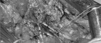
The main method of treating an aneurysm of any part of the aorta is surgical. The point of the method is to replace the dilated section of the aorta in order to prevent its further stretching and rupture. To replace the aorta, two methods are used - the endovascular (intravascular) method using a special intravascular prosthesis (stent graft), and open surgery - aortic replacement.
Each method has its own indications, and each of them has its own advantages and disadvantages.
The advantages of the surgical method lie in its versatility, that is, the ability to correct all disorders associated with an aortic aneurysm, regardless of the department and nature of the lesion. For example, in case of an aneurysm of the ascending aorta and damage to the aortic valve, aortic and aortic valve replacement is performed in combination with coronary bypass surgery.
To perform surgery on the ascending aorta and its arch, it is necessary to use artificial circulation, systemic hypothermia, and often complete circulatory arrest.
Indications for surgical treatment
- transverse size of the aneurysm,
- aneurysm growth rate;
- the formation of complications of this disease.
For each section of the aorta, there is a cutoff limit for the transverse size of the aorta, after which the risk of aortic rupture statistically significantly increases. Thus, for the ascending and abdominal aorta, the transverse diameter of the aneurysm is 5 cm dangerous in terms of rupture, for the thoracic aorta - 6 cm. If the diameter of the aneurysm increases by more than 6 mm in 6 months, then this is also an indication for surgery. Also threatening in terms of rupture and dissection of the aorta are the saccular form of the aneurysm and expansion of the aorta, which is smaller than the diameter that is the indication for surgery, but accompanied by pain at the site of expansion and dysfunction of the presenting organs. Dissections and completed ruptures of aneurysms are absolute indications for emergency surgery.
What is the aorta and where is it located?
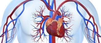
The aorta is considered the largest vessel of the body and has a key role in maintaining normal hemodynamics. It is from here that a large circle of blood circulation begins, which supplies oxygen-rich blood to all structures of the body. It departs from the left ventricle of the heart, is mostly located along the spinal column and ends, diverging into two branches: the right and left iliac.
Structure and departments
It belongs to the elastic type of arteries; histologically its wall is formed by three layers:
- Internal (intima) – represented by endothelium. It is he who is most susceptible to pathological processes, including atherosclerosis. This lining forms the aortic valve.
- Middle (media) - mainly consists of elastic fibers, which, when stretched, increase the lumen of the bed. This allows you to maintain stable blood pressure. It also contains a small amount of smooth muscle fibers.
- External (adventitia) - consists mainly of connective tissue elements with a low content of elastic fibers and high collagen, which gives the vessel additional rigidity, despite the small thickness of the wall.
Topographically, the artery consists of three main parts: the ascending section, the arch and the descending section.
The ascending section begins in the area of the third intercostal space, along the left edge of the sternum. The aortic valves are located at the point where the vessel exits the heart. Their second name is “crescentic” because they resemble curved pockets, consisting of three leaflets and preventing the reverse flow of blood after the aorta has left the ventricle. There are also small bulges - sinuses, in which the coronary arteries that supply the myocardium begin. In the same place there is a short expanded section - the bulb. Opposite the articulation of the second right rib with the sternum, the ascending aorta passes into the arch.
The arc turns to the left and ends near the fourth thoracic vertebra, forming the so-called isthmus - a place where the artery is somewhat narrowed. Posterior to this is the tracheal bifurcation (the point at which the breathing tube divides into two bronchi). Branches extending from its upper side supply the upper part of the body:
- brachiocephalic trunk;
- left general carotid;
- left subclavian.
The descending section is the longest part of the vessel, consisting of the thoracic (thoracic) and abdominal (or abdominal) sections. It originates from the isthmus of the arch, is mostly located in front of the spine and ends near the fourth lumbar vertebra. At this point, the aorta diverges into the right and left iliac branches.
The thoracic region is located in the thoracic cavity and extends to the aortic opening of the respiratory muscle of the diaphragm (opposite the 12th vertebra). Along its entire length there are branches that supply blood to the organs of the mediastinum, lungs, pleura, muscles and ribs.
The final, abdominal part, provides blood supply to the abdominal and pelvic organs, the abdominal wall, and the lower extremities.
Normal vessel size indicators
Determining the diameter of the aorta is very important in the diagnosis of many of its pathologies, especially aneurysms or atherosclerosis. This is usually done using x-ray (for example, computed tomography or magnetic resonance imaging) or ultrasound (echocardiography) studies. It is important to remember that this value is very variable, as it changes depending on age and gender.
| Department | Diameter, cm | |
| Men | Women | |
| Root | <4 | <3,7 |
| Rising | 3,4-3,6 | 3,0-3,5 |
| Arc | 3,0-3,5 | 2,7-3,4 |
| Thoracic | 2,5-3,0 | 2,4-2,6 |
| Abdominal | 1,9-2,4 | 1,8-2,1 |
Blood flow, speed and pressure
Also, to study the work of the aorta, its functional indicators are examined. This is usually done using ultrasound or Doppler ultrasound.
Systolic pressure in the vessel is about 120-140 mmHg. Art., diastolic – 70-90 mm Hg. Art. The value may vary slightly depending on age and individual characteristics.
The linear speed of blood flow in the initial sections is about 0.7-1.3 m/s. Distally (i.e. further from the beginning) it decreases to 0.5 m/s.
The normal flow rate is 4-5 liters per minute.
Types of open surgical operations for aortic aneurysms:
- Bentalla-De Bono operation (replacement of the ascending aorta with a valve-containing conduit with a mechanical prosthetic aortic valve);
- David's operation (replacement of the ascending aorta while preserving the native aortic valve);
- Supracoronary aortic replacement;
- Prosthetics of the ascending aorta and its arch (Borst technique, the use of oblique aggressive anastomosis and other techniques);
- Thoracic aortic replacement;
- Abdominal aortic replacement.
Endovascular interventions
They allow you to dramatically reduce the amount of surgical trauma, shorten hospitalization periods and reduce the inevitable suffering of the patient associated with surgical approaches. One of the main disadvantages of the method is the need for repeated interventions.
Types of endovascular operations for aortic aneurysm:
- implantation of a stent graft into the abdominal aorta,
- implantation of a stent graft into the ascending (thoracic) aorta.
The most modern method of treating aortic aneurysm is a hybrid method, which allows achieving optimal treatment results with the least surgical trauma.
Hybrid surgeries combine the advantages of open and endovascular interventions.
To prevent the development of aortic aneurysms, the most important thing is the need to control risk factors, namely arterial hypertension. In addition to arterial hypertension, the most significant risk factors are age (over 55 years), male gender, smoking, the presence of aneurysms in direct relatives, and high cholesterol levels.
What treatment and correction methods exist and are considered effective?
A peculiarity of aortic pathologies is that their treatment mainly involves invasive surgical intervention. Conservative therapy is used only to support vital signs and relieve symptoms, which allows for safe surgery.
There is now a trend towards minimally invasive endoscopic operations, which are safer and more effective.
Today the following surgical treatment methods are used:
- resection with anastomosis – used for small aneurysms or coarctation;
- prosthetics;
- coronary artery bypass grafting (creation of bypass circulatory pathways) – for occlusive diseases, ischemic heart disease or heart attack;
- implantation of artificial valves, balloon valvuloplasty,