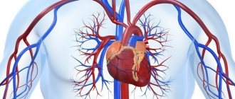
The main purpose of ultrasound of the abdominal aorta is to identify an aneurysm or atherosclerotic lesion of the aorta and damage to its large branches: splenic, hepatic, renal and mesenteric arteries. Ultrasound of the abdominal aorta is a non-invasive diagnostic method that has high value in identifying many vascular diseases. The results of ultrasound are very dependent on the examination technology, the quality of the equipment and the qualifications of the specialist. Today, the low price of ultrasound of the aorta and abdominal organs, the simplicity and safety of the study make it indispensable for the primary detection of abdominal aortic aneurysm.
The innovative vascular center specializes in vascular surgery, so we strive to diagnose vascular pathology at the highest level. Our center has excellent stationary and mobile ultrasound machines that allow us to study in detail the structure of the aortic wall and its branches, as well as identify aneurysms. The specialists of our medical center are dedicated to working specifically with vascular pathology, therefore they identify it much better than general ultrasound doctors.
Preparing for the study
To successfully perform an ultrasound of the abdominal cavity, preparation is necessary aimed at reducing the amount of gases in the intestines that interfere with seeing the retroperitoneal organs, including the aorta. The preparation rules for examining the abdominal aorta and its branches using ultrasound are simple:
- 2 days before the examination, exclude legumes, potatoes, cabbage, melon, dairy products, soda and all foods high in carbohydrates from the diet. These products may cause increased gas formation.
- On the eve of the ultrasound, start taking medications to improve bowel function. For these purposes, taking espumisan or regular activated carbon is suitable.
- It is advisable to skip dinner the evening before the procedure, and breakfast in the morning.
Aneurysms of the abdominal and thoracic aorta
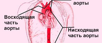
The aorta is the largest artery in the human body, through which oxygenated blood flows from the left ventricle. Branches extend from it to various internal organs and tissues, and at the level of the pelvis it is divided into two iliac arteries, which supply blood to the lower extremities. The aorta can be divided into thoracic and abdominal sections, located in the corresponding cavities. Sometimes, under the influence of various factors, the wall of this vessel becomes thinner, and due to blood pressure, a protrusion (aneurysm) is formed. Since various complications can develop, patients need to receive medical attention.
Etiology
An abdominal aortic aneurysm in most cases occurs against the background of atherosclerotic damage to its wall, which is observed in more than 80% of cases. Much less often, the cause of thinning of the muscle layer is inflammatory diseases (tuberculosis, rheumatism, syphilis), accompanied by vascular damage. Nonspecific aortoarteritis occurs against the background of other infectious diseases.
Congenital pathology of the connective tissue structure (fibromuscular dysplasia, Marfan syndrome) also plays a large role in the development of this pathological process.
Due to the development of vascular surgery in recent decades, aneurysms are increasingly caused by operations on the abdominal aorta:
- angioplasty, when using a special balloon to increase the diameter of the artery;
- removal of blood clots and atherosclerotic plaques along with the inner membrane (endarterectomy);
- thinning of the wall at the junction during aortofemoral replacement.
However, in most cases of complications of surgical intervention, a false aneurysm is formed, the walls of which are made not of the vessel, but of the surrounding tissues. Risk factors for the formation of an abdominal aneurysm are smoking, obesity, male gender, family history (the presence of similar diseases in close relatives) and hypertension.
Manifestations and complications
Symptoms of an aneurysm may be absent for quite a long time and appear only with a significant increase in its size, or with the development of various complications.
In the first case, patients complain of a feeling of pulsation in the abdomen, bulging of the anterior abdominal wall in a horizontal position. This is especially true for thin people. The pain syndrome is usually mistaken for renal or hepatic colic. In addition, dysfunction of internal organs caused by external compression may be observed:
- aneurysm dissection. When an aneurysm dissects, a false tract is formed, through which there is no blood flow;
- digestive processes are disrupted (heartburn, deterioration of peristalsis, constipation);
- from the excretory system, hematuria and urinary retention often develop;
- when exposed to vessels leading to the testicles, varicocele or ischemia may develop;
- when nerve roots and plexuses are compressed, symptoms of sciatica and radiculitis appear, characterized by pain in the lower back and lower extremities.
One of the most common complications is dissection of the aortic wall with the formation of a second lumen, which is usually blind, that is, it lacks adequate blood flow. As a rule, this is a continuation of a defect that arose at the level of the thoracic region.
A dissecting aneurysm is characterized by the following symptoms:
- Deterioration of blood flow to various internal organs. In this case, their function is disrupted, for example, when the mesenteric arteries are damaged, intestinal necrosis develops, and the renal arteries develop acute renal failure. If the blood supply to the lower extremities is impaired, intermittent claudication occurs.
- The pain is usually quite intense and is associated with an effect on sensitive nerve receptors located in the vessel wall and in organs that have been subjected to ischemia. It is localized in the anterior abdominal wall, but can also radiate to the back.
- Neurological disorders may appear in the form of paresis and paralysis, as well as peripheral polyneuropathy.
Rupture of an abdominal aortic aneurysm is accompanied by a sudden decrease in high blood pressure, sweating, pain in the abdomen and lumbar region, signs of motor agitation and weakness appear. If emergency measures are not taken, the patient dies within a short time from massive hemorrhage into the abdominal cavity.
Aneurysms of the thoracic aorta usually develop as a result of degenerative changes in its wall and are more often classified as true aneurysms. According to etiology, aneurysms are divided into: 1) non-inflammatory (atherosclerotic, traumatic, postoperative, iatrogenic); 2) inflammatory (syphilitic, rheumatic, with nonspecific aortoarteritis, mycotic); 3) congenital (with Marfan syndrome, cystic medianecrosis, coarctation of the aorta, congenital tortuosity of the aortic arch). Based on localization, they are distinguished: 1) aneurysms of the sinuses of Valsalva; 2) aneurysms of the sinuses of Valsalva and the ascending aorta; 3) aneurysm of the ascending aorta; 4) aneurysms of the ascending aorta and aortic arch; 5) aneurysms of the aortic arch; 6) aneurysms of the aortic arch and descending aorta; 7) aneurysm of the descending aorta; thoracoabdominal aneurysms. By type, aneurysms are divided into true, false and dissecting, and by shape - into saccular and diffuse (spindle-shaped).
Thoracic aortic aneurysms are observed predominantly in men. The clinical picture is poor. Sometimes an aneurysm is an incidental finding. Symptoms depend on the location, size and extent of the aneurysm. The leading symptom in 1/3 of patients is pain behind the sternum, in the neck, back, and between the shoulder blades. In another 1/3 of patients, the leading symptom is shortness of breath, stridor, and dry cough. Almost half of the patients have arterial hypertension. Sometimes hoarseness, bradycardia, Horner's symptom, and ischemic disorders of the brain are observed. Vascular murmurs are detected in 75% of patients.
Diagnosis of aneurysms at the prehospital stage is difficult. 40-50% of patients are hospitalized with an erroneous diagnosis. An accurate topical diagnosis is provided by the x-ray method, which reveals the expansion of the mediastinal shadow, calcification of the aneurysm wall, and deviation of the esophagus when it is contrasted with barium. The most valuable research methods are computed tomography and magnetic resonance imaging, which help identify the location and size of the aneurysm. Aortography confirms the diagnosis. Differential diagnosis includes tumors and mediastinal cysts.
The prognosis for untreated aneurysms is unfavorable. Within three years after diagnosis, 37.7% die, after 5 years - 54% of patients. Causes of death: rupture of aneurysm (35-40%), heart failure (20-36%), compression of the tracheobronchial tree, pneumonia (14-20%).
How is an ultrasound of the abdominal aorta and its branches performed?
The principle of ultrasound scanning of the abdominal cavity is to process the ultrasound signal reflected from the organs. This allows you to evaluate the structure and size of this organ. Ultrasound examination of the abdominal aorta is not dangerous and does not cause any discomfort. The diagnostic process itself includes only a few steps:
- In the ultrasound diagnostic room, the patient sits on the couch to the right of the doctor and opens his stomach.
- The doctor lubricates a special sensor and the patient’s abdomen with a transparent gel, which reduces tissue resistance and helps the ultrasound wave penetrate inside.
- The specialist then slowly moves the sensor along the abdominal wall and records the results of the observations.
The capabilities of ultrasound devices allow not only to assess the size, shape and structure of the vessel wall, but also to study the characteristics of blood flow. This is achieved using Doppler ultrasound and color Doppler mapping. Dopplerography is based on recording the ultrasonic signal from moving red blood cells and changing the speed of this signal depending on the approach or distance of these particles to the sensor. The speed of blood flow, determined by Doppler ultrasound, is an indicator of the patency of blood vessels below the study site or in areas of narrowing of the vessel. In narrowed places it is significantly strengthened; if there is an obstacle to the outflow of blood below the test site, then the speed decreases.
The incidence of glomus tumors (GO) is less than 2% among all soft tissue tumors [1–3]. As a rule, HO is a single formation, less often accompanied by multiple lesions of different parts of the body [3-6].
Cases of atypical localization have also been described: in the thyroid gland [7], trachea [8], lungs [9], bones [10], esophagus [11], stomach [12], intestines [13], kidney [14], peritoneum [15], intraneurally [16] and intravenously [17].
Characteristic symptoms of HO are severe pain, increased sensitivity to cold and local pain on palpation of the formation. With a typical localization of the formation, these symptoms make it possible to make a diagnosis in almost 90% of cases. Of the additional research methods, MRI has the greatest sensitivity. Most HOs are characterized by a benign course, but rare examples characterized by malignant potential have been described. Thus, A. Folpe et al. [18] developed criteria for malignancy: deep location of the tumor, size more than 20 mm, presence of atypical mitoses, a combination of moderate and high degrees of nuclear and mitotic activity, more than five mitoses per 50 fields of view.
Treatment of patients with intradermal HO is surgical. Radical operations lead to complete cure. The relapse rate, according to various authors, ranges from 12 to 33% [18–26].
In 25 years, this is the first case of retroperitoneal HO in our clinic.
We present a clinical case.
Patient G
., 59 years old, during an examination at the place of residence, a retroperitoneal tumor was identified, and therefore laparoscopic tumor removal was performed in one of the Moscow clinics. After 3 months, a follow-up examination revealed a recurrence of the tumor, and therefore she contacted the Russian Cancer Research Center named after. N.N. Blokhina. Immunohistochemical examination revealed that the formation has the structure of G.O. A computed tomography (CT) scan of the chest and abdomen revealed that the patient had essentially continued tumor growth. The tumor is located para-aortically on the left at the level of vertebrae LII—LIV, measuring 8x5x7 cm. The tumor infiltrates the left ureter, psoas muscle, mesentery of the sigmoid colon, circularly covers the aorta from the left renal artery to the bifurcation, infiltrates the lumbar arteries, and is closely adjacent to the inferior vena cava (Fig. 1).
Rice.
1. Computed tomogram of the abdominal cavity. On November 25, 2015, the patient was operated on. During the audit, no distant metastases were detected in the abdominal cavity.
At the first stage, the right and left halves of the colon were mobilized in an acute manner, the inferior vena cava, aorta, and ureters were visualized. Next, resection of the sigmoid colon was performed. The aorta is highlighted above the tumor, the left renal artery and vein are visualized. The latter are bandaged and crossed. Then the right and left iliac arteries were isolated, the infrarenal segment of the aorta was mobilized along with the tumor, and the lumbar arteries were ligated (Fig. 2).
Rice. 2. The stage of isolating the infrarenal aorta, the common iliac arteries are taken on holders. 1 - right common iliac artery; 2 - left common iliac artery; 3 - tumor.
The tumor was removed en bloc with the aorta, left kidney and sigmoid colon (Fig. 3). Prosthetics of the aorta and common iliac arteries was performed (Fig. 4). The ischemia time of the lower extremities was 20 minutes. Operation time 3 hours. Blood loss 400 ml.
Rice. 3. Removed macropreparation. 1 - tumor; 2 — lumen of the infrarenal aorta; 3 - left kidney; 4 - sigmoid colon.
Rice. 4. View of the surgical field after vascular reconstruction.
When carrying out immunohistochemical studies, antibodies to vimentin, aSMA, H-caldesmon, calponin, desmin, myogenin, CD10, CD31, CD34, D2−40, pancytokeratin AE1/AE3, EMA, CD56, CD57, CD68, CD117, S100 protein, GFAP were used , HMB45, Melan A, to estrogen and progesterone receptors, Ki-67.
Expression was detected in the majority of tumor cells: vimentin (+++), N-caldesmon (+++) and calponin (+++), and in some tumor cells expression of desmin, estrogen receptors (5%) and progesterone (10%) was noted ). The proliferation index in Ki-67 tumor cells was 1% (Fig. 5).
Rice. 5. Histological signs of malignant glomus tumor. Hematoxylin and eosin staining. a, b — atypical glomus cells surround dilated blood vessels, ×100; c — the tumor consists of round cells with a pronounced nucleolus, with high mitotic activity, ×400; d — tumor grows into the aortic wall, ×50.
Reactions to other immunohistochemical markers in tumor cells are negative [6–15].
It was established that the formation is a malignant HO of low degree of differentiation FNCLCC (2/1/0 =3/8 points) with damage to the aortic wall, not reaching 0.2 cm to its intima, para-aortic tissue and mesentery of the colon, growing into the wall of the left ureter, with metastases in 2 of 6 lymph nodes and a metastasis (size 5x4x3 cm) in the mesentery of the colon. No elements of tumor growth were found in the wall of the colon, in the left kidney, para-aortic lymph nodes, the edges of aortic resection and the greater omentum.
A follow-up examination 6 months after surgery revealed no signs of disease progression.
Based on the published data, the prognosis of benign HO is satisfactory; with a follow-up period of 4 to 60 months, not a single patient with benign HO of the lung died from disease progression [19].
The prognosis for malignant HO is naturally worse, which is due to the progression of the disease at various times after surgery. Under G.O. In the lung, metastases most often appear in the lungs, and in case of gastric tumors - in the liver. According to the literature, the following chemotherapy regimens were used in the treatment of patients with HO, both in the adjuvant and therapeutic regimens: adriamycin/CDDP, doxorubicin [7, 27]. After 4 courses of chemotherapy with doxorubicin, a patient with malignant tumor in the upper lobe of the right lung and multiple metastases experienced complete regression of the tumor. The patient lived for 15 months after diagnosis of systemic progression and initiation of chemotherapy [27]. The Adriamycin/CDDP regimen was used in the adjuvant setting in a patient with malignant thyroid tumor; the relapse-free period was 30 months [7].
Below we provide summary data on relapse-free survival and localization of metastases in patients with malignant tumors of atypical localization (see table).
Relapse-free survival and localization of metastases in patients with malignant tumors of atypical localization
The only treatment method for patients with locally advanced glomus tumor is radical surgical treatment.
There is no conflict of interest.
Interpretation of ultrasound data of the abdominal aorta
During an ultrasound of the aorta and abdominal vessels, the doctor examines the size of the aorta and its lumen, how evenly it is colored during Doppler mapping. Dilatation of the abdominal aorta of more than 4 cm speaks in favor of an aneurysm. Ultrasound diagnostics allows you to assess the condition of the arteries of the kidneys, intestines, liver and stomach. It is possible to detect dilation of the arteries of internal organs. With complicated aneurysms, it is possible to detect a retroperitoneal hematoma due to rupture, fluid in the abdominal cavity, thrombosis of the aorta or visceral arteries. Often, a good ultrasound scanner can detect dissection of the vessel wall, its tears and atherosclerotic plaques. Ultrasound of the abdominal aorta and visceral branches lasts about 25-40 minutes. After the examination, you can immediately return to normal activities.
Aortic aneurysm - symptoms and treatment
Treatment of aortic aneurysms is aimed at preventing its rupture. It can be conservative or surgical.
Conservative treatment
The main purpose of conservative therapy is to reduce blood pressure and the force of heart contraction, as well as the correction of concomitant diseases, such as coronary heart disease, diabetes, kidney disease, etc. [1]
Surgery
Modern surgical techniques for treating aortic aneurysms are divided into endovascular and traditional surgical interventions.
The endovascular method is the remote implantation of special tubular prostheses (stent grafts) into the affected area of the aorta from the inside through a catheter installed in the femoral artery. This method of treatment is performed under local or regional anesthesia, it is low-traumatic, low-painful and reduces hospital stay, and also reduces mortality. Increasingly, in the practice of vascular surgery, minimally invasive endovascular technologies are used in the treatment of aneurysms of the thoracic and abdominal aorta, although it is still too early to discount open surgical interventions [4].
Major operations for thoracic aneurysms are among the most difficult in medicine. They are performed in specialized cardiac surgery centers with the heart turned off during artificial circulation. The technology of the operation consists of replacing the dilated section of the aorta with a special tubular prosthesis, which is sewn into place of the aneurysmal sac [5][10]. The surgeon makes an incision to provide access to the aneurysm, excises the affected part and replaces it with a prosthesis. But in some cases, for example after injury, the aorta can be sutured end to end without the use of a prosthesis.
Abdominal aortic aneurysms with a diameter of more than 5.5 cm in men and 5.2 cm in women have a high risk of rupture, and therefore require immediate consultation with a vascular surgeon to determine indications for surgical treatment. It is performed in the vascular departments with access through the anterior abdominal wall (laparotomy) and also aims to replace the affected segment of the aorta with a prosthesis. Some medical centers have mastered laparoscopic treatment of aneurysms, which does not require incisions [4][7].
During surgical treatment of such severe diseases (as with other therapeutic and diagnostic interventions), there is a possibility of complications. It is also worth noting that not all regional, regional and republican medical centers have specialists who work on open heart surgery.