- home
- general surgery
- Abdominal adhesions
Interview with Professor Puchkov on the First Medical Channel on the topic: Adhesive disease
As a rule, the internal organs and tissues of the body have a smooth surface. Normally, organs do not stick together and glide easily. When the surface of an organ, tissue, or body cavity is damaged, inflamed, or has a scar, there is a risk of adhesions between organs (adhesions). The organs are connected to each other by fibrous fibers and “stick” to each other. This phenomenon is called adhesive disease.
Prices for services
Initial appointment with a gynecologist + ultrasound (assessment of complaints, medical history, examination in a gynecological chair, pelvic ultrasound, consultation)
Primary appointment – visiting a doctor of a specific specialty for the first time. Make an appointment
1800 ₽
Repeated appointment with the gynecologist
With the exception of repeated appointments with doctors: Blatsios N.D., Dzhashiashvili M.D. Make an appointment
1200 ₽
Ultrasound of the pelvis in women (uterine cavity, ovaries)
Make an appointment
1500 ₽
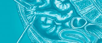
To understand the occurrence of peritoneal adhesions, it is necessary to know the structure of the peritoneum: the histological structure of the peritoneum is quite complex, it includes a number of morphologically different layers of heterogeneous structure. The most superficial layer is the mesothelium - under physiological conditions there is a continuous change of mesothelial cells. There is always fluid in the abdominal cavity, which is released in certain areas of the peritoneum by ultrafiltration from the vessels.
Thanks to the movements of the diaphragm and peristalsis, fluid moves freely in the abdominal cavity and enters areas of the peritoneum where it is absorbed. The presence of fluid in the abdominal cavity greatly facilitates the peristaltic movements of the intestines and eliminates friction between the serous membranes of the abdominal organs.
Causes of adhesive disease:
- Reasons leading to gluing of the peritoneal layers.
- Reasons leading to soldering of the omentum to areas of damaged peritoneum.
- Loss of fibrin, which, falling out on the peritoneum, gradually turns into connective tissue cords.
- Numerous adhesions.
All these reasons lead to the formation of planar or strand adhesions, which can cause the development of acute intestinal obstruction. All these reasons give rise to the development of adhesive disease, which sometimes occurs with the clinic of OKN. Some authors interpret adhesive disease as the obligatory presence of OKN, but this is not so. The main reason for the formation of adhesions is trauma to the peritoneum.
From the first minute, a serous-fibrinous exudate containing various cellular elements appears at the site of peritoneal damage. Fibrin will fall out of the exudate, and the damaged surface of the peritoneum will become covered with fibrin. At the end of the 2nd day, tender fibrous formations can be observed on the peritoneum. When damaged areas of the peritoneum come into contact, they bond together due to fibrin threads. However, in the future, with shallow damage to the peritoneum, such fibrin deposits may undergo resorption and the adhesive surfaces may separate under the influence of peristalsis. If the damage to the peritoneum was deeper, involving layers deeper than the limiting membrane, then healing of the peritoneum occurs according to the type of secondary intention.
In these cases, well-vascularized granulation tissue is formed on the surface of the peritoneal defect; collagen fibers appear between the fibrin threads, located according to the direction of tension. Numerous anastomoses of venous vessels and nerve fibers appear in the adhesions. The adhesions formed in this way do not undergo resorption. Particularly powerful adhesions are formed when the parietal and visceral layers of the peritoneum are damaged and the wound surfaces come into contact. After surgical trauma, serous surfaces with a damaged mesothelial layer in some cases may be adjacent to one another, and the resulting postoperative intestinal paresis maintains the direct contact of these surfaces, which makes it possible for the process of development of adhesions to develop quietly within 2-3 days. The peristaltic movements of the intestines that occur on the 3rd day are no longer able to separate the glued surfaces and the adhesions become persistent and strong.
Adhesions and adhesions in the abdominal cavity can also develop as a result of inflammatory processes in the abdominal cavity. One of the main reasons here is acute purulent peritonitis. Chronic inflammatory process - tuberculosis, can also cause adhesions. In acute purulent peritonitis, pus accumulates in the abdominal cavity, the parietal peritoneum (and especially the visceral) sharply swells and becomes edematous, so the peritoneal mesothelium, even under the influence of a minor injury, easily sloughs off, exposing the deeper layers of the peritoneum.
The presence of an inflammatory process in the abdominal cavity leads to the cessation of peristalsis, due to which sections of the intestinal loops can come into contact with each other for a long time and thereby create conditions for gluing. In addition, deposited fibrin can also cause adhesions. The greater omentum (the policeman of the abdominal cavity) is soldered to the inflamed peritoneum of the loops, further causing the formation of cord adhesions.
The omentum entangles the intestinal loops, which leads to the formation of conglomerates of intestinal loops. Most often, in acute peritonitis, adhesions form in the lower sections, since exudate accumulates there. In most cases, after diffuse purulent peritonitis, gluing of the loops of the small intestine to each other is observed.
Chronic tuberculous peritonitis: strand and planar adhesions may occur, sometimes entire conglomerates of intestines are formed, which are difficult to separate. The omentum plays an important role in the formation of adhesions, which adheres to the tuberculous tubercles on the intestinal serosa; various types of conglomerates of intestinal loops that arise give rise to the development of OKN, and with tuberculous peritonitis, the surgeon is sometimes forced to perform surgical intervention for emergency reasons.
Presence of foreign bodies in the abdominal cavity. Even medicinal drugs increased the formation of adhesions. Of particular importance is the ingestion of tiny talc powder, which, if it gets on the peritoneum, leads to the formation of granulomas on the peritoneum. In this case, talc has not only a mechanical, but also a chemical effect - in these places an aseptic inflammatory process occurs, which has a chronic proliferative nature.
Experimental studies have shown that after introducing talc into the abdominal cavity, wide planar adhesions develop between the omentum and the parietal peritoneum, and flat adhesions between the loops of the small intestine.
Surgeons should always remember this, since in most surgical clinics, talc is widely used when putting on gloves: you should never wear gloves near the surgical field; you should change gloves when they tear.
When ligatures are applied to vessels, intestines, etc., suture material remains, which is also a foreign body. Catgut is especially undesirable in this regard; nylon and lavsan are used instead.
The introduction of drugs into the abdominal cavity causes an increase in the formation of adhesions. In the past, the technique of leaving a microirrigator in the abdominal cavity to administer antibiotics was widely used. However, now this is considered not entirely appropriate: after 1-2 days, the peritoneum sticks together and a canal forms around the microirrigator, and antibiotics do not enter the abdominal cavity. Moreover, the effect of antibiotics on an object is through its absorption into the blood, and then exposure. Local administration of antibiotics is controversial.
According to the majority of surgeons, adhesions that arise after a deep injury to the peritoneal integument most likely do not resolve, but undergo restructuring. Adhesions that arise against the background of an acute inflammatory process in any part of the abdominal cavity in a number of people undergo reverse development.
The possibility of resorption of inflammatory adhesions is indicated by the fact of resorption of the appendiceal infiltrate. If during the period of an acute inflammatory process the infiltrated omentum is fused over a large area with the appendix and adjacent intestinal loops, then after the infiltrate is reabsorbed, very small adhesions of the omentum and the appendix often remain, and all other adhesions are resolved.
From these observations it was concluded that in the immediate period after recovery from purulent peritonitis, it makes sense to use various physiotherapeutic procedures that cause the resorption of adhesions. The use of these procedures in a later period, when adhesions have already formed, will be poorly justified.
The adhesive process develops mainly after operations performed in the lower parts of the abdominal cavity and after appendectomies, which apparently should be explained by the greater frequency of this operation. Most often, the adhesive process after laparotomy develops in patients aged 20-30 years, so indications for surgical intervention at this age, especially in women, should be made very reasonably. In vain, an appendectomy performed at this age can lead to the development of adhesive disease. Therefore, prophylactic appendectomies are not justified.
The development of adhesive disease largely depends on the constitution of the body. In some cases, after one laparotomy, a significant number of adhesions develop, in other cases, after a number of laparotomies, no adhesions are formed.
The scale of the adhesive process can be different: from total to the formation of individual strands fixed at two points. As a rule, the adhesive process is more pronounced in the surgical area. Often, loops of intestines are soldered to the postoperative scar, or fixed to the walls of the postoperative hernial sac.
Therefore, when there is an operation for a ventral postoperative hernia, especially a strangulated one, it is very easy to damage the swollen loops of intestines when the hernial sac is opened.
Make an appointment
Causes
The formation of adhesions is facilitated by the proliferation of connective tissue, which leads to the adhesion of serous membranes due to disruption of the synthesis and breakdown of a high molecular weight, non-globular protein known as fibrin. Another cause of pathologies in the intestines are injuries in the abdominal cavity caused by mechanical or radiation. Intestinal adhesions can become a complication of inflammatory processes in the gastrointestinal tract or genitourinary system, and can also be congenital due to abnormalities of intrauterine development.
Adhesive Disease Clinic
Adhesions formed in the abdominal cavity, regardless of the cause of their occurrence, cause a disorder of normal intestinal motility, which leads to difficulty in emptying the contents of the intestinal loops, causing pain in the abdomen and constipation. Bloating of the intestinal loops creates tension on the fixed omentum, which also gives rise to pain. When the adhesions are pulled, the nerves present in them can also contribute to increased pain. Sometimes adhesions create a constriction of the intestinal loop and cause acute intestinal failure. Due to the fact that the adhesive process can be located in different parts of the abdominal cavity, various organs can be involved in it.
Taking into account the complaints of patients, two clinical forms of adhesive disease can be distinguished:
- Adhesive disease with pain in the abdominal cavity.
- Adhesive disease with periodically recurring attacks of OKN.
Painful sensations during adhesive disease depend, on the one hand, on the irritation of the nervous apparatus of the intestinal loops, and on the other hand, on the irritation of the nervous elements. With adhesive disease, patients may experience pain in various parts of the abdomen, depending on the location of the adhesions, but the patient’s main complaint will be abdominal pain. In this group you can find patients with a relatively calm course of this disease - they have a history of 1-2 laparotomies.
Many patients begin to aggravate due to addiction to narcotic drugs. Abdominal pain can be small, aching in nature, in most cases it is constant pain, sometimes periodically intensifying. The pain often intensifies with physical stress or with errors in diet.
Increased pain forces patients to resort to using heating pads, after which the pain decreases and disappears completely. Along with abdominal pain, patients experience dyspeptic symptoms: nausea, constipation, bloating, etc. Patients of this type do not lose their ability to work, but constant aching pain forces them to often go to the clinic. Prescribing physiotherapeutic procedures in the form of diathermy and iontophoresis improves the condition and reduces pain. Patients can use HBOT, sulfur mud baths, which brings relief for some time.
A number of patients, against the background of constant abdominal pain, periodically experience severe painful attacks that require the administration of drugs. During such attacks, patients are admitted to hospitals, where they undergo a new laparotomy. The appearance of severe pain attacks is associated with great physical stress of the patient or with the consumption of large amounts of food, after nervous disorders.
Patients with frequently recurring attacks of pain become irritated, they develop psychasthenia, lose weight, decrease appetite, and often become drug addicts. Often these patients are harsh, rude, and their ability to work is usually reduced. Contact with such a patient is quite difficult. An objective examination reveals several postoperative scars; upon palpation outside of an attack, the abdomen is soft, usually painless; During a painful attack, a sharp pain is detected in certain parts of the abdomen, and there may be muscle tension. Various painkillers and physiotherapy provide only temporary relief.
The omentum sometimes adheres to the postoperative scar on the anterior abdominal wall and this often leads to pain. The tension of the omentum attached to the scar significantly increases pain when extending the body backwards. If the patient is asked to bend forward, the pain decreases. If there are positive symptoms of omental tension, patients are subject to surgical intervention, the purpose of which is to cut off the greater omentum and its resection.
In adhesive disease with periodically recurring attacks of OKN, along with abdominal pain and constipation, attacks of acute intestinal obstruction with typical clinical symptoms are observed: cramping abdominal pain, vomiting, impaired passage of gases, bloating, postoperative scars on the abdominal wall, palpation reveals some tension in the abdominal muscles, pain in the areas of bloating (P.N. Napalkov said: “the intestines stand like a stake”). Patients in these conditions are excited, Valya's symptom is determined, with a slight shock a splashing noise is detected - Sklyarov's symptom.
Due to the presence of scars on the abdominal wall, intestinal peristalsis is usually not observed. Hepatic dullness may be pushed aside by distended intestinal loops. Auscultation reveals bowel sounds of varying intonation. When examining the rectum, there may not be anything typical: sometimes the ampulla can be swollen, sometimes collapsed (the Obukhov hospital symptom appears quite late).
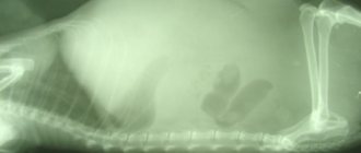
An X-ray examination reveals Kloiber's cups; with a pronounced adhesive process, Kloiber's cups do not move in different positions - a symptom of fixation. In this kind of patients, quite often the symptoms of obstruction are relieved after using the usual measures: a heating pad, a cleansing enema, but if we cannot completely exclude OKN, we need to observe the patient over time - radiologically trace the passage of barium suspension through the gastrointestinal tract. Usually, when a patient is admitted with an unclear clinical picture of OKN, the patient undergoes a survey X-ray of the abdominal cavity and after that they drink about 200 ml of barium suspension and, at an interval of 3 hours, the patient undergoes an X-ray of the abdominal cavity. To speed up the movement of barium, sometimes the suspension is made in very cold water (since cold water enhances peristalsis).
In this type of patient, it is often possible to stop the obstruction and they are discharged, and after a few days they can be admitted again. Such obstruction is often interpreted as dynamic intestinal obstruction of a spastic nature. The use of sedatives eliminates intestinal spasm, thereby restoring its patency. According to many authors, conservative treatment in this group of patients eliminates the symptoms of obstruction in 75% of cases. Surgical intervention for this type of obstruction without the use of conservative measures will be a surgeon’s mistake. At the same time, the surgeon faces a difficult task: is there a mechanical obstruction in this case that cannot be eliminated by conservative measures? A barium test helps here.
To eliminate mechanical obstruction, surgical intervention is necessary. The earlier the intervention is applied, the better the prognosis. We must always remember about the possibility of developing mechanical intestinal obstruction during adhesive disease. To eliminate mechanical intestinal obstruction, emergency surgical intervention is used, the volume of which varies (depending on the amount of intestinal necrosis).
Differential diagnosis of mechanical and dynamic intestinal obstruction is possible with the mandatory use of x-ray examination.
Clinic of adhesive disease in tuberculous peritonitis: as a rule, young people suffer; patients can palpate intestinal conglomerates; during an attack, loud peristalsis can be heard; the onset of pain is often preceded by an abdominal injury or sudden muscle tension. The clinical picture during an attack resembles acute intestinal obstruction with characteristic cramping pain and other symptoms. Some discrepancy between the existing phenomena of obstruction with a clearly expressed dysfunction, and the absence of peristalsis can help in diagnosis. The presence of a tumor formation with a smooth surface and fibrous encysted chronic peritonitis in the abdomen provides significant assistance for recognition - but this does not always happen.
Laboratory data do not provide anything pathognomonic for adhesive disease: ESR may be accelerated, leukocytosis may appear when a painful attack occurs, and the same can be observed with the development of acute intestinal obstruction. To establish a diagnosis of adhesive disease, it is necessary to conduct an X-ray examination of the gastrointestinal tract, since the presence of laparotomies in the anamnesis does not indicate the presence of adhesions in the abdominal cavity. Until recently, laparoscopy was contraindicated, as there is a high risk of damage, but modern endoscopists use laparoscopy for adhesive disease.
X-ray diagnostics is based on the detection in a polypositional study of various types of deformations, unusual fixation, adhesions with the abdominal wall; they study the state of the relief of the mucous membrane, the elasticity of the intestinal walls, especially in the deformation zone: the folds of the mucous membrane, although deformed, do not break off, unlike the tumor process. The adhesive process is not characterized by rigidity of the intestinal wall, which is characteristic of a malignant tumor.
Conservative treatment of adhesive disease: usually adhesive disease has a chronic course, only periodically giving attacks - exacerbation of pain. Therefore, conservative treatment in remission is modified when a painful attack occurs. A painful attack with some gas retention can be stopped with a cleansing enema, heat on the stomach, and antispasmodics. In a hospital setting, epidural blockade of trimecaines has a positive effect. Previously, lumbar blockades according to Vishnevsky were widely used, however
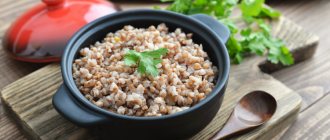
If you have constipation, it is recommended to eat foods that enhance intestinal motility, but not too much. If constipation continues, mild laxatives should be used and regular meals are necessary. You should not eat food that can cause severe bloating - soy food, large amounts of cabbage, milk, etc. Physiotherapeutic procedures should be periodically applied - solar plexus diathermy, paraffin or ozokerite applications on the stomach, iontophoresis, mud therapy can be recommended. Physiotherapeutic procedures must be combined with a diet. If the diet is not followed, physiotherapeutic procedures are ineffective. Heavy physical work should be avoided; muscle tension increases pain. By following a diet, monitoring regular bowel movements, and periodically applying physiotherapeutic procedures, patients with adhesive disease can live tolerably for a long time. But a violation of this lifestyle immediately leads to an exacerbation of adhesive disease.
Make an appointment
Why are adhesions bad?
Nature made sure that in our harmonious body the organs were equipped and arranged clearly and correctly, like in Tetris. They occupy the entire internal space and touch each other with suitable sides, like a carefully fitted puzzle. If you consider all the organs separately from the body, you will be amazed at how much space they take up and how they fit inside us! It is precisely because postoperative scars and adhesions disrupt this initial harmony that they affect our body.
What is the negative impact of adhesions? They:
- interfere with the mobility of the organ, which affects its function. Moreover, both external mobility, which depends on the movements of the diaphragm, suffers, as well as internal mobility, which is active and does not depend on the movement of the diaphragm;
- disrupt blood circulation in the affected organ;
- disrupt the innervation of the organ;
- contribute to the occurrence of pain and spasms in the organ.
Sometimes the adhesion is so powerful that it can disrupt the anatomically correct position of the organ. All of the above reasons lead to other disorders in the body. And yet, which at first glance are not related to the affected area. Adhesions and scars that arise after abdominal surgery can cause pain in various parts of the spine, joints, lead to changes in posture and disruption of the body’s position in space, etc.
Surgical treatment of adhesive disease
Treatment is a very difficult task - you can never be sure that a laparotomy performed for adhesive disease will be the last for the patient and will eliminate the process that caused the adhesive process. Therefore, it is always worth considering the feasibility of a particular operation and drawing up a clear plan based on a clinical examination. Only in emergency cases should this scheme be abandoned. The question of the soldering of small intestinal loops to the scar remains open. Therefore, during laparotomy, the old scar should not be excised - the incision is made 2-3 cm away from the scar.
When separating intestinal adhesions, it is advisable to use hydraulic preparation with novocaine. Deserosed areas of the intestinal walls must be carefully sutured. The soldered sections of the gland should be crossed between the applied ligatures. In cases where the intestinal loops form very adherent conglomerates, and it is not possible to separate them, it is necessary to apply a bypass anastomosis between the adductor and efferent intestines (as if to shunt), since the separation of this conglomerate will take a lot of time, and secondly, it will cause additional trauma to the peritoneum . Before deciding on planned surgical intervention, patients require a high-quality X-ray examination. During surgery, releasing intestinal loops from adhesions is a rather difficult task, which, according to Noble, takes about 90% of the operation time. In 1937, Noble proposed an operation, which was called Noble enteroplication of the intestine.
The essence of the operation is that after separation of the adhesions, the intestinal loops were laid horizontally or vertically and sutured together in the area of the mesenteric edge with a continuous thread. Thus, the intestinal loops were fixed in a certain position, and later they grew together. Recurrences of intestinal obstruction were observed after surgery - 12-15%, so this operation was treated with caution. In addition, stitching the intestinal loops takes a lot of time, then the loops begin to peristalt worse.
In 1960, this operating principle was modified by Chalds and Phillips, who proposed to perform enteroplication not by suturing intestinal loops, but by suturing the mesentery of the small intestine with a long needle. Surgery using this method provides better peristalsis and an easier postoperative period. In addition, less time is spent on this operation.
In 1956, White and in 1960, Deder proposed fixation of intestinal loops with an elastic tube inserted into the intestinal lumen by enterostomy. Dederer proposed performing a microgastrostomy, through which a long tube with many holes was inserted throughout the entire length of the small intestine. This method is not bad at all due to the fact that the tube was a frame for intestinal loops and the loops were fixed and fused in a functionally advantageous position. But opening the stomach cavity (Dederer) or intestine (White) was unfavorable with regard to infection of the abdominal cavity. However, during operations for intestinal obstruction, the tube is passed transnasally, bringing it almost to the ileocecal angle.
The tube is fixed to the wing of the nose; later, during the period of intestinal paresis, the contents of the intestine are drained through this tube; nutrients can be introduced into this tube. But basically it is removed a few days after surgery, after reliable restoration of peristalsis, since long-term removal of intestinal contents can cause electrolyte imbalances.
Treatment of adhesions
Treatment of postoperative adhesions is carried out using conservative and surgical methods. There is no rush to resort to surgical treatment, since repeated insertion into the abdominal cavity can provoke an even greater development of the adhesive process. The operation is mandatory if critical changes occur in the abdominal cavity. The severity of adhesions can only be determined during surgery - for example, performed for intestinal obstruction. The most effective method for removing adhesions that have formed around the fallopian tubes is laparoscopic surgery. The recovery process after such treatment proceeds quickly and with minimal risk of complications. Conservative treatment of adhesive disease is based on diet (limiting coarse fiber foods) and physiotherapeutic methods.
The doctor decides how to treat adhesions after appendectomy - this can be surgery or conservative treatment methods (diet, physical therapy). In the early stages of the development of the adhesive process, it can be influenced by conservative methods. Drug treatment for adhesions can also be prescribed in gynecology - for adhesions that occur in the area of the appendages, vaginal suppositories are used, but such therapy is not very effective.
If adhesions occur after removal of the uterus, treatment is carried out extremely rarely, since they do not cause a feeling of discomfort, and reproductive function is lost irretrievably in any case. In this case, the doctor determines the advisability of treating the adhesive process based on the patients’ complaints.
More detailed information about the pathology, including the causes of adhesions in the chest cavity, can be obtained on our website https://www.dobrobut.com/.
Related services: Physiotherapy Family doctor consultation
Prevention
Prevention of adhesive disease consists of timely surgical intervention for acute diseases of the abdominal organs, without harsh actions, without stopping tampons (there are indications for installing tampons - unstoppable bleeding, when an abscess opens in the abdominal cavity), using tubes made of erective materials. Sanitation of the abdominal cavity is important, which must be carried out using an electric suction, using gentle methods and only drying in hard-to-reach places with tampons.
After suffering peritonitis, the patient must remain under the supervision of a surgeon for a long time. Very early after surgery, it is necessary to stimulate intestinal peristalsis; this is facilitated by the placement of an epidural catheter, HBOT, proserin, and hypertensive enemas. To prevent the occurrence of adhesions, the introduction of anticoagulants, novocaine, prednisolone with novocaine has been proposed. The positive effect of intra-abdominal influence of fibrinolysin with hydrocortisone has been proven. However, all these methods are not reliable. Examination of disability. Adhesive disease reduces the ability to work, causing them some kind of disability. After surgery, patients are referred to VTEK. More often, a decrease in working capacity allows them to be assigned to group 3 disability. Patients should be transferred to work with physical stress. Abdominal injuries in patients with adhesive disease can often lead to intestinal rupture, since the intestinal loops are fixed and cannot move with a direct blow.
Causes of adhesions in the abdominal cavity
The pathological condition in question can develop for a variety of reasons. There are three main groups of factors that can provoke the formation of adhesions:
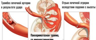
- Mechanical injuries. These are injuries to the abdomen resulting from a fall from a height, a bullet or knife wound, or as a result of surgery.
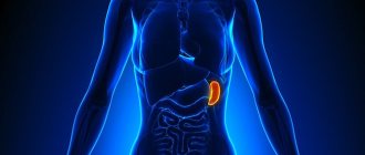
- Inflammatory diseases of the abdominal organs. Thus, adhesive disease can develop as a result of current or after previous cholecystitis (inflammation of the gallbladder), enteritis (inflammation of the small intestine), adnexitis (inflammation of the ovaries) and so on.
- Chemical damage to the abdominal organs. Most often observed when bile or stomach contents leak (for example, when a perforated gastric ulcer occurs).
Often the adhesive process occurs in the pelvis. It can develop rapidly, with characteristic symptoms, or be asymptomatic. Thus, adhesions after a cesarean section often do not appear at all. They can be diagnosed when a woman consults a doctor about the inability to become pregnant again, since adhesions in the pelvis can provoke female infertility.