Internal bleeding is accompanied by the outpouring of certain volumes, at which critical changes occur in the body. Relatively mild forms are quickly corrected under the supervision of doctors and leave no consequences. On the other hand, it is not always possible to detect problems until the extreme moment.
Symptoms are nonspecific. If the patient does not know about the existence of bleeding, a late call to the ambulance is likely.
Recovery is very difficult, partly because of the need to act quickly. There is no time for reasoning and thorough diagnosis.
Therapy is carried out mainly by surgical methods. The task is hemostasis, stopping the pathological process. Prognosis largely depends on the moment of initiation of therapy. Its qualities, the main reason.
Classification
{banner_banstat0}
The internal bleeding unit is a topic for another discussion. The disorder is typified by a variety of methods. The first criterion is the moment at which the change occurs.
- Early form. It is accompanied by a momentary violation of the anatomical integrity of the vessels, their destruction, and the release of a large amount of liquid connective tissue into the internal cavities of the body.
From a clinical point of view, this is a classic variety. It occurs most often in the practice of doctors, which is why it is assumed first.
- Delayed or late form. The pathological process develops after some time. From several hours to 3-5 days. This is mainly due to the destruction of the blood clot, purulent melting of the vessel.
The disorder is extremely insidious, since the patient may not even suspect that there is a problem or that trouble is approaching.
According to the etiology of the disorder, the reasons that became the basis of the nature of the condition are called:
- Mechanical form. Most common. Becomes the result of trauma, tissue damage. It is necessary to stop internal bleeding as quickly as possible using medical and/or surgical methods.
- Arrosive bleeding. It occurs as a result of infectious and inflammatory processes, during the disintegration of large tumors and necrotic destruction of tissue. In some other situations, it is accompanied by the gradual development of the main provoking factor.
With a sufficient amount of foresight, the doctor can predict a possible complication in the early stages.
- Diffuse variety. A characteristic increase in the permeability of the vascular wall. As a rule, such bleeding does not pose a great danger in the early stages, since the volumes of liquid tissue are still small. Gradually, the pathology worsens; the condition cannot be left without treatment.
Depending on the location, the following types of process are called:
- Cavity. Generalized name. In fact, we are talking about such forms as intra-abdominal, pleural bleeding and others.
- Parenchymatous. Liquid pours into the space between the tissues, accumulates and fills them, causing compression and disruption of the functional activity of organs.
There are many more of them, since there can be many localizations. An example is effusion into the brain, liver, spleen, kidneys, pancreas. Signs of parenchymal bleeding of this type are obvious, accompanied by a state of shock and sudden jumps in blood pressure.
There is a more detailed criterion based on this. In this case, they are called gastrointestinal, pulmonary, articular, intracranial and other varieties. Read more about stomach bleeding in this article.
According to the severity of the pathological process, three forms can be distinguished:
- Easy. Accompanied by loss of up to 500 ml of blood. Provided that the victim is an adult. Percentage based assessment is more effective. In this case, this figure is 10-15%.
- Average. About 15-20% of the total amount of liquid connective tissue.
- Severe or fatal. From 20 to 30% blood. Accompanied by severe symptoms, it poses an enormous danger to life.
There is also an absolutely lethal variety. If the loss is more than 30%. In this case, it is almost impossible to return the patient to normal due to the extremely short time frame suitable for correction.
The type of vessel affected is called:
- Venous. The blood flows out slowly; upon puncture examination and drainage, a dark cherry tint of liquid connective tissue is noticeable. The intensity of the pathology is low, so there is time for treatment. However, in some localizations, the correction time is extremely limited.
- Arterial. Accompanied by sharp, jerky releases of blood. The patient dies quickly; at best, there are a few minutes for first aid, then urgent resuscitation measures are required.
- Capillary type. As a rule, such small vessels are localized in organs. With massive damage to structures, large volumes of blood enter the interstitial space. Therefore, it is not as safe as it might seem.
A mixed type is also possible, when several types of vessels are damaged.
Classification is carried out on other grounds. For example, according to the degree of obviousness of the pathological process. Explicit, noticeable immediately or after some time based on symptoms and objective signs, and hidden forms.
There are varieties in which blood loss occurs regularly, for example, against the background of diseases of the blood vessels and hematopoietic system. Also acute types caused by an intense third-party factor. Whether it is a mechanical impact or an infectious lesion, etc.
All classifications are used equally to clarify the process and its nature. But the opportunity to thoroughly study the conditions arises after first aid. There is no time for long thoughts, descriptions and evaluations.
About the disease
Gastrointestinal bleeding is an extremely dangerous sign. We are talking about the leakage of blood that comes out of damaged vessels and enters the organs of the gastrointestinal tract. Bleeding is considered a complication of inflammatory and chronic diseases. It is often caused by alcohol and drug intoxication of the body.
The sources of hemorrhage are the stomach, intestines, and esophagus. The disease is as common as appendicitis. Often this phenomenon occurs as a complication of duodenal ulcers, chronic gastritis, and the appearance of neoplasms.
We recommend diagnosing gastrointestinal bleeding at the medical institution “KDS Clinic”. All examinations are carried out using modern technical equipment and innovative techniques. The disease will be diagnosed even at an early stage.
Signs of internal bleeding of varying severity
{banner_banstat1}
The clinic is determined by the severity of the pathological process.
With a mild disorder, when there is little bleeding, the following manifestations are found:
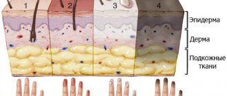
- Paleness of the skin and mucous membranes. Occurs immediately after the development of an abnormal condition. The body acquires a whitish tint, as do the gums, which is clearly noticeable upon visual assessment.
- Increased sweating. Hyperhidrosis.
- Feeling cold. Reduced skin temperature. With an objective study using a thermometer, the indicators, as a rule, are at normal levels or are slightly reduced. Restoration does not give a clear result, the problem persists. When palpating the fingertips, a decrease in local temperature is noted.
- Slight drop in blood pressure. Up to 10 mmHg above or below. It is possible to change diastolic and systolic at the same time.
Usually the patient does not feel these problems with a small amount of blood loss. There are no neurological deficits or headaches. Although options are possible, depending on the individual characteristics of the body.
- Dyspnea. Violation even without physical activity. The patient is short of air. It persists throughout the entire period until specific treatment is carried out.
- Tachycardia at 95-100 beats per minute. Not always noticeable to the victim. Moreover, the intensity of the heartbeat decreases, the force of the beats, despite the number, remains the same or decreases.
- Thirst. Strong desire to drink.
- Darkening in the eyes. Due to insufficient circulating blood volume. Ischemia of brain tissue occurs. Compensatory mechanisms do not work.
The average degree of bleeding is determined by the same manifestations. But a little more intense. Plus, there are a number of additional, subjective signs:
These include:
- Dry mouth. Changes in the hydration of mucous membranes.
- Fainting conditions. Syncopal phenomena occur one or more times, depending on the nature of the insufficiency of trophism of nerve tissues. The brain always suffers, the only difference is the severity of the disorder.
- Dizziness. The patient cannot navigate even in a familiar environment and falls. Forced to take a sitting or lying position. To alleviate the severity of the disorder.
- Nausea. Without vomiting or with a single episode of it.
- Weakness and drowsiness. To the point of being unable to concentrate on anything.
The severe form is accompanied by critical signs:
- Rapid drop in blood pressure. 30-40 mm Hg or more. Up to functionally impossible indicators, which are fraught with cardiogenic shock and death of the patient.
- Tachycardia. Increased heart rate up to 120-160 beats per minute or even more. The strength of contractions and pumping ability are paradoxically reduced. The pulse is palpable weakly, despite the nature of the rhythm disorder.
- Severe shortness of breath.
- Cloudiness of consciousness. The person practically does not understand the situation.
- Apathy. The patient does not respond to words addressed to him; any influence, be it painful or mechanical, is practically useless. There is no response to light or sound stimuli.
- Loss of consciousness is almost inevitable.
The patient is in critical condition. Symptoms of severe internal bleeding are easily recognized and are included in the list of cardiac and neurological abnormalities.
There is often pain in the place where the anatomical integrity of the vessels has been violated. For example, for kidney injuries, liver injuries, hemorrhagic stroke, lung rupture, and other diseases and conditions.
Signs of internal bleeding also depend on the location of the violation of the anatomical integrity of the vessel. Generally speaking:
- Cavity types are accompanied by the described manifestations. At the same time, there are specific moments during palpation: symptoms of irritation of the abdominal wall, sounds during auscultation.
- Tissue varieties are noticeable by the main manifestations of the affected organ.
- The effusion of blood into the joints leads to severe pain, an increase in size up to a significant, unnatural one. There can be many options.
Recognizing internal bleeding when the problem is minor is not so easy. With severe disorders, everything becomes obvious almost immediately. It is necessary to evaluate the signs of the disorder so as not to miss the moment to start therapy.
Complications
Hemorrhagic shock is an acute condition characterized by stopping hemoperfusion (arterial blood flow) of vital organs, a drop in blood pressure up to the absence of diastolic pressure, the development of multiple organ failure and the rapid onset of clinical death.
Disseminated intravascular coagulation syndrome (DIC) is an extremely dangerous complication characterized by the formation of multiple blood clots in the microvasculature, which results in depletion of coagulation factors and platelet activity (consumptive coagulopathy), and additional sources of bleeding are formed. The prognosis is unfavorable.
Repeated bleeding - develops as a result of ineffective surgical treatment, the presence of uncorrectable pathologies of the blood coagulation system, advanced cancer, liver cirrhosis, etc.
Causes
{banner_banstat2}
There are many main provoking factors. It is worth mentioning only the most common ones. In generalized form, the list is represented by the following points:
- Mechanical factors. Injuries, physical damage.
- Infectious pathologies in which a purulent process is observed. As a result, a melting of arteries, veins, and capillaries develops.
- Effect of ionizing radiation on tissue. Rarely seen. As a result of man-made disasters, inadequate selection of dosage during radiotherapy.
- Natural processes that affect the body in a negative way. There are more of them than might seem at first glance. This includes, for example, pregnancy, difficult childbirth in women, and intense physical activity.
- Vascular diseases. Increased fragility as a result of vitamin C deficiency is a classic case.
- Pathologies of the hematopoietic system. Hemophilia, problems with the synthesis of coagulation factors (thrombocytopenia), myeloproliferative disorders.
- Thrombocytopathies of various types. Platelet synthesis is normal, but they are not complete and do not clot blood effectively.
- Chemical hazards. Poisoning with certain substances. Vapors of acids, alkalis and other components. Especially often, as a result, pulmonary bleeding develops with the release of pink, foamy sputum.
- Destructive pathological processes. Cancerous tumors of any location with infiltrative growth and decay. Also tuberculosis, syphilis in the advanced phase, etc.
- Rupture of aneurysms of any vessels. In the case of the aorta, death occurs within 3-5 minutes.
The causes of bleeding can be mechanical or organic; depending on the situation, each specific factor creates certain risks.
Etiology
The causative factors for internal bleeding are divided into 3 large groups:
1. Mechanical damage:
- cut, stab, chopped wounds;
- amputation injuries (severation of a finger, limb);
- falls from a height (rupture of the spleen, liver);
- animal bites, etc.
2. Arrosive bleeding - occurs due to necrosis of the vascular wall (for example, during the disintegration of a malignant tumor, gangrene of a limb, burns, poisoning with acids or alkalis).
3. Diapedetic bleeding (hemorrhage) - soaking of tissues with blood without damaging the vessel wall. Most often observed in the microvasculature in genetic diseases with coagulation disorders (hemophilia, thrombocytopenia), acute conditions (disseminated intravascular coagulation syndrome, sepsis).
First aid for internal bleeding
{banner_banstat3}
No one except specialists can effectively help the victim. There is no point wasting time, there is already so little of it.
It is important to call the emergency team first. Next, before the doctors arrive, it is worth assessing the situation.
- If bleeding into the chest cavity or lungs is suspected, be sure to sit the person down. This way there is a greater chance of maintaining respiratory function.
- In other situations, you need to give the body a horizontal position.
- Place an ice pack or cold water at the site of the suspected hemorrhage. This will lead to vasoconstriction and partially slow down blood loss.
Resuscitation measures in case of critical condition, cardiac arrest, loss of consciousness, as a rule, do not make sense; you must first stop the bleeding.
In case of syncope (fainting), you need to turn your head to the side so that if vomiting occurs, choking and asphyxia (suffocation) do not occur.
Upon arrival of specialists, it is recommended to briefly describe the nature of the violation and, if possible, accompany the victim to the hospital.
Prohibited actions
{banner_banstat4}
What you should absolutely not do:
- Give enemas.
- Give any medications. It is not safe. The use of medications is impossible; a sharp deterioration in the condition is likely.
- Under no circumstances should you heat the affected area. This will lead to the dilation of blood vessels and will cause a sharp increase in the intensity of the outpouring of liquid connective tissue.
- Also, you should not move the patient again, so as not to provoke increased bleeding.
Attention:
Self-action in such a serious situation is the best way to ensure death for the patient. Everything must be within the minimums described above.
Diagnostics
{banner_banstat5}
When admitted to a hospital, there is usually very little time for examination. Because the pathological process is dangerous and acute. It is accompanied by a pronounced clinical picture and gets worse every second. First aid for internal bleeding is necessary to eliminate the disorder.
The minimum diagnostic program includes:
- Visual assessment of body condition.
- Blood pressure measurement. Indicators against the background of bleeding are always reduced. To what extent is a question determined by the situation.
- Listening to pulmonary sounds. Auscultation.
- Heart rate study.
- Palpation of the abdominal cavity.
If the patient is conscious, it is important to briefly question him about his state of health and determine the probable cause of the disorder.
Next, a set of primary measures is carried out to stop the bleeding.
Upon completion, endoscopic diagnostic procedures may be prescribed. Blood test (general analysis). Specific techniques, depending on the case.
Symptoms of neoplasms
Advanced neoplastic diseases are indicated by:
- underweight;
- enlarged, hard liver with a lumpy structure;
- palpable tumor in the abdominal cavity;
- enlarged and hard lymph nodes.
Other symptoms:
- Subcutaneous emphysema in a patient with persistent vomiting indicates Boerhaave syndrome (esophageal perforation). A prompt consultation with a surgeon is necessary.
- Telangiectasia on the mucous membranes may indicate Randu-Osler-Weber disease.
Further treatment
{banner_banstat6}
Combined methods are mainly used.
First of all, it is necessary to eradicate the source of the disorder. This problem is solved by surgical intervention methods.
The vessel and tissues are sutured, and the damaged area is eliminated (if necessary, doctors perform a resection and remove part of the altered structures). Mandatory measures are also taken regarding the consequences of the pathological process.
In parallel, transfusion is necessary to restore lost blood volume and the introduction of saline solutions to correct the balance of electrolytes. The patient's condition is constantly monitored in a hospital setting.
Forecast
{banner_banstat7}
Controversial. Depends on the situation. There are two main factors. This is the degree of the disorder (its severity), as well as the moment of initiation of therapy. With timely treatment, if the disorder is relatively harmless, there is every chance of full recovery.
Internal bleeding is a life-threatening process that is not always obvious at first glance. Without proper correction, it is often fatal.
Specialists eliminate the causes of the anomaly using surgical methods. Medication methods are also used. The forecasts remain vague. When assessing prospects, you need to proceed from the situation.


