An examination by a doctor and the medical history collected during an appointment are often not enough to make an accurate diagnosis and prescribe treatment. Especially when it comes to the abdominal cavity. The location of internal vital organs does not make it possible to make an accurate diagnosis, or confirm it without instrumental diagnostics. To obtain the most informative results from the study, it is necessary to properly prepare for the examination procedure. We tell you how to do this.
For what indications is abdominal ultrasound prescribed?
The reason for prescribing an ultrasound of the abdominal cavity may be the patient’s complaints about:
- cutting, pulling, aching pain and severe discomfort in the abdomen, as well as pain radiating to the scapula, sacrum, shoulder, etc.;
- vomiting for no apparent reason;
- dyspeptic disorders;
- bitterness in the mouth;
- yellowing of the sclera and skin;
- flatulence not associated with eating foods that cause increased gas formation;
- suspected neoplasm of the abdominal organs.
Injuries in the peritoneal area without extensive wound surfaces are an indication for an emergency ultrasound of the abdominal cavity. Doctors recommend that patients suffering from chronic diseases of the liver, gallbladder, and pancreas undergo an ultrasound of the abdominal organs at least once every 6-12 months.
An abdominal ultrasound may be prescribed as part of preparing the patient for surgery, as well as during therapy to assess the effectiveness of treatment.
What does an abdominal ultrasound show?
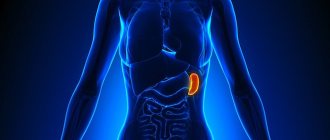
During the study, the abdominal organs are visualized and assessed for compliance with physiological standards:
- liver;
- gallbladder;
- bile ducts;
- pancreas;
- spleen;
- adrenal glands;
- abdominal lymph nodes;
- vessels of the peritoneal organs.
If there are indications, as part of the examination, the doctor can assess the functional state of the organs of the retroperitoneal space. Using the ultrasound method, a sonologist (a doctor conducting an ultrasound examination) has the opportunity to assess the structure of parenchymal organs, its disturbances, identify a foreign body, stones in the gallbladder or its ducts, determine the presence and extent of spread of various neoplasms in the peritoneal organs.
Ultrasound of the abdominal organs in combination with other diagnostic methods allows us to identify a whole range of diseases, including pancreatitis, hepatitis, cholecystitis, benign and malignant neoplasms, abscesses, cysts, adhesions and many other dangerous diseases.
Ultrasound is the method of choice for diagnosing cholelithiasis. Using an ultrasound examination, the doctor can detect not only the presence of stones, but also their size, position, and origin.
For maximum precision in performing a medical procedure, under the control of an ultrasound machine, a puncture biopsy of tissue of the abdominal organs or aspiration of fluid accumulation in ascites can be performed.
Basic rules of preparation
Ultrasound examinations of the ABP should be done on an empty stomach, and are prescribed both in the morning (on an empty stomach) and in the afternoon. It is necessary to adhere to some rules and recommendations before an ultrasound examination:
- Follow a diet 2-3 days before the procedure.
- You must not smoke for two hours before the examination.
- It is not recommended to conduct ultrasound examination after FGDS, FCS.
- If studies were carried out with the introduction of a radiopaque substance into the body - a barium preparation, for example, during irrigoscopy, fluoroscopy of the esophagus, stomach, then an ultrasound scan should be performed two days later.
This is a reminder for those who are preparing for an ultrasound scan of the OBP.
Preparing for an abdominal ultrasound
The informativeness and effectiveness of the study largely depends on preliminary preparation for the procedure, which does not cause any particular difficulties for patients. Before an abdominal ultrasound, the following rules must be observed:
- Three to four days before the examination, the patient should adhere to a gentle neutral diet and avoid overeating. It is necessary to exclude from the food consumed foods that stimulate increased gas formation in the intestines: legumes, cabbage, baked goods, sweet and carbonated drinks, fresh milk without heat treatment, fermented milk products, fruits with a high sugar content. Intestinal gases can distort the result of an abdominal ultrasound.
- If a gentle diet does not bring the desired result, and gases accumulate in the intestines, it is recommended to take enterosorbents or enzyme preparations that reduce gas formation.
- Patients suffering from constipation must have a bowel movement the day before the test. For these purposes, you can use laxatives or give a cleansing enema.
- The time interval between the last meal and the ultrasound examination should be at least 6-8 hours, and if the doctor aims to examine the condition of the liver or abdominal aorta, then 8-12 hours. The study is carried out strictly on an empty stomach: after eating, intestinal motility is activated, which reduces the diagnostic information. Some relief in restricting food intake is given to patients with diabetes mellitus and pregnant women. These categories of patients can eat a few crackers and drink sweet tea in the morning. If an ultrasound is prescribed for a child who is breastfed, the procedure must be performed no earlier than 3 hours after the last feeding. For children aged 1-3 years, the recommended break in eating before an ultrasound scan should be at least 5 hours.
- If the patient must systematically take medications (antihypertensive drugs, glucocorticosteroids, statins, etc.), the doctor must be informed about this.
- An hour before the ultrasound, you must stop smoking. Nicotine and tar cause spasms of the gastrointestinal tract, which can negatively affect the diagnostic information.
- Contrast-enhanced X-ray studies performed shortly before an abdominal ultrasound can significantly distort the results. The break between studies must be at least five days.
Factors influencing ultrasound results?
In addition to diet and cleansing, the following points also affect the correctness of the results:
- Tobacco smoking on the day of screening. You should give up cigarettes a couple of hours before the scan.
- Chewing gum, caramel, lollipops. Avoid using them on the day of your appointment.
- Parallel radiography. The interval between different types of screening should be at least 3 days.
- The degree of fullness of the bladder. When the kidneys are scanned, the urine should be full. In others the situation is devastated.
- Excess body weight. When referring for ultrasound screening, the weight and condition of the patient should be taken into account, since excessive obesity complicates ultrasound penetration.
How is an abdominal ultrasound performed?
Patients who are about to undergo an abdominal ultrasound for the first time are often interested in how the examination is performed.
Without going deeply into technical details, we can say that the diagnostic method is based on the ability of ultrasound sent by the sensor of an ultrasound machine to be reflected from organ tissues with different acoustic resistance and return to the sensor again. As a result, a two-dimensional image is obtained on the device’s monitor in real time. The study visualizes the functioning of organs over time, so the doctor can detect even minor deviations from the norm. Ultrasound of the abdominal cavity does not cause discomfort for the patient, is tolerated painlessly even by small children, and does not last long. To carry out the procedure, the patient must undress to the waist and lie down on his back on a medical couch. If the doctor needs to visualize the kidneys, he may ask the patient to turn onto his stomach.
After this, the doctor performing the ultrasound applies a special acoustic gel to the skin. This is necessary so that the contact of the sensor with the skin is as tight as possible, and the ultrasonic waves are better absorbed by the tissues of the internal organs. In some cases, holding your breath for a short time may be necessary.
After applying the gel, the doctor moves the sensor along the abdominal wall to visualize the organ being examined as clearly as possible. At the end of the procedure, the remaining gel is removed with a napkin. Depending on the indications, the procedure lasts no more than 15-30 minutes.
Ultrasound in a medical setting is performed on an outpatient basis in a room specially designed and equipped for this purpose. During an ultrasound examination, the doctor reads out loud the parameters of the abdominal organs and identified deviations from the norm, establishing a preliminary diagnosis. The nurse enters the study data into a special form, and the ultrasound protocol with the conclusion is given to the patient. The sonologist's conclusion is not a final diagnosis. Making a final diagnosis is the prerogative of the doctor who issued the referral for ultrasound. In particularly complex diagnostic cases, a council of specialized specialists may be convened.
Diet before ultrasound examination
You need to prepare for an ultrasound three days in advance. During this time, it is necessary to reduce the load on the mucous membrane and gradually normalize the function of the digestive tract. Diet is the basic preparation, but it is not limited to it. Avoid foods that cause gas and reduce fiber intake.
The number of meals should be at least five times a day in small portions, every 3.5–4 hours. The last dose is no later than 4 hours before bedtime. Liquids should be consumed 1.5–2 liters per day, a little less for children. If the examination is scheduled for dinner, then the last meal taken in the evening is a light dinner. If the procedure is in the afternoon, then a light breakfast, including a couple of eggs and green tea. You can have breakfast depending on the appointed time at 8–10 am (6–8 hours before the procedure). After this, it is advisable not only not to eat, but also not to drink. You should not eat candy or chew gum during this time.
specialist
Our doctors will answer any questions you may have
Chikov Sergey Vladimirovich Ultrasound Doctor
Ask a Question
Prohibited Products
Gas formation in the intestines greatly interferes with ultrasound examination. Therefore, three days before the procedure, you need to exclude from your diet a number of foods that cause flatulence and minimize fiber intake.
Legumes
The fruits of legumes are rich in vegetable protein and complex carbohydrates. This combination leads to intense gas formation. Peas, vetch, beans, lentils, and beans are prohibited when preparing for an ultrasound.
Carbonated drinks
Carbonated drinks contain carbon dioxide, also known as carbon dioxide. Sometimes it is formed as a result of the natural fermentation process, for example, in kvass. Therefore, all these drinks are prohibited.
Flour products and pasta
These foods contain gluten as well as polysaccharides, which when combined cause bloating. This also includes semolina.
Whole grain cereals
These include corn, rice and millet cereals. All of them are rich in fiber and can cause gas formation.
Onion and garlic
These foods contain fructans, a polymer of fructose. Even in small quantities, onions and garlic can cause bloating.
Raw vegetables
It is recommended to exclude cabbage, as well as radishes, radishes, daikon and watercress. These foods irritate the intestinal walls, contain a lot of fiber and can cause gas.
Dairy
Flatulence can be caused by milk. It contains milk sugar - lactose. A lack of the lactase-breaking enzyme can cause gas.
Alcoholic drinks
Alcohol irritates the walls of the stomach and intestines and provokes an inflammatory reaction. Beer also contains fermentation products, including carbon dioxide.
Fruits and berries
First of all, fruits cause bloating. Containing large amounts of simple sugars - fructose. These include grapes, melon, bananas. Berries and fruits with sourness cause gas formation to a lesser extent.
Sweets
Candies, chocolate and other sweets containing simple carbohydrates that promote gas formation are excluded from the menu.
On the eve of the study, you should also refrain from fatty foods, including rich soups with meat broth, fatty fish, lard, and fatty meat.
Authorized Products
Three days before the ultrasound, it is recommended to switch to food with easily digestible proteins and other components that do not cause putrefactive processes in the intestines and gas formation. All dishes should be eaten in moderation.
The following dishes are recommended:
- porridge made from buckwheat and oatmeal cooked in water (rolled oats);
- low-fat broths, soups cooked with vegetables;
- dietary meat with easily digestible protein (turkey, chicken, rabbit);
- boiled or steamed vegetables (carrots, cucumbers, pumpkin, zucchini);
- quail and chicken eggs;
- low-fat fish (any river fish, cod species);
- mashed potatoes.
The following drinks are allowed the day before:
- still water (mineral or artesian);
- lightly brewed tea;
- dried fruits compote;
- fruit drinks;
- herbal infusions;
- fruit juices.
Contraindications
As with almost any medical procedure, there are a number of contraindications for performing an abdominal ultrasound. These include:
- hyperthermia;
- infectious diseases in the acute phase;
- open wounds in the peritoneum;
- skin diseases with exudation localized in the area under study;
- severe general condition of the patient.
Pregnancy, contrary to popular belief, is not a contraindication to the procedure. The study is completely safe for the health of the woman and fetus and cannot cause deterioration in well-being.
Constant improvement of diagnostic equipment has made it possible to carry out the procedure within pediatric practice over the past two decades. If pathology of the abdominal organs is suspected, the examination can be carried out even on newborn children.
Ultrasound of the abdominal organs in a medical setting provides the opportunity to most accurately identify the cause of the existing pathology, make the correct diagnosis and, as a result, receive prescriptions for effective therapy.
Why is an ultrasound prescribed?
At the first suspicion of intra-abdominal diseases, doctors write out a referral for an ultrasound scan. This allows you to detect anomalies such as:
migration of cancerous processes – metastasis;- tumor processes in the pancreas;
- liver cancer;
- cystic formations;
- tissue degeneration, abscesses, internal hemorrhages;
- benign neoplasms;
- pathological accumulation of fluid in the retroperitoneal space.
In order to “catch” the disease at an early stage and quickly take the necessary therapeutic measures, it is necessary to undergo regular examinations, including with the help of ultrasound.
