Why is fluorography necessary?
The main goal of diagnosis is to identify the pathological process at an early stage and begin treatment before specific symptoms appear. Here are the diseases that fluorography diagnoses:
- Pneumonia;
- Pleurisy;
- Tuberculosis;
- Malignant tumors of the lungs, mediastinum;
- Emphysema.
If a health problem is detected early, the chances of a favorable clinical outcome increase. This is what can be seen on fluorography:
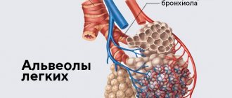
- Lungs;
- Heart;
- Less often - bones.
This screening examination shows the inflammatory process, foreign bodies, tumor formations, accumulation of infiltrate, cavities of a non-physiological nature (various cysts). Fluorography even shows if the patient smokes. The result is not the main criterion for making a diagnosis; it allows a more accurate assessment of a specific clinical case.
Fluorography is the main method for diagnosing tuberculosis
25.Mar.2021
On this day in 1882, Dr. Robert Koch announced that he had discovered the bacterium that causes tuberculosis, making the diagnosis and treatment of this disease possible. Tuberculosis is an infectious disease
, which is transmitted by airborne droplets and occurs, as a rule, in a latent form, but in some cases takes on an open, active form. Most often, Koch's bacilli affect the lungs, but sometimes also the nervous, genitourinary, skeletal, digestive systems, brain, skin, and eyes. Common symptoms of pulmonary tuberculosis are cough, sometimes with sputum and blood, chest pain, weakness, weight loss, fever and night sweats. The risk of developing tuberculosis in infected people is 5-15%. However, it is significantly higher in people with weakened immune systems, diabetes, malnutrition, and smokers.
In recent years, the Russian Federation has seen a decrease in the incidence of tuberculosis, however, this disease remains a serious problem for domestic health care. According to WHO classification, the Russian Federation is still a country unfavorable for tuberculosis.
Like any disease, tuberculosis is easier to cure and prevent its spread in the early stages, therefore, in medical institutions to prevent tuberculosis, screening programs are used (detection of the disease in its latent form, including fluorography), medical examinations and vaccinations are carried out. For the adult population, a fluorographic examination of the lungs is provided, for children - skin tests (Mantoux test, Diaskintest).
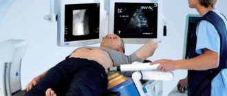
In the X-ray department of the Pyt-Yakh District Clinical Hospital, a modern computerized fluorographic apparatus is used to screen the population for tuberculosis. It is an effective and safe examination method for the patient. A fluorogram appears on a digital camera immediately after the photo is taken. This allows you to reduce the appearance of uninformative images to zero and avoid the need for repeated fluorography. It is very important to know that the radiation dose during the study is reduced by 4-5 times. Such a small dose makes it possible to expand the age group for X-ray prevention of tuberculosis and other lung diseases. In 2020, a new digital device was put into operation in the X-ray room, the main advantage of which is high-quality images with a lower radiation dose for the patient. The launch of a new fluorograph made it possible to significantly increase the throughput of the fluorographic examination room and eliminate queues.
Fluorography is mandatory for all citizens over 15 years of age once a year. The results of fluorography are absolutely necessary when visiting a clinic, undergoing various medical examinations, or anywhere else; you should not refuse it. It is often only thanks to mass fluorography that it is possible to identify new cases of tuberculosis, especially since the examination is carried out free of charge.
Preventive fluorography in Pyt-Yakh in 2021 covered 98% of the population subject to examination, which is 27,400 people. Thanks to fluorography, in 2021 it was possible to identify 6 cases of active tuberculosis, 4 tumors and 91 pneumonia.
However, medical workers at the Pyt-Yakh District Clinical Hospital state from year to year that part of the adult population has not undergone fluorography for more than 2 years. Practice shows that untimely detection of diseases leads to a protracted course.
Prepared by zav. X-ray department of the Pyt-Yakh District Clinical Hospital Nikolay Ogorelkov.
What does fluorography give and when to do it?
For prevention, persons over 17 years of age are recommended to undergo examination once every 2 years. There are also categories of people who are exposed to x-rays 1 and 2 times a year.
Here's what you can see on fluorography when the picture is already in hand:
- Focal shadows in the lung area;
- Compaction, expansion of roots, as a sign of pulmonary pathologies;
- Fibrous tissue as a result of surgery, trauma, infection;
- Aperture changes;
- Branching of the vascular pattern as a symptom of bronchitis, pneumonia, early stage of oncology;
- Change in the usual shadow of the mediastinum.
Based on the results, it is recommended to contact a phthisiatrician, who, in addition to decoding, will help determine the clinical picture, get examined more quickly, and prescribe treatment.
What is a chest x-ray
Chest X-ray is a projection radiographic examination of the chest, revealing destructive changes in the organs of the chest cavity and nearby anatomical structures. A chest X-ray is the most common radiographic examination.
Indications for a chest x-ray:
- suspected lung cancer or tuberculosis;
- chest pain, shortness of breath;
- hemoptysis;
- attacks of suffocation;
- traumatic injuries to the chest.
To obtain an image, one type of ionizing radiation is used - x-rays. As a rule, x-rays are taken in frontal and lateral projections.
What pathologies does X-ray of the lungs reveal?
- neoplasms;
- bronchitis;
- tuberculosis;
- pneumothorax;
- pleurisy;
- chronic obstructive pulmonary disease;
- fibrosis.
Congestive heart failure, cardiomegaly, and fluid accumulation in the pericardial sac may also be detected.
A radiologist interprets the images. This process requires in-depth knowledge in the field of x-ray anatomy, so patients are strictly not recommended to engage in self-diagnosis.
If fluorography is bad, what could it be?
The transcript of the study is ready the next day, within 5 minutes after the study. If the result is poor, the person is referred to a TB specialist. It is too early to make a final diagnosis; a comprehensive diagnosis is carried out. Doctors may recommend repeat FLU. For the most part, the transformation of lung tissue is caused by the development of connective tissue in the lungs, which grows and layers.
Depending on the location and shape of the pathology focus, the doctor diagnoses fibrosis, sclerosis, adhesions, radiance, heaviness, shadows, and scar transformations. Based on the results, it is possible to diagnose oncology and emphysema, cysts, abscesses, calcifications, and other neoplasms and cavities.
Diagnostics can be done in every city, both in a private medical center and in a city clinic. The result obtained cannot be trusted 100%; an integrated approach to the health problem is needed.
How does a CT scan differ from an X-ray of the lungs?
The fluorography method was patented in the 1940s. The first images were printed on film containing silver. The research was expensive and was not as widespread and widespread as it is today. However, in the middle of the 20th century, fluorography of the chest organs proved to be excellent in the diagnosis of tuberculosis, oncological tumors, and bone fractures. When images began to be displayed on a screen instead of film and digital fluorography appeared, the method became more accessible, images became more convenient to store, and it became possible to enlarge them and adjust the quality.
CT scanning was not invented and patented until the 1970s, but it revolutionized modern medical diagnostics and scientists Godfrey Hounsfield and Allan Cormack were awarded the Nobel Prize for their discovery. The novelty consisted of a fundamentally important and new opportunity to study internal organs, bones and the vascular bed “in volume”, that is, on slice images in three projections. Computer processing made it possible to combine multiple images obtained as a result of scanning the body with dynamically rotating sensitive sensors into a detailed and detailed 3D model.
Computed tomography (CT) differs from x-rays in a number of ways:
1. Result of lung examination. A fluorogram is a planar two-dimensional image in a frontal projection. Digitization of the image allows you to enlarge the image, but the information content of lung fluorography is lower due to an incomplete two-dimensional view and possible artifacts (distortions). Inaccuracies are possible due to the overlapping of shadows of organs on some areas of tissue. The result of a CT scan of the lungs is recorded on a DVD, which stores many cross-sectional scans (up to 1 mm thick) of the chest in three projections: horizontal, frontal, sagittal, as well as a 3D model of the respiratory tract, skeleton and vascular bed (if the study was carried out with contrast).
2. Technique. Both fluorography and CT of the lungs are performed quickly (1-3 minutes) and do not require preliminary preparation. An exception is a CT scan of the lungs with contrast - such an examination takes 10-15 minutes; there are recommendations that should be followed before the examination. Modern multislice tomographs (in particular, our Siemens somatom go.now device) scan the chest in dynamics - the patient moves on the tomograph table to the gantry (ring-shaped frame), and sensitive sensors located in several rows move in a spiral and take pictures. To obtain a fluorographic image, the patient needs to press his chest against the screen and hold his breath.
3. Image clarity. The clarity of chest CT images is improved. Make the picture more contrast, better examine the vessels and soft tissues, possibly by intravenous administration of an iodine-containing drug (CT scan with contrast). Thanks to the special technique of the X-Ray procedure, the rays obtained during tomography have a higher penetrating power.
4. Radiation dose. Radiation exposure during fluorography is minimal - 0.05–0.5 mSv. CT scan of the lungs shows 2–2.9 mSv. This is a harmless radiation exposure. It is believed that it is safe to do up to 5 CT scans within a year.
5. Availability. The cost of computed tomography is higher, and not all medical institutions have special equipment. Meanwhile, fluorography is suitable for annual preventive screening (if there are no special indications for CT) and various medical commissions.
Pregnancy and other contraindications
Contraindications to manipulation are pregnancy and childhood. It is believed that the minimum threshold is 15 years, since before this time it will be difficult for children to tolerate increased radiation exposure.
Among the relative contraindications, which are sometimes ignored when the benefit exceeds the possible harm, are:
- claustrophobia;
- dyspnea;
- general serious condition.
But sometimes even pregnancy, which is a contraindication for almost all types of diagnostics, does not become an obstacle to the procedure. It should be done only after preliminary consultation with your doctor. Control must be carried out by a specialist throughout the entire manipulation.
An important factor in the success of diagnosis and safety for the fetus is the appointment of an examination after 25 weeks. During this period, it had already formed its basic systems.
The reasons for “photographing” the respiratory system of pregnant women and women during the breastfeeding period should be as compelling as possible. Preventive X-ray examinations are not carried out for pregnant women and children under 15 years of age.
Main indications
The main objective of the diagnostic method is to identify diseases of the lungs or bronchi. It is not for nothing that the technique is considered a preventive approach that prevents serious complications for diseases of the respiratory system.
As a rule, a local therapist or pulmonologist gives a referral for testing when a patient comes to them with characteristic symptoms:
- prolonged persistent cough;
- dyspnea;
- pain syndrome in the chest area.
When the above signs are detected, the chance of detecting a pulmonary or bronchial deviation increases significantly. Often the study allows you to establish:
- inflammatory process even at the initial stage of development;
- pneumothorax;
- neoplasms of malignant and benign nature;
- pleural damage;
- emphysema.
But most often, even during a preventive examination, tuberculosis is detected, so annual mandatory examinations are recommended, which help prevent the disease, which currently leads in a high percentage of deaths.
In addition to standard indications, there are two more categories for examination: family members living with pregnant women or newborns in order to minimize the risk of illness in infants and persons of military age.
The second category will include unscheduled fluorography if there is suspicion of:
- foreign body in the lungs;
- inflammation;
- tuberculosis;
- tumors of the mediastinal organs;
- diseases of the heart, large vessels.
Such precautions make it possible to recognize concomitant diseases such as fibrosis or sclerosis. Also, using a small image, it is possible to determine the localization:
- cysts;
- cavern;
- abscesses;
- infiltrates;
- accumulations of gases.
Despite the productivity of the technique, fluorography can hardly be called the main argument for diagnosing pulmonary pathologies. It is often necessary to use auxiliary tests, including sputum analysis, radiography, and MRI with contrast.
Stereotypes and differences from radiography
When undergoing an examination, it is important to pay attention to the direction issued by the attending physician. If it says that you need to undergo fluorography, you do not need to change the order yourself, relying on a classic X-ray to better show the condition of your lungs. Such ignoring of specialist advice will only lead to unreasonable radiation exposure to the body. In medicine, radiation dose limits (1 mSv/year) are established only for healthy people when conducting preventive studies (for example, when applying for a job). Limits on radiation doses required for diagnostic purposes (in sick people) are not established by law. It only states that the doctor should strive for a minimum level of radiation, but not at the expense of the quality of diagnosis. In general, a reasonable postulate, but how is it implemented in practice? But in practice in Russia the average “medical” radiation dose turns out to be several times higher than in the UK, USA and France. At the same time, in some of our regions the average “medical” radiation dose exceeded 3 mSv. Fluorography provides less information than radiography. But the World Health Organization does not recommend the use of traditional film fluorography, even in underdeveloped countries. The solution to the problem was the transition to digital fluorography, which, with high information content, reduces the radiation dose several times. Here are the approximate radiation doses (mSv) that a person receives when examining the chest using various methods:
- radiography (in two projections) – 0.25;
- fluorography (in one projection) – 0.6–1.6;
- fluoroscopy – 3.0;
- digital fluorography – 0.01–0.06;
- computed tomography – 3.5–5.0.
So, if nothing bothers you, and you are referred for an X-ray for prevention, then it is better to undergo digital fluorography or radiography (“image”) instead of conventional fluorography. You should definitely refuse to perform fluoroscopy (when the doctor looks at the patient behind a screen): not only does the patient receive a large dose, but his image will not remain, and the conclusion will be solely on the conscience of the doctor who conducted the study.