Chronic myeloid leukemia is a malignant blood disease in which there is excessive formation and impaired maturation of granulocytes in the bone marrow. A unique feature of this type of leukemia is the presence in tumor cells of a specific marker - the Philadelphia chromosome. It is formed as a result of the t(9;22) translocation, in which one fragment of chromosome 22 changes places with a fragment of chromosome nine. As a result, a chimeric gene is formed that disrupts the process of division and maturation of myeloid cells.
- Causes of development of chronic myeloid leukemia
- Pathogenesis of chronic myeloid leukemia
- Stages of chronic myeloid leukemia
- Symptoms and signs of chronic myeloid leukemia
- Diagnosis of chronic myeloid leukemia
- Treatment of chronic myeloid leukemia
- Prognosis and prevention of chronic myeloid leukemia
Pathogenesis of chronic myeloid leukemia
The Philadelphia chromosome is the result of a reciprocal translocation between chromosomes 9 and 22. In this case, the ABL oncogene from chromosome 9 is transferred to chromosome 22 and attached to the BCR gene. As a result, a hybrid BCR-ABL gene is formed, which regulates the synthesis of a special oncogenic protein - tyrosine kinase bcr-abl. This oncoprotein disrupts the process of cell division, protects tumor cells from programmed death (apoptosis) and disrupts their adhesion to the bone marrow stroma, due to which immature cells enter the bloodstream.
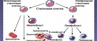
Most often, chronic myeloid leukemia develops as a result of a mutation in a pluripotent hematopoietic stem cell, which is located in the bone marrow, but there are cases when the primary focus is in the liver or spleen.
In chronic myeloid leukemia, the pathology of the granulocytic lineage mainly predominates, but all lines of hematopoiesis can suffer - erythrocyte lineage, monocytes, etc. Healthy stem cells are preserved and can give rise to new hematopoiesis after chemotherapy.
What is a chromosome?
It may also appear in other types of leukemia. The Philadelphia chromosome is a truncated chromosome belonging to the group of small acrocentrics. Each normal female cell of this group contains 2 pairs of them - 21 and 22, while a male cell includes not four, but five of these chromosomes, because in addition to 21 and 22 pairs, the Y chromosome is also included.
Without G-banding, that is, with a normal color, the Ph chromosome is detected in almost every patient suffering from CML, namely in 95-98 percent of cases. On chromosomes that are differentially stained, it is clear that one of the 22 pairs is shortened.
Stages of chronic myeloid leukemia
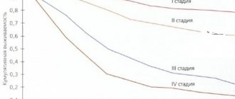
During chronic myeloid leukemia, 4 stages are distinguished, which reflect the progression of the pathology. In this case, the disease can be detected on any of them.
Chronic or preclinical stage - has no clinical symptoms; the diagnosis can be suspected by a general blood test, which patients can take either as part of a medical examination or for the diagnosis of another disease. In most cases, chronic myeloid leukemia is detected at this stage.
Acceleration phase, or progression stage. During this period, the number of tumor granulocytic cells increases, and various symptoms appear, for example, weakness or bone pain. Objectively, there is an increase in the number of blasts in the blood or bone marrow up to 15-29%, the number of basophils increases (more than 20%), thrombocytopenia or thrombocytosis is detected (more than 1000×10 9 ).
Blast crisis is a phase during which there is a sharp increase in the number of blast cells (more than 30%) and the disease in its course resembles aggressive acute leukemia.
The patient is in serious condition. There is an increase in temperature, persistent infections, bleeding, and leukemic skin lesions. At this stage, leukemia is difficult to treat.
The phase of chronic myeloid leukemia is necessarily assessed when making a diagnosis and then rechecked as the pathology progresses and the need to change treatment.
Symptoms and signs of chronic myeloid leukemia
The symptoms of chronic myeloid leukemia are characterized by a variety of clinical manifestations and depend on the aggressiveness of the course and stage of the disease. In general, there may be several syndromes:
- Tumor intoxication syndrome. It manifests itself as weakness, weight loss, and loss of appetite that is inadequate to the current state. There may be fever, increased sweating, itchy skin, and bone pain.
- Tumor proliferation syndrome. It develops with an active increase in the number of malignant cells infiltrating the liver and spleen. In this case, patients note pain and heaviness in the left side.
- Anemic syndrome - develops with a decrease in the number of red blood cells and hemoglobin levels. Manifested by weakness, shortness of breath, increased fatigue during routine physical activity. A decrease in pressure, pallor of the skin and mucous membranes, dizziness, and tachycardia may be observed.
- Disorders of the blood clotting system - thrombosis and hemorrhage (bleeding). The cause of thrombosis is most often thrombocytosis (increased platelet level above 1000 × 10 9 / l). In this case, thrombophlebitis, heart attacks, and strokes may occur. Hemorrhagic manifestations are characterized by an increase in bleeding time after injury, as well as the formation of a petechial hemorrhagic rash. They develop against the background of a critical decrease in platelet levels.
Diagnosis of chronic myeloid leukemia
In most cases, chronic myeloid leukemia is an incidental finding that is discovered during examination for another reason. It can be suspected by a general blood test, in particular by an increase in the number of leukocytes and the predominance of granulocytic hematopoiesis in the formula. In this case, not only the number of neutrophils, but also basophils and eosinophils may increase. There may be mild anemia or abnormal platelet counts.
If the doctor suspects chronic myeloid leukemia, the patient is referred for further examination - puncture and bone marrow biopsy. To confirm the diagnosis, it is necessary to conduct a standard cytogenetic study of the bone marrow for the presence of the Philadelphia chromosome. At least 20 metaphases are studied. If cytogenetics is not possible, fluorescent in situ hybridization is used to identify the chimeric gene. The expression of the chimeric gene in peripheral blood cells is also determined by PCR. If a typical transcript is not detected, and there are clinical and hematological signs of chronic myeloid leukemia, detection of rarer mutations is indicated - BCR-ABLp190, p230.
In the blast crisis phase, immunophenotyping of blast cells and cytological and biochemical examination of the cerebrospinal fluid are performed. When chronic myeloid leukemia is detected in the activation phase or blast crisis, the search for the Philadelphia chromosome can be carried out by sequencing the genetic material of blood cells.
Its role in the progression of CML
Deletion of the 9q+ marker plays an important role in the progression of CML, but is still not fully understood. When the coding sequences of the ABL and/or BCR genes are lost due to deletion, this results in the expression of only one chimeric BCR-ABL gene, but no expression of the ABL-BCR gene. This event probably also plays an important role in the progression of leukemia. Moreover, researchers are discussing the possibility of inactivating as yet unknown suppressor genes that are localized in the chromosomal deleted region.
Protein with mol. m. 210 thousand is encoded by the chimeric gene BCR-ABL, which has greater protein kinase activity than the product of the normal ABL proto-oncogene (H145). During leukemia, which is caused by the Abelson virus in mice, a protein, the product of the gag/abl hybrid gene, which has high protein kinase activity, has oncogenic activity. The experiment involved cutting out the gag/abl gene, after which the virus could no longer cause leukemia in mice. That is, the Philadelphia chromosome is a marker of this disease.
Treatment of chronic myeloid leukemia
At the initial stage, before obtaining cytogenetic confirmation of the diagnosis (as we already know, chronic myeloid leukemia is diagnosed in the presence of the Philadelphia chromosome), symptomatic therapy with hydroxyurea is prescribed. Its goal is to reduce the overall level of white blood cells and platelets. If there is intolerance to the drug or insufficient reduction in platelet levels, anagrelide can be used. If there are signs of leukostasis (encephalopathy, visual disturbances, renal dysfunction), leukapheresis is performed.
After cytogenetic confirmation of the diagnosis, specific antitumor therapy is prescribed. The main drugs are tyrosine kinase inhibitors (TKIs). The dosage is selected depending on the level of leukocytes. At the initial stage, to prevent tumor lysis syndrome, enhanced hydration is necessary (additional fluid administration in a volume of 2-2.5 l/m2, if there are no contraindications from the cardiovascular system) and the administration of allopurinol.
The goal of specific treatment for chronic myeloid leukemia is to suppress the tumor cell clone, reduce the risk of pathology progression and prolong the patient’s life to values comparable to general population indicators. With the introduction of TKIs into practice, these tasks became quite feasible and not only made it possible to increase the overall survival of such patients several times, but also to eliminate lifelong medication use and transfer to dynamic observation in patients with a good molecular tumor response.
Currently, according to accepted treatment protocols, TKIs should be prescribed to all patients with newly diagnosed chronic myeloid leukemia. Their mechanism of action is based on the blockade of the ATP-binding pocket of the pathological molecule BCR-ABL, which deprives this protein of tyrosine kinase activity, which stimulates excessive division of tumor cells. With constant use of TKIs, the tumor clone undergoes reduction, which makes it possible to restore normal hematopoiesis.
In Russia, the following drugs from the TKI group are registered for first-line therapy:
- imatinib,
- nilotinib,
- dasatinib.
Imatinib
Imatinib has selective activity against BCR-ABL tyrosine kinase and several other tyrosine kinases. It is prescribed in long courses and must be taken daily. The initial dosage is 400 mg per day for the chronic phase of myeloid leukemia, and 600 mg/day for the accelerated phase or blast crisis. The dosage does not depend on the patient’s height, gender and body weight. The drug is available in tablet form or capsules. Can be used on an outpatient basis. If the result of therapy is unsatisfactory, the dosage can be increased, and if toxic complications develop, it can be reduced.
Nilotinib
Nilotinib is a highly selective BCR-ABL tyrosine kinase inhibitor. It was developed based on the imatinib molecule and modified to increase affinity for BCR-ABL tyrosine kinase. Available in capsules. The dosage for chronic phase therapy is 600 mg/day, for acceleration phase therapy - 800 mg/day. The drug is taken 2 times a day with an interval of 12 hours strictly on an empty stomach, since food increases the bioavailability of the drug, which increases its concentration in plasma and can provoke the development of toxic complications. If the therapeutic effect in the treatment of the chronic phase is insufficient, the dosage may be increased to 800 mg/day. If complications develop, the dose is reduced.
Dasatinib
Dasatinib has activity against many tyrosine kinases, including mutant BCR-ABL. Penetrates the blood-brain barrier. Available in forms for oral use. The recommended dosage is 100 mg/day for the chronic phase and 140 mg/day for the accelerated phase and blast crisis. If chronic phase therapy is insufficiently effective, the dosage may be increased to 140 mg/day. If toxic complications develop, it is reduced to 80 mg/day.
Bosutinib
Bosutinib is a relatively new drug, registered in Russia in 2014 and is used for second and subsequent line therapy in cases of intolerance or ineffectiveness of the above drugs. The standard daily dose is 500 mg, which can be increased to 600 mg if necessary.
The choice of first-line drug is carried out individually for each patient. This takes into account the phase of leukemia, the sensitivity of the tumor clone with individual mutations, the toxicity profile of each drug and the presence of concomitant diseases in the patient.
Determination of the spectrum of mutations is carried out when the pathology manifests itself in the acceleration phase or blast crisis, or when therapy with the selected drug is ineffective and there is a need to change the drug, since there is a possibility of the emergence of resistant clones. For example, mutations F317L/V, T315A, V299L cause low sensitivity to dasatinib, so it is changed to nilotinib. Mutations Y253H, E255K/V, F359V/C, on the contrary, make tumor cells resistant to nilotinib, so dasatinib is indicated for such patients.
Mutations E255K/V, G250E, V299L cause resistance to bosutinib. The presence of the T3151 mutation determines resistance to all types of TKIs, so allogeneic hematopoietic stem cell transplantation is recommended for such patients. Ponatinib therapy is also a possible option, but it has not yet been registered in Russia.
The effect of first-line therapy can be classified into one of three groups:
- Optimal answer. This is a good result, which allows us to hope for a long period of disease-free survival (7-8 years or more). The criteria for achieving optimal response are complete hematologic response within 3 months, complete cytogenetic response within 6 months, and major molecular response within 12-18 months.
- Warning. In the presence of warning factors, careful monitoring of the patient's condition and readiness to change the treatment regimen are required. Prevention factors include a high-risk group for chronic myeloid leukemia, a more than 10-fold increase in the transcription level of the mutant gene, and the presence of additional chromosomal abnormalities in cells with the Philadelphia chromosome.
- Therapy failure. This includes the progression of the disease, the emergence of new mutations, and the appearance of additional chromosomal abnormalities in cells with the Philadelphia chromosome. A change in therapy is required.
Other treatments for chronic myeloid leukemia
Donor stem cell transplantation is indicated for patients who have failed second-line therapy and for patients with the T315I mutation. Patients intolerant of TKIs and unable to undergo transplantation can be treated with hydroxyurea, interferons and cytostatics.
Causes and risk factors
It is currently unknown what exactly influences induce blood germs to malignant mutations, but oncologists have well studied the factors that contribute to the development of acute myeloid leukemia.
- Exposure to carcinogens. Certain chemical compounds can induce cells to change. These are a number of products formed during tobacco smoking and incomplete oxidation of fats, many types of industrial emissions, some medications, etc.
- Radiation. X-rays and other radioactive, ionizing radiation change the hereditary apparatus of living cells.
- Genetic factor. The risk of the disease increases several times for people who have close relatives with leukemia.
- Diseases. Some diseases increase the likelihood of blood cancer. These are Down syndrome, congenital anemia and thrombocytopenia, neurofibromatosis, etc.
Prognosis and prevention of chronic myeloid leukemia
There are no specific methods of prevention, since the reasons for the formation of mutations that cause chronic myeloid leukemia are unknown. As for the prognosis, it all depends on the patient’s age, response to treatment and the possibility of allogeneic transplantation. In general, the situation is quite favorable and allows us to hope for a life expectancy comparable to general population indicators. In some patients, it is possible to achieve stable remission and avoid lifelong use of TKIs with regular follow-up.
At the Euroonco clinic, the treatment of myeloid leukemia complies with all modern treatment protocols. In difficult cases, the decision is made by a council of specialists, and when remission is achieved, we pay special attention to regular monitoring of the patient, which also allows us to improve treatment results.
Book a consultation 24 hours a day
+7+7+78
Kinds
Oncohematologists distinguish several dozen types of acute myeloid leukemia, which are grouped according to similar characteristics:
- with typical genetic mutations;
- with dysplasia that causes cell changes;
- arising as a result of treatment of other diseases;
- with the proliferation of myeloid germ against the background of Down syndrome;
- myeloid sarcomas;
- dendritic cell plasmacytoid tumors.
Depending on the form of the disease, the doctor chooses treatment tactics, since the duration of remissions and the overall prognosis for different types have serious differences.