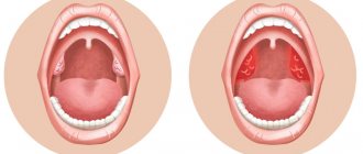
Enamel hypoplasia is a non-carious lesion of teeth that occurs before tooth eruption during the development of its tissues. The term “enamel hypoplasia” is conditional, since changes are also observed in other tooth tissues - dentin and pulp.
Terminology of dental enamel hypoplasia
Dental enamel hypoplasia is a qualitative and quantitative violation of tooth enamel. This definition of the disease is most often found in Russian-language literature. In international sources, enamel hypoplasia (including prenatal and neonatal) refers only to its quantitative changes - thinning, pits, grooves. Qualitative changes (changes in color, transparency) in foreign literature are opacity, hypomineralization, dismineralization and non-endemic mottling of enamel (ICD-C).
Enamel hypoplasia in children
Enamel hypoplasia in children can develop in utero, during the newborn period, before and after the first year of life, up to the age of three. Despite the period of its occurrence, the mechanism for the appearance of changes in enamel is the same. Initially, the function of ameloblasts during the formation or secretion of enamel substance is reduced or impaired. As a result, the construction of the enamel protein matrix and its mineralization are damaged. In the later and most severe stages of the disease, vacuolar changes in ameloblasts and their destruction are detected. The cells can no longer function and amelogenesis stops.
Possible complications
In the early stages, the disease manifests itself in the form of small cosmetic defects without causing serious discomfort. If the spots are clearly visible, this indicates more intense destruction of the enamel. In the second case, treatment must be carried out as quickly as possible, since ignoring the problem can cause serious consequences, such as:
- Increased wear of incisors.
- Tissue destruction.
- Deformation and loss of damaged teeth.
- Development of malocclusions.
Causes of enamel hypoplasia
There are several groups of causes of enamel hypoplasia. Depending on the period of their exposure, temporary or permanent teeth are affected.
Enamel hypoplasia of primary teeth
In the occurrence of enamel hypoplasia of primary teeth, the key etiological factors are:
- Causes of prenatal hypoplasia (the main period of exposure to a negative factor is pregnancy):
- Maternal diseases: hormonal disorders, epilepsy, rubella, toxoplasmosis, alcoholism;
- Physical factors (irradiation);
- Insufficient intake of vitamins, micro- and macroelements from food.
- The causes of neonatal hypoplasia (in the neonatal period - the first 56 days of a child’s life) can be prematurity, birth trauma, asphyxia, hemolytic disease of the newborn.
Enamel hypoplasia of permanent teeth
Hypoplasia of the enamel of permanent teeth is most often associated with diseases of the child that disrupt the metabolism in the body.
These are diseases:
1) Central nervous system: the mineral metabolism of phosphorus and calcium is disrupted, the amount of magnesium and potassium in the blood and bones decreases;
2) Endocrine system:
- Hyperthyroidism promotes the supply of calcium and phosphorus to the teeth and bones. With hypothyroidism, these elements are washed out.
- Against the background of insufficiency of the parathyroid glands, the content of calcium and phosphorus in the blood increases, decreases in the bones, nails, hair, and the lens are also affected;
3) Toxic dyspepsia and other diseases of the digestive system (due to insufficient absorption of calcium and phosphorus);
4) Hypovitaminosis C, D, E (up to rickets);
5) Acute infectious diseases;
6) Allergic diseases;
7) Insufficient nutrition.
Also, the quality of the enamel of permanent teeth depends on the condition of their temporary predecessors. Chronic apical periodontitis, mechanical trauma and removal of primary teeth with trauma to the follicle of a permanent tooth can lead to its hypoplasia.
Symptoms
Signs of pathological development of the surface layer of the dental crown:
- violation of the integrity of the enamel in the form of grooves, ridges, lines, spots, up to the complete absence of tissue;
- pigmentation of white, yellow, yellow-brown shade;
- deformation - shortening of roots, curvature of the crown;
- increased sensitivity to irritants.
At the same time, the enamel can retain its smoothness and shine, and the defects present during cutting are not prone to growth. However, by the appearance of the defect one can also judge some characteristics of the damaging factor:
- Localization on certain teeth depends on at what stage of development the body suffered a negative impact. If the child was ill at the age of 5–6 months, the edges of the front incisors and the cusps of the “sixes” will be affected on the permanent teeth; at 8–9 months, the marginal incisors and the cutting surface of the canines will be affected.
- The size of the defect determines the duration of exposure. If the disease lasts throughout the preschool period, hypoplasia evenly covers the entire surface of the teeth.
- The number of pathogenic structures within one tooth depends on the frequency of the pathogenic influence. For example, if hypoplastic changes are uneven and have a wave-like structure, this indicates a history of infections or chronic diseases with periods of remissions and exacerbations.
Classifications of enamel hypoplasia
The most common classification of enamel hypoplasia is the classification of M.I. Groshikov. It is based on different etiologies and the number of teeth affected. Based on this, methods of treatment and prevention of various forms of hypoplasia differ.
Systemic enamel hypoplasia
Systemic enamel hypoplasia is a disorder in the structure of all teeth, but more often in groups associated with close periods of formation and eruption.
Such diseases in the ICD-C (1995) as prenatal enamel hypoplasia, neonatal enamel hypoplasia, enamel hypoplasia, non-endemic mottling are nothing more than “systemic enamel hypoplasia” according to M.I. Groshikov.
Features of the clinical manifestation of defects in systemic enamel hypoplasia:
- appearance from the moment of eruption;
- symmetrical, of the same size on teeth of the same name;
- localized parallel to the chewing surface or cutting edge, most often on the tubercles or vestibular surface.
There is also a relationship between the defect and the action of the damaging factor:
- The type of defect (qualitative or quantitative change in enamel) depends on the intensity of the factor;
- Localization of the defect - depending on the time of its exposure;
- The width of the defect depends on the duration;
- The number of defects indicates the frequency of action of the damaging factor.
What do patients complain about?
The most common form of hypoplasia is mild. The integrity of the enamel is not compromised, but it may have spots, pits, grooves, and irregularities. There is no pain; patients are only concerned about the aesthetic defect. In a more severe form of hypoplasia - aplasia - the enamel is absent in some area or on the entire tooth. Where there is no enamel, the deeper tissue of the tooth - dentin - is exposed. It is sensitive, so the patient's main complaint is pain from touch, chemical and temperature irritants.
Forms of enamel hypoplasia
Clinically, the following forms of enamel hypoplasia are distinguished: spotted, cup-shaped (erosive), grooved (wavy) forms, thinning or aplasia of the enamel.
The spotted form is spots and stripes, most often white or yellow, with clear or fuzzy contours. Their surface can be smooth and shiny or rough and dull. Shiny smooth enamel means demineralization of its subsurface layer, dull and rough – changes in the surface layer at the end of the enamel formation process.
Cup-shaped, grooved forms, thinning, aplasia of enamel are manifested by areas of hypoplasia through which dentin is visible, grooves, aplasia (complete absence) of enamel. The edges, walls and bottom of the defects are sometimes yellow-brown pigmented and smooth.
Separately, it is necessary to mention molar-incisal hypomineralization. Its characteristic feature is damage to one to four permanent molars, often combined with damage to the incisors. Clinically, these are cloudy spots of white, yellow or brown color, sometimes covering the entire crown of the tooth. Children may be bothered by teeth chipping and sensitivity. Because of this, they may refuse to brush their teeth, which soon leads to the development of caries. Parents may be concerned about the unaesthetic appearance of their teeth.
Teeth of Hutchinson, Fournier and Pflueger
Also manifestations of systemic enamel hypoplasia are the teeth of Hutchinson, Fournier and Pfluger. They are characterized by a change in tooth shape. The main reason is late congenital syphilis.
Local enamel hypoplasia
Local enamel hypoplasia (Turner's tooth) is a violation of the development of enamel (sometimes dentin) of individual permanent teeth. As a result, the tooth changes color: it becomes white or yellow-brown, and areas of hypoplasia appear on it. Turner's tooth is directly related to the periapical inflammatory process of the primary tooth.
Focal enamel hypoplasia
With focal enamel hypoplasia (regional odontodysplasia), underdevelopment of all dental tissues is observed. Typically, the process involves several teeth located nearby. These temporary, and subsequently permanent, teeth are characterized by late development and eruption. After eruption, the teeth are yellowish, with a rough surface. The characteristic name for such teeth is “ghost teeth,” which is also due to their special appearance on an x-ray. The enamel and dentin are thin, their density is reduced, the pulp chamber is large, the roots are wide and short, with open apexes.
Symptoms
Depending on the form and extent of the disease, the following symptoms may be observed:
- Change in tooth color.
- White, symmetrically located spots of different shapes.
- Partial or complete absence of enamel.
- Dips in the teeth.
- Pain when eating.
- Hypersensitivity of teeth to chemical and thermal irritants.
Reference! Some signs of hypoplasia (waves, dots, grooves) become clearly visible only as the disease progresses, but the dentist can detect them in the initial stages.
Treatment of enamel hypoplasia
Several methods are used to treat enamel hypoplasia. The choice of each depends on the violation of aesthetics, type, depth, area of the defect, degree of enamel mineralization, motivation of the child and parents, and technical capabilities.
The essence of the conservative method is to increase the mineralization of hard tooth tissues. This is both endogenous and exogenous use of vitamins, preparations containing fluorine, calcium, phosphorus. It can be used both independently and as an initial step before other methods.
Microabrasion and/or bleaching is carried out after the mineralization of the tooth is completed. The technique consists of etching the enamel, followed by grinding it with a minimally abrasive bur and polishing it with a rubber cup. This method is effective if the defect is in the surface layer of the enamel or when it is clouded.
The surgical method is preparation and then filling. It is also carried out either after conservative therapy or after mineralization of the tooth is completed. Used for the deepest defects in the enamel. Options include filling with GIC (followed by replacement with composite), composite materials, veneers, laminates and crowns.
Another treatment tactic for focal hypoplasia. The optimal approaches to restoring teeth in this case are covering the teeth with crowns soon after eruption or removing them with prosthetics.
Prevention of enamel hypoplasia
The main directions of prevention of enamel hypoplasia:
- Prevention of diseases in a pregnant woman, her balanced diet;
- Prevention and treatment of somatic diseases in young children;
- Sanitary and educational activities of a dentist in a antenatal clinic, a children's clinic;
- Treatment or removal of temporary teeth with complicated caries;
- Prevention of trauma to temporary teeth;
- Atraumatic removal of a temporary tooth.
The article was written by Titenkova O.V. especially for the OHI-S.COM website. Please, when copying material, do not forget to provide a link to the current page.
Diagnostics
Patients with aplasia usually come to the dental clinic with complaints of increased tooth sensitivity. During the examination, the cause of this phenomenon is revealed.
With hypoplasia there may be no complaints. If the defects are minor, they are usually an accidental discovery. But in most cases, parents pay attention to stains on the child’s enamel and make an appointment with a dentist.
To carry out an accurate diagnosis, the doctor needs to collect an anamnesis, obtain detailed information about how the pregnancy proceeded, how the child developed in the first year of life. Studying medical records will provide detailed information about past illnesses. It is important for the dentist to know when defects appeared on the teeth. If the patient is a school-age child, the specialist will clarify whether a similar problem occurred on baby teeth. The dentist will also ask if this phenomenon has occurred among close relatives; perhaps the damage to the enamel is hereditary.
After the conversation, the dentist proceeds to examine the oral cavity. This makes it possible to establish the size, prevalence and nature of the defects, as well as to carry out a differential diagnosis with other lesions, such as caries.
Prevention
You need to think about preventing non-carious dental diseases and, in particular, hypoplasia even before the birth of the child. This is due to the fact that enamel malformations in most cases occur during fetal development.
To minimize the likelihood of the formation of this pathology, you need to follow these tips:
- Every trimester of pregnancy, a woman should be examined by a dentist.
- Good nutrition is the key to normal metabolism in mother and baby.
- During pregnancy, you should never take medications without first consulting a doctor.
- The baby needs to be breastfed for at least a year and ensure that it does not injure the oral cavity.
- If the first signs of pathology develop, you should immediately consult a doctor.