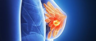
A breast cyst is a manifestation of fibrocystic disease (mastopathy), accompanied by hardening of gland tissue, dilation of the milk ducts and a disorder of hormonal metabolism.
A cyst is a cavity, usually filled with light-colored fluid. It is mainly formed from parts of the mammary gland or milk ducts. Due to excessive enlargement of the surrounding tissues, as well as the tissue of the ducts, stagnation of mammary gland secretion occurs in the dilated ducts. The volume of secretion increases over time and a breast cyst forms. This formation occurs very often, mainly in nulliparous women aged 35–55 years.
Basically, the cyst capsule includes benign cells, but there may also be malignant ones. The size of the cyst can be several millimeters and reach several centimeters. Cysts can be grouped or single.
With polycystic disease, numerous cysts of different sizes merge and form multi-chamber clusters. The cyst can cause pain as it grows and compresses the surrounding breast tissue, where nerve fibers may be located. In addition, there are fatty cysts that form when the sebaceous glands are blocked and can appear in any part of the breast. Such formations are safe, but with inflammation they can greatly increase.
Cystic formations can have an oval, round, irregular shape. A typical cyst has even and smooth internal walls, while the atypical type is characterized by growths on the walls and protruding into the cystic cavity.
What types of cysts are there and what is a “large cyst”?
Cysts that can only be detected by ultrasound or mammography are considered small. A large cyst is the formation that is revealed by palpation. As a rule, these are superficial formations of more than 1.5 cm, or deep-lying ones with a diameter of 2 or more centimeters. Breast cysts are formed from lobules or milk ducts and can be single or multiple (multi-locular).
As already mentioned, detecting a cyst is very difficult until it acquires an impressively large size. There are cases when it causes severe pain in a woman in the breast area, especially during the periods before and during menstruation.
As practice shows, when a breast cyst occurs and develops, symptoms and any clinical manifestations may be absent. Such a formation, invisible to the patient herself, is detected during an ultrasound examination. Sometimes the cyst manifests itself sharply with a burning sensation or painful tugging, aching, pulling sensations in the chest area. Such pain occurs due to the growth and enlargement of the formation and compression of the surrounding tissues, which may contain nerve endings. When compressed, swelling of the nerve fibers occurs, causing pain of various types.
Typically, a woman can find lumps in her mammary gland that are soft or soft-elastic in consistency, easily dislodged, and moderately painful. They appear several days or even weeks before menstruation. Cysts often appear after overheating: baths, saunas, exposure to hot climates. In such situations, a woman should immediately consult a mammologist and undergo an ultrasound examination of the mammary glands, mammography, and pneumocystography.
Causes of breast cysts
The appearance of a cyst in the mammary glands is due to the occurrence and manifestation of mastopathy, or fibrocystic disease. It is accompanied by expansion of the milk ducts, thickening of the glandular tissue and obligatory disruption of hormonal metabolism, when an imbalance of sex hormones and thyroid hormones occurs.
A woman’s hormonal levels are disrupted in the following cases: the presence of stressful situations, ovarian dysfunction, thyroid disease, polycystic ovary syndrome, etc. Such an imbalance, in turn, provokes not only inflammatory diseases of the reproductive system (acute and chronic adnexitis, endometritis), but also disrupts the activity of breast tissue. The result is the appearance of foci of fibrosis and cysts.
Other causes of breast cysts include the number of births and timing (late or early), previous abortions, IVF, taking contraceptives containing hormones, and genetic predisposition. Of no small importance is a history of mastitis and surgical interventions on the chest.
Classification
Cystic formations can degenerate into malignant tumors. The probability of malignancy of the mastopathy lesion ranges from 5–30% and is determined based on the results of morphological analysis of the biopsy specimen. The existing typology of cysts indicates an atypical intensity of proliferation of epithelial tissues (proliferation).
First-degree formations do not have signs of proliferation, and there is no likelihood of developing a malignant neoplasm. Cells of second-degree cysts are prone to atypical growth, but have an unchanged morphological structure. In this case, the likelihood of their malignancy remains moderate. The third degree of pathology is characterized by aggressive proliferation of atypical cells prone to the formation of malignant tumors. The cyst accounts for 3–5% of clinically diagnosed breast cancer cases.
Women's theme
The mammary gland (breast gland) is a paired glandular organ. It functions only in women, producing milk after childbirth. The mammary gland is an organ of increased vulnerability, as it is under fire from many hormones. During the menstrual cycle, pregnancy, and lactation, the mammary glands are influenced by 15 hormones. The breast is susceptible to changes when at least one of them is produced in the wrong quantity. This is where women's problems begin.
Every woman over 17 years of age should undergo regular preventive examination by a mammologist. The frequency of visiting a mammologist depends on various factors: age, heredity, the presence of gynecological diseases, the presence of complaints. Women over 30 who have no complaints need to visit a mammologist every 1-1.5 years. Women over the age of forty are recommended to have a mammogram. Timely diagnosis and examination make it possible to detect various mammological diseases in the early stages. And this, in turn, will prevent breast cancer.
Survey
After conducting an independent minimal examination of the breast, the woman herself is able to confirm the suspicion of a neoplasm in the mammary gland. But only a mammologist makes an accurate diagnosis, determining the type of cyst or possible other formation. For a complete diagnosis, mammography alone is not enough. A comprehensive examination is required. Ultrasound examination and pneumocystography will give an unambiguous result in determining the diagnosis. Sometimes the liquid inside the formation becomes very cloudy and thickened due to the pus formed in it. This makes it difficult to establish an accurate diagnosis. With such manifestations, it is necessary to make sure that there are no malignant formations and conduct an examination of the walls. Such an examination cannot be postponed, since in 10-17% of all cases the cyst may degenerate into breast cancer.
Our services
The administration of CELT JSC regularly updates the price list posted on the clinic’s website. However, in order to avoid possible misunderstandings, we ask you to clarify the cost of services by phone: +7
| Service name | Price in rubles |
| Needle biopsy (without the cost of histological examination) | 2 500 |
| Ultrasound of the mammary glands | 3 000 |
| Mammography | 3 000 |
All services
Make an appointment through the application or by calling +7 +7 We work every day:
- Monday—Friday: 8.00—20.00
- Saturday: 8.00–18.00
- Sunday is a day off
The nearest metro and MCC stations to the clinic:
- Highway of Enthusiasts or Perovo
- Partisan
- Enthusiast Highway
Driving directions
Should a breast cyst be treated?
There can be only one answer - it is necessary to treat.
Appropriate treatment measures are taken depending on the size and number of formations in the breast. Conservative treatment is indicated when multiple small cysts are detected. If a large formation in the mammary gland is diagnosed, the mammologist punctures the mammary gland cyst and removes fluid from it. The collected material must be sent for cytological examination. Punctures are performed when there are no signs of epithelial growth inside the cyst. After the puncture, the walls of the cyst stick together.
Relapses are rare - re-filling of the cavity with liquid, as a rule, does not occur. But if the formation occurs again, then additional examination is necessary. Then the question of surgical intervention is raised. Puncture of a breast cyst is accompanied by the introduction of air into the formation cavity. This procedure accelerates the gluing of its walls, transforming the cyst into a small scar, almost invisible during ultrasound or mammography.
Breast cyst is a common pathology in women of reproductive age. Modern diagnostic methods in mammology make it possible to detect it at an early stage of development. Timely diagnosis and treatment methods of mammology, in turn, allow women to get rid of cysts and their consequences with maximum effect.
Surgical removal of cysts
When cysts grow and the pills are no longer effective, surgical intervention becomes necessary. There are several methods for removing tumors:
- Puncture - performed in cases where the cyst consists of vesicles connected to each other by connective tissue and filled with liquid inside. During the manipulation, the cyst is punctured and all the secretion is removed from it.
- Laser removal is a modern and safe, but not a cheap method. Excision of a cyst with a laser is carried out quickly and painlessly, without scarring. LED exposure destroys only atypical cells. Over the next two months, they are completely replaced by healthy glandular cells.
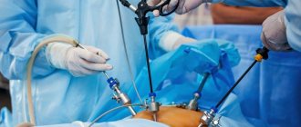
- Surgical intervention is necessary for large cysts or high-density neoplasms. The operation is carried out with high precision; upon completion, the tissue at the incision site is stitched together in layers. A sick leave after surgery is issued by the doctor for the entire recovery period.
Treatment methods
If a woman is diagnosed with growths in a cyst (cystadenopapilloma), then the cystic growths are removed surgically. In the presence of a benign formation, mammologists prescribe conservative treatment of breast cysts. It provides resorption and anti-inflammatory therapy.
In any case, treatment should be aimed at restoring the proper functioning of the endocrine glands, as well as curing diseases of the female genital organs.
Specialists at Alla Fedorovna Kartasheva’s clinic select an individual therapy regimen for each patient and provide the most effective treatment.
Rules for disease prevention
- Annual visit to a mammologist, regular mammography or ultrasound of the mammary glands.
- Regular self-examination.
- Taking care of the mammary gland.
- Take COCs only as prescribed and under the supervision of a physician.
- Rejection of bad habits.
- Selecting and wearing the right bra.
CELT clinic specialists will help you choose the appropriate treatment and maintain your health. The latest equipment, advanced techniques, professional doctors - CELT offers complete patient support, from diagnosis to recovery.
At an appointment with a mammologist
Consultation and diagnosis of women begins with a survey. The mammologist must find out the patient’s complaints and identify unfavorable factors that can affect the normal functioning of the mammary glands. Next, a careful examination of the breast is necessary - palpation of all areas of the mammary gland, lymph nodes, zones of lymphatic drainage, nipples and areolas.
Only comprehensive diagnostics can guarantee accurate diagnosis. It includes a clinical examination of the patient with palpation of the mammary glands, ultrasound, mammography (X-rays taken on special devices with a low dose of radiation, mammographs). If formations are detected, puncture and biopsy are performed. If there is a suspicion of a connection between breast disease and pathology of other organs and systems of the body, the woman is prescribed appropriate tests. The mammologist will also indicate that consultations with a gynecologist, endocrinologist and other specialists necessary for your case are mandatory.
Visits at the clinic are conducted by doctors of the highest category - mammologist, mammologist-oncologist. Experienced surgeons at our clinic provide surgical treatment of the mammary glands: removal of intraductal papilloma, removal of skin lesions, sectoral resection, removal of benign lesions, as well as conservative treatment of all breast diseases. Equipping the clinic with the most modern medical equipment allows us to diagnose and treat mammological diseases using the most modern minimally invasive techniques.
In our clinic you will be greeted by experienced, highly qualified specialists. Consultations and explanations from the clinic’s doctors will not leave you with any questions. Our clients are surrounded by the care and attention of the best mammologists in the capital. We will always help you and bring back the joy of life.
Medicines
Photo: medistok.ru
Depending on the type, size, characteristics of the course of the cyst, as well as disorders detected according to laboratory and instrumental studies, the doctor may prescribe the following groups of drugs:
- Hormones . Since hormonal imbalances are considered one of the reasons for the appearance of cysts, the specialist will recommend adjusting the levels of estrogen and prolactin.
- Anti-inflammatory and painkillers . Necessary for signs of inflammation and severe pain.
- Vitamin complexes . Helps replenish the lack of nutrients, activate metabolic processes, and improve immunity.
- Antibiotics . Required for suppuration of cysts.
- Other means . Taking into account the etiology of the disease, iodine preparations, sedatives, dopamine agonists, etc. can be used.

