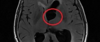
Cysts of the pineal gland (epiphysis) are common, asymptomatic, and in most cases are an incidental finding. Cysts, especially large and atypical-looking ones, are difficult to distinguish from cystic tumors, so patients with suspicious changes should undergo long-term observation. On MRI or MRI, a pineal gland cyst appears as a single-chamber fluid formation with the density of cerebrospinal fluid or with a signal intensity similar to that of cerebrospinal fluid. Peripheral contrast enhancement is typical for most cysts, and rim-shaped calcifications are found in 25% of cases.
Get an MRI of the pineal gland in St. Petersburg
Symptoms of a pineal gland cyst
The vast majority of pineal gland cysts are small in size (in 80% of cases less than 1 cm) and are characterized by an asymptomatic course. Cysts that cause symptoms mainly occur in women in the second half of life. Larger cysts can cause a volumetric effect on the quadrigeminal plate, leading to compression of the superior colliculus and the occurrence of Parinaud's syndrome. When the cerebral aqueduct located between the third and fourth ventricles is compressed, obstructive hydrocephalus may develop. Rarely, if the cyst bleeds, it can quickly increase in size. This condition is called pineal cyst apoplexy.
A pineal gland cyst can cause headaches, blurred vision, and the inability to look up or down. Rare symptoms: ataxia, emotional disorders, disturbances in mental activity, dizziness, sleep disturbances, nausea, hormonal imbalance (early puberty, secondary parkinsonism).
general information
The pineal gland (pineal gland, pineal gland) is located deep in the brain tissue . The main function of the organ is the production of the hormone melanin. Melanin performs the following functions in the human body:
- Regulates puberty, sleep and wakefulness.
- Affects the pituitary gland and hypothalamus.
- Activates the immune system.
The appearance of a cyst may be asymptomatic . When the formation interferes with the circulation of blood and spinal secretions, the synthesis of melanin is disrupted, and pathological symptoms of a physical and psychopathic nature appear.
A true gland cyst is covered from the inside with connective tissue cells which may contain calcified deposits. A false cyst does not have an internal cellular layer; it occurs in severe cases of hypertension, rheumatism, and tetanus.
Structure
The wall of a pineal cyst consists of three concentric layers:
- The inner layer is represented by fibrillary glial tissue, often with inclusions of hemosiderin
- The middle layer is the parenchyma of the pineal gland with or without calcifications
- The thin outer layer consists of connective (fibrous) tissue
Hormonal changes are thought to play a role in the formation of cysts, as they are most often found in young women. With age, cysts first increase in size and then “shrink.” In men, these formations remain in a stable state for a long time. Cysts usually contain proteinaceous fluid that differs from cerebrospinal fluid on CT scans; Sometimes blood is found in them. The walls of the cyst intensively accumulate contrast.
Prevention
To prevent the development of this disease, there are only general recommendations:
- prevention of cerebrovascular diseases
- reducing the risk of stroke
- reducing the risk of head trauma during childbirth
- excluding contact of a pregnant woman with patients with infectious diseases or bacteria carriers
- reduce the likelihood of fetal hypoxia in a pregnant woman (excluding the use of cigarettes, alcoholic beverages, and drugs)
- reducing the risk of traumatic brain injury
- maintaining personal hygiene and maintaining a healthy lifestyle
A very common disease is asymptomatic and does not have a significant impact on a person’s quality of life. However, you should remember the possible risks of an epiphysis cyst and be able to recognize its presence in time.
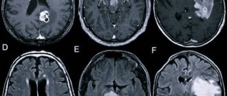
What does a pineal gland cyst look like on CT and MRI?
Computed tomography visualizes a fluid-dense mass with well-defined edges and peripheral calcifications found in 25% of patients. In many cases, there is also a peripheral accumulation of contrast in the cyst in the form of a thin and smooth “rim”. The cyst disrupts the course of the internal veins of the brain, displacing them upward.
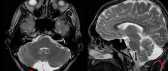
Pineal cyst with a typical hyperintense signal on MRI (T2 WI) (marked with a blue arrow). Left: axial tomogram, right – sagittal
The following signs are observed on MRI:
- T1 VI
- Typical iso- or hypointense signal compared to brain parenchyma
- The signal is usually uniform
In 55–60% of cases, the signal is hyperintense compared to the cerebrospinal fluid
- High intensity signal
- High intensity signal that is often not completely suppressed
- There is no diffusion restriction
- Approximately 60% of cysts accumulate contrast
Reasons for appearance and development
There are several reasons for the development of this pathological condition, but the main ones are:
- Blockage of the pineal gland excretory duct In this condition, the secretion of the pineal gland, which contains hormones and should be released into the blood, cannot leave the gland due to obstruction of the outflow.
The reasons for this may be:- tumor growths compressing the excretory duct from the outside
inflammatory diseases of the brain substance (encephalitis)
- autoimmune processes
- blockage of the excretory duct by a water bubble (for example, with metabolic disorders)
- anatomical features of the body (winding or narrow duct)
- production of too viscous secretion by the pineal gland
- Development of parasites There are a number of parasites, for example, Echinococcus, the larvae of which move through the bloodstream throughout the body and can settle in some organs. When a larva enters the pineal gland, it forms a capsule around itself and is gradually filled with metabolic products of the worm. This version of the cyst is more dangerous, since in this case the increase in the size of the formation occurs much faster. Fortunately, pineal echinococcosis is quite rare.
- Stroke A cavity in the epiphysis can form during a hemorrhagic stroke. Most often, this occurs in people suffering from arterial hypertension and having problems with the blood vessels of the brain (for example, atherosclerosis). In this case, the vessel ruptures and blood begins to flow into the surrounding brain tissue. Afterwards, the blood clot is replaced along the periphery by connective tissue, forming a bubble with liquid. A similar mechanism of cyst formation is observed in traumatic brain and birth injuries.
- Congenital cysts In very rare cases, a cyst may appear in utero. This is possible in case of abnormal development of the fetus, after infectious diseases in a pregnant woman, or in case of fetal hypoxia.
In all these conditions, the lumen of the duct decreases, which causes the formation of a cyst. It is worth noting that the size of the tumor remains quite small.
How to distinguish a pineal cyst from a tumor?
A pineal cyst, especially one with nodular contrast enhancement, cannot be differentiated from a cystic pineocytoma based on imaging techniques alone. Other formations may also occur in the pineal area: papillary tumor, germinoma, embryonal cancer, choriocarcinoma, teratoma, epidermoid cyst, arachnoid cyst, vein of Galen aneurysm, metastases.
Although such cases are rare, it is always best to have the results examined by an experienced neuroradiologist. Rechecking CT or MRI images will help you more accurately understand whether a tumor is suspected. Today this can be done remotely using Second Medical Opinion services, such as the National Teleradiological Network.
Possible contraindications
Magnetic resonance imaging is considered absolutely safe for the body only if the patient has no contraindications. Therefore, before making an appointment for an MRI, you need to visit a doctor and check if you have one of the serious contraindications:
- The presence of a pacemaker or metal structures in the body that cannot be removed independently;
- Diseases of the central nervous system or serious psychological disorders in which the patient is unable to control his body and lie still;
- The patient's weight is over 250 kg.
- Severe fear of confined spaces.
If the doctor recommended undergoing magnetic resonance imaging with contrast, you need to make sure there is no pregnancy, lactation, renal failure, kidney problems or allergies to the components included in the drug.
Treatment of pineal cyst
In almost all cases there is no need for treatment; in most cases, small cysts do not require follow-up studies. Depending on the size and location of the cyst, as well as symptoms, treatment may consist of stereotactic removal of the cyst, aspiration of its contents, creation of a connection with the cerebrospinal fluid spaces, and shunting. If the cyst recurs after treatment, radiation therapy is possible.
Get an MRI of the pineal gland in St. Petersburg
Methods for surgical removal of tumors
Unfortunately, a cyst on the pineal gland, independent of echinococcal infection, is difficult to treat therapeutically. Therefore, in extreme cases, it is necessary to resort to surgical intervention: if the cyst grows rapidly, affects the cardiovascular system, and provokes hydrocephalus.
- Endoscopy. Through a special tube inserted into the cranial cavity, a drainage is installed into the cyst to drain fluid. The capsule of the cystic formation is emptied and falls off.
- Shunting. It involves draining fluid from the neoplasm into a cavity where the accumulation of cerebrospinal fluid is not dangerous to human health. For example, through a special catheter - from the skull into the abdominal cavity.
- Trepanation of the skull. The operation is highly traumatic, but also highly effective. After opening the skull, the cyst is completely removed from the body of the pineal process. If it is not possible to separate the cystic formation, the surgeon may remove the pineal gland completely.
After the cyst is eliminated, its contents are sent for histological examination to ensure that it is benign.
Important! You cannot refuse therapeutic measures; sometimes supportive therapy is enough to curb the growth of the cyst.
Sometimes surgical intervention is contraindicated, despite the threatening size of the capsule and its negative impact on the epiphysis of the brain. For example, in people with low blood clotting, pregnant women, elderly patients. Then doctors recommend conservative treatment. But usually it does not help reduce the size of the cystic formation, but eliminates the signs of the disease: headache, nausea, blurred vision.
What else do you need to know?
If you are about to undergo magnetic resonance imaging for the first time, we recommend that you familiarize yourself with some features:
- If loud noises scare you, be sure to take earplugs or special noise-isolating headphones with you. The tomograph makes a loud noise while taking images.
- There is lighting and ventilation inside the tomograph.
- A microphone and speakerphone are provided to communicate with a specialist.
- If you feel unwell, you can use the panic button, which will stop the research process.
- It is important to remain completely still. Even minimal body movements can lead to blurry pictures.
How to properly prepare for the procedure
To obtain clear and informative images, it is recommended to follow a few simple rules:
- Do not eat for 7-8 hours before the examination if an MRI with contrast is planned;
- Do not drink a lot of liquid immediately before the procedure, so as not to experience the urge to urinate;
- Tell your doctor if the test worries you. If you are experiencing severe panic, your doctor may recommend taking a sedative;
- Take with you a change of clothes without metal fittings;
- To avoid wasting time in the diagnostic room, remove all metal-based jewelry and accessories in advance.