The adrenal glands are a “factory” of hormones
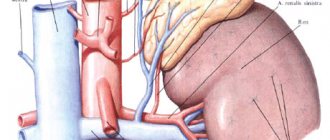
The adrenal glands are small paired organs located, as the name suggests, above the kidneys. Their main task is to produce hormones - substances that regulate all vital processes in the body. For example, glucocorticoid hormones are responsible for metabolism, mineralocorticoid hormones are involved in water and salt metabolism, androgens and estrogens are analogs of sex hormones, adrenaline and norepinephrine are stress hormones. The adrenal glands are surrounded by a dense connective tissue capsule and are embedded in adipose tissue. Bundles of the capsule (trabeculae) penetrate into the internal parts of the gland, forming partitions that divide the organ into layers and zones. The adrenal glands are, in addition, surrounded by the renal fascia, to which they are connected very firmly. The right adrenal gland has the shape of a truncated cone, flattened from front to back. From the front it resembles a triangle with rounded corners - “Napoleon's tricorne”. The apex of the left adrenal gland is flattened and is shaped like a crescent. Front view of isolated right and left adrenal glands
How is ultrasound of the adrenal glands performed?
The procedure for scanning the adrenal glands using ultrasound is simple.
First, you will need to free the abdominal area from clothing, and then take the position on the couch required for the diagnostician. This may be a lying position on your stomach or back, perhaps a standing or side position. When the patient is ready, the doctor applies a contact gel to the area being examined, and the same composition is applied to the device’s sensor. Next, the diagnostician determines the location of the right adrenal gland and begins the ultrasound scanning procedure. In order to get a good scan of the adrenal glands, the patient will need to take a deep breath and hold it while lying on their side. Then the patient will need to lie on the other side, and the diagnosis will be repeated on the other side. On ultrasound, the right adrenal gland is better viewed from the anterior approach, and the left adrenal gland is better viewed from the side. It is not always possible to detect the adrenal glands on both sides. The right one is detected almost always, but the left one is detected only in half of the cases. During ultrasound diagnostics of healthy glands, visualization is zero, since their structure is no different from the retroperitoneal tissue, therefore, it is possible to identify only the area where the adrenal glands are localized. The glandular tissue itself can only be detected in children or thin young people. Author: Telegina Natalya Dmitrievna
Therapist with 25 years of experience
>
All types of diagnostics are important - from examination to ultrasound
The diagnostic complex for studying the pathology of the adrenal glands should include: clinical examination, laboratory (clinical, biochemical, hormonal), instrumental and pathomorphological examination methods. A clinical examination and interview of patients is carried out to identify signs characteristic of various symptom complexes of adrenal pathology. In laboratory diagnostics to identify pathology of the adrenal glands, the levels of adrenocorticotropic hormone, cortisol, aldosterone, dehydroepiandrosterone sulfate (DHEA sulfate), the activity of renin and angiotensin in the blood plasma, and the daily excretion of adrenaline, norepinephrine and vanillylmandelic acid in the urine are most often determined. For the purpose of topical diagnosis, ultrasound with color Doppler scanning, computed X-ray tomography (), magnetic resonance imaging (MRI) is used; in complex cases, for the purpose of differential diagnosis - angiography or selective venography with separate blood sampling. In some cases, to verify the morphological structure of adrenal formations, aspiration puncture with a thin needle is performed under ultrasound or CT control with cytological examination.
Kidney ultrasound
- Cost: 2,700 rub.
More details
Why do “flies” fly before my eyes?
Clinical manifestations of primary hyperaldosteronism (PHA): arterial hypertension (headache, dizziness, appearance of “floaters” before the eyes); disorders of neuromuscular conduction and excitability (muscle weakness, parasthesia, convulsions, bradycardia); changes in renal function (polyuria, polydipsia, nocturia). These signs are not always present simultaneously; An oligosymptomatic and even asymptomatic course of the disease is often observed. Approximately two-thirds of all cases of PHA are caused by aldosteroma. Another possible cause of PHA is nonadenomatous bilateral adrenal hyperplasia, which occurs in one third of patients with symptoms of idiopathic primary hyperaldosteronism. The main treatment method for aldosteroma and nodular adrenal hyperplasia is surgery - removal of the altered adrenal gland along with the tumor (adrenalectomy). Performing organ-saving operations (tumor enucleation, adrenal resection) by most surgeons operating on endocrine organs is considered inappropriate due to the high rate of relapse of tumors in the remaining tissue (up to 50%). Already in the immediate postoperative period, in most cases, normalization of blood pressure, a decrease in the concentration of aldosterone in plasma, a gradual increase in renin activity, and normalization of potassium are noted.
Acute adrenal insufficiency. Symptoms
Dysfunction of the adrenal cortex causes a lack of some hormones and an excess of others. Primary and secondary acute adrenal insufficiency is a disruption of the cortical layer of the glands. This condition is very dangerous; if it occurs, it is necessary to begin immediate treatment in a hospital. The cause of this pathology may be the sudden withdrawal of tableted glucocorticosteroids, hemorrhage into the gland tissue, or a sharp deterioration in the course of chronic adrenal insufficiency due to stress. In the neuropsychic form of acute adrenal insufficiency, the patient experiences convulsions, severe weakness, and headache. After some time, a disturbance of consciousness begins (stupor, delirium, coma). The cardiovascular form of acute adrenal insufficiency is manifested by circulatory disorders. There is pale, cold skin on the hands and feet, weak pulse, lack of spontaneous urination, bluish lips, and a drop in blood pressure.
"Crisis" tumor
Pheochromocytoma is a tumor of chromaffin cells that produces excess amounts of catecholamines (adrenaline, norepinephrine and dopamine). Pheochromocytoma may develop from chromaffin tissue of the adrenal medulla (90%) and may be non-adrenal in location. The disease occurs at any age. The main symptoms of the disease in the vast majority of patients are arterial hypertension, hypermetabolism and hyperglycemia. The classic clinical picture is considered to be periodic rises in blood pressure, accompanied by some vegetative symptoms, reminiscent of the effects of the administration of adrenaline or norepinephrine. In the initial period of the disease, these attacks, or crises, occur rarely - once every few months or even years. Over time, the frequency, duration, and severity of attacks usually increase. The onset of a crisis is quite often characterized by the appearance of unaccountable fear, sometimes a feeling of chilliness, paresthesia, marbling or pallor of the skin. Sometimes, on the contrary, there is pronounced redness of the facial skin, shiny eyes, dilated pupils, and frequent urge to urinate. A crisis can begin with paresthesia, convulsions, and vasoconstriction of the extremities. The crisis ends as suddenly and quickly as it began. Blood pressure returns to its original values, the pallor of the skin gives way to redness, and sometimes profuse sweating and excessive secretion of the salivary glands are observed. After an attack, general weakness and weakness persist for a long time. The diagnosis of pheochromocytoma is established when elevated amounts of catecholamines or their metabolites are detected in 24-hour urine. The most reliable is urine analysis collected within 3 hours after the attack. The accuracy of the method reaches 95%. It is recommended to conduct such studies several times. Tumor detection is usually carried out using ultrasound, CT and MRI. If the diameter of the tumor is more than 10 mm, then the sensitivity of these methods approaches 100%. Ultrasound is important in establishing the location of pheochromocytoma. However, its effectiveness largely depends on the experience of the specialist; Moreover, for tumors of extra-adrenal localization, the method is not very informative. Treatment of tumors that produce catecholamines is only surgical. However, surgical interventions for pheochromocytoma are classified as very complex, primarily due to the high degree of surgical risk associated with the likelihood of severe hemodynamic disturbances. In this regard, the cooperation of the surgeon and anesthesiologist and the choice of the most rational method of preoperative preparation and anesthesiological care are of particular importance. During preoperative preparation, the main attention should be paid to the prevention and relief of hypertensive crises. After surgery, hormone replacement therapy is often prescribed - a constant intake of adrenal hormones.
Extra pounds, hump and stretch marks...
The main and only treatment method for hypercortisolism caused by a tumor of the adrenal cortex or nodular hyperplasia of the adrenal cortex (Cushing syndrome) remains removal of the affected adrenal gland with the tumor. The clinical picture of hypercortisolism is very specific, therefore, in a significant proportion of patients, nosological diagnosis does not present any great difficulties. obesity, a “hump” on the back of the neck, wide purple “striae”, arterial hypertension, impaired carbohydrate metabolism and sexual function, osteoporosis and hypokalemia allow us to suspect overproduction of cortisol by the adrenal glands. At the next stage of diagnosis, especially in the absence of clear external clinical signs, the most important, and sometimes decisive, importance is the assessment of laboratory hormonal parameters. Ultrasound examination of the adrenal glands has relatively high sensitivity, it is non-invasive, relatively harmless, and accessible. The sensitivity of the ultrasound method in detecting adrenal tumors depends on their size and averages 50-90%. Corticosteroma is detected as a round formation of various sizes, with an indistinct capsule. Acoustic density appears to be slightly reduced compared to normal adrenal gland. CT is highly effective in diagnosing diseases of the adrenal glands. For adrenal adenomas, the sensitivity of the method reaches 98%. With benign corticosteromas, the prognosis is favorable. Already in the first 1.5-2 months after tumor removal, a gradual regression of clinical symptoms is noted: the patient’s appearance changes, metabolic processes are normalized, blood pressure decreases to normal, stretch marks and the face turn pale, sexual function is restored; diabetes mellitus observed before surgery disappears. In the first months after surgery, patients' body weight decreases significantly (sometimes by more than 20 kg); hirsutism usually disappears after 3-8 months. X-ray signs of bone tissue restoration are recorded after 10-12 months, but bone pain disappears within 1-2 months after surgery.
Surgical treatment of adrenal diseases:
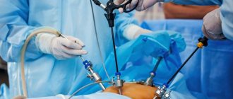
Currently, operations on the adrenal glands can be performed using traditional “open” access, or using high-tech methods (endoscopic operations). The standard access for adrenalectomy is most often lumbotomy or laparotomy - quite traumatic and time-consuming interventions. Such approaches as transdiaphragmatic, subdiaphragmatic, transthoracic can also be used. The Center for Endocrine Surgery widely uses endoscopic techniques in adrenal surgery, which can be either laparoscopic or extraperitoneal access. Endoscopic techniques are less traumatic compared to “open” surgery: in endoscopic operations there are 3 or 4 punctures of 1 cm each, patients spend less time in hospital treatment, recovery time is reduced by 2-3 times. The type of surgery is most often determined by the size of the tumor.
When a woman turns into a man...
Androsteroma is a tumor of the adrenal cortex that produces excessive amounts of androgens. These rather rare tumors (1-3% of all adrenal tumors) are detected in all age groups, but mainly in women under the age of 40 years. The rarity of androster detection in men may be associated with less distinct signs of virilization in them; Some of these tumors imitate hormonally inactive adrenal tumors. Symptoms of androsteroma are characterized by rapid virilization. Girls experience male-pattern pubic hair growth, enlargement of the clitoris during puberty, lack of breast growth and menstruation, increased muscle development, and the appearance of a male voice. Boys show signs of premature puberty. In women, the subcutaneous fat layer decreases, the muscles become more prominent, their mass increases, the voice becomes coarser and deepens. There is hair growth on the face, body and limbs, and hair loss on the head. Menstruation soon stops. The mammary glands become smaller. The clitoris is significantly enlarged. In most cases, there is increased libido. CT and MRI data are of decisive importance in making the diagnosis of androsteroma. Ultrasound helps to clarify the topographic relationship of the tumor with the kidney, aorta, inferior vena cava, spleen and liver, which is necessary for choosing surgical tactics. Androster treatment is only surgical - removal of the adrenal gland affected by the tumor. If the tumor is successfully removed, the symptoms of the disease quickly reverse. In advanced cases and in patients with inoperable tumors or distant metastases, treatment is symptomatic. The prognosis for early diagnosis and timely surgical treatment of benign androsterae is favorable. However, quite often children remain short in stature due to early closure of growth plates. In case of malignant androsteromas and the presence of distant metastases, the prognosis is unfavorable.
What can be seen on an ultrasound of the adrenal glands?
Ultrasound diagnostics of the adrenal glands will show the following pathologies of this organ:
- Causes of adrenal hyperplasia, leading to various disorders of the endocrine system at an early age. This pathology is characterized by early hair coverage of the axillary and groin areas, late menstrual cycle, inhibition or even cessation of growth development. The presence of such a disease in a child can be diagnosed using an echographic ultrasound of the adrenal glands.
- Ultrasound of the adrenal glands and kidneys in infants shows hematomas of the glands, which sometimes occur as a result of trauma at birth.
- Adrenal cysts are a rarer ultrasound finding. Typically, this disease begins during embryonic development. Identifying glandular cysts is a complex matter and takes a long period of time, since the difference between an adrenal cyst and a subcapsular kidney cyst can only be detected by ultrasound of the kidneys and adrenal glands.
| Service | Price according to Price | Promotion Price |
| Ultrasound of the abdominal organs | 1500 rub. | |
| Ultrasound of the abdomen and kidneys | 1700 rub. | |
| Ultrasound of the abdomen, kidneys and bladder | 2000 rub. | |
| Ultrasound of the adrenal glands | 800 rub. | |
| Kidney ultrasound | 800 rub. | |
| Comprehensive ultrasound (ultrasound of the abdominal organs + ultrasound of the kidney + ultrasound of the thyroid gland) | 2400 rub. | 1999 rub. |
| Comprehensive ultrasound (ultrasound of the abdominal organs + ultrasound of the kidneys + ultrasound of the thyroid gland + ultrasound of the pelvis with an abdominal probe + ultrasound of the mammary glands) | 4200 rub. | 2999 rub. |
| Comprehensive ultrasound (ultrasound of the abdominal organs + ultrasound of the kidneys + ultrasound of the thyroid gland + ultrasound of the prostate gland with an abdominal probe) | 3300 rub. | 2499 rub. |
| Comprehensive body diagnostics (MRI of the thoracic spine, MRI of the lumbar spine, ultrasound of the abdominal organs, ultrasound of the kidneys, ultrasound of the bladder, consultation with a neurologist, consultation with a therapist) | 11700 rub. | 7000 rub. |
| Service | Price according to Price |
| Ultrasound of the bladder with determination of residual urine | 800 rub. |
| Ultrasound of the pelvis in women and men with an abdominal sensor | 1200 rub. |
| Ultrasound of the pelvis in women with a vaginal probe | 1300 rub. |
| Comprehensive pelvic ultrasound with abdominal and vaginal probe | 1400 rub. |
| Ultrasound of folliculogenesis | 750 rub. |
| Ultrasound cervicometry | 1000 rub. |
| Ultrasound of the prostate gland with a rectal probe | 1500 rub. |
| Ultrasound of the scrotum (testicles, appendages) | 1000 rub. |
| Ultrasound of the prostate and bladder with an abdominal probe | 1900 rub. |
| Service | Price according to Price |
| Ultrasound of the mammary glands | 1400 rub. |
| Ultrasound of the thyroid gland | 1500 rub. |
| Ultrasound of lymph nodes (one zone) | 1000 rub. |
| Ultrasound of soft tissues of one area (face, neck, arms, legs, abdomen, groin) | 800 rub. |
| Service | Price according to Price |
| Ultrasound of one small joint | 1000 rub. |
| Ultrasound of one large joint | 1500 rub. |
| Ultrasound of two small joints | 2000 rub. |
| Ultrasound of two large joints | 2500 rub. |
| Service | Price according to Price | Promotion Price |
| “Healthy Legs” program (duplex scanning of the veins of the lower extremities with assessment of valves and perforating veins and duplex scanning of the arteries of the lower extremities) | 3200 rub. | 3000 rub. |
| Triplex scanning of N/C veins with assessment of valves and perforating veins | 2500 rub. | |
| Triplex scanning of the veins of the upper extremities | 2000 rub. | |
| Triplex scanning of the arteries of the lower extremities with measurement of the brachial-ankle index (BAI) | 2000 rub. | |
| Triplex scanning of the arteries of the upper extremities | 2000 rub. | |
| Duplex scanning of the arteries of the aorto-iliac segment | 1900 rub. | |
| Duplex scanning of the abdominal aorta and visceral arteries | 1900 rub. | |
| Duplex scanning of the lower half of the vein | 1700 rub. | |
| Duplex scanning of the iliac vein | 1700 rub. | |
| Duplex scanning of the renal arteries | 1700 rub. | |
| Intracranial duplex of cerebral vessels | 2500 rub. | |
| Ultrasound of neck veins | 2500 rub. | |
| Ultrasound of the brachiocerebral arteries (BCA) | 2500 rub. | |
| Heart ultrasound or echocardiography for adults | 2400 rub. | |
| Comprehensive head diagnostics (MRI of the brain, MRI of cerebral vessels, ultrasound of neck vessels, consultation with a neurologist) | 10900 rub. | 7500 rub. |
| Comprehensive neck diagnostics (MRI of the cervical spine, MRI of neck vessels, ultrasound of neck vessels, ultrasound of the thyroid gland and soft tissues of the neck, consultation with a neurologist) | 13200 rub. | 9500 rub. |
... and a man into a woman
Corticoestroma is a hormonally active tumor of the adrenal cortex, emanating from the zona reticularis and zona fasciculata, excessively producing estrogens, and in some cases, glucocorticoids. Corticoestromas are most often malignant with pronounced expansive growth. Clinically, corticoestroma is characterized in males by the appearance of bilateral gynecomastia, redistribution of fatty tissue and female-type hair growth, testicular hypotrophy, increased timbre of voice, decreased potency up to its loss. Oligospermia is often observed. Some patients experience pigmentation of the areolas of the mammary glands and even discharge from the nipples when pressing on them. In boys, signs of the disease include gynecomastia and premature bone maturation. A feminizing tumor of the adrenal cortex in girls is accompanied by a clinical picture of premature puberty: enlargement of the mammary glands and external genitalia, pubic hair growth, accelerated body growth, premature skeletal maturation, vaginal bleeding. In women, this tumor does not manifest itself in any way and can only be accompanied by an increase in the level of estrogen in the blood. Adrenal tumors that manifest only pure feminization are extremely rare. Treatment is only surgical (removal of the tumor along with the affected adrenal gland). To prevent possible adrenal insufficiency in the postoperative period, glucocorticoid replacement therapy is prescribed.
In what cases should it be done?
- In case of injury to the kidneys or adrenal glands;
- If you have pigmentation on your skin;
- If you regularly experience lower back pain;
- If you feel constant weakness;
- You are obese, but the cause of its occurrence is unknown;
- If a woman is diagnosed with infertility;
- If you have a lot of stretch marks on your skin;
- You have increased blood pressure for unknown reasons.
No special preparation is required.
Incidentally discovered tumors
With the introduction of ultrasound examination and high-resolution radiological methods of topical diagnostics, such as computed tomography and magnetic resonance imaging, into medical practice, formations in the adrenal glands began to be detected in patients examined for various reasons, which had not previously manifested themselves clinically. In most cases, we are talking about relatively small neoplasms ranging in size from 0.5 to 6 cm in diameter. These tumors became known as incidentalomas. The incidental discovery of an adrenal tumor should prompt the physician to more carefully examine the patient for symptoms of adrenocortical, adrenomedullary disease, or malignancy. Tactics regarding adrenal incidentaloma depend, firstly, on whether it is a source of excess production of any hormone, and, secondly, whether it is a malignant tumor. Hypersecretory formations of the adrenal glands require specific therapy and, most often, surgical intervention. In general, the approach to adrenal incidentalomas is such that hypersecretory and malignant formations require removal, as well as tumors of large sizes (more than 5 cm) and suspicious for malignancy, while formations that have been proven to be benign, such as a simple adrenal cyst, myelolipoma and Adrenal hematoma requires only regular (every six months) CT monitoring.
Salvage adrenalectomy
In 2002, it was 10 years since Michel Gagner first described the experience of successful laparoscopic adrenalectomies in periodicals. In one decade, this minimally invasive surgical technique has rapidly spread throughout the world, developing and improving. Currently, due to its safety and practicality, endoscopic adrenalectomy has become firmly established in the arsenal of adrenal surgery. During the period from November 1996 to May 2004, we performed 152 endoscopic operations in patients with adrenal pathology. Over the past few years, we have preferred extraperitoneal (“posterior”) adrenalectomies, which are more difficult to perform for the surgeon, but much easier to tolerate by the patient. We have extensive experience in these operations. Most of our patients are discharged from the clinic 3-4 days after surgery, and after 2-3 weeks they are allowed full physical activity, which is impossible with “open” operations. Add to this the absence of a large scar, possible weakening of the muscles of the lumbar region in the postoperative period, and the advantages of endoscopic adrenalectomy will become undeniable. After one of the adrenal glands is removed, the second one takes on the entire load. In this case, the patient usually requires lifelong hormone replacement therapy, which should be prescribed by an endocrinologist. In the future, it is necessary to undergo a follow-up examination every six months and consult with a doctor, who will adjust the therapy depending on the test results and the patient’s health condition. Patients who have undergone adrenalectomy also need to avoid physical and mental stress, and not drink alcohol or sleeping pills.