- home
- general surgery
- Adrenal tumor
- Aldosteroma
For a free written consultation, in order to determine the type of adrenal tumor, its localization to the main structures of the organ and indications for surgery, as well as choosing the correct surgical treatment tactics, you can send me a complete description of the abdominal ultrasound, CT or MSCT data with contrast, results of hormonal examination (in daily urine - metanephrines and catecholamines, in blood - cortisol, aldosterone), examination by an endocrinologist, indicate age and main complaints. Then I will be able to give a more accurate answer to your situation.
Primary hyperaldosteronism (Conn's syndrome)
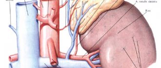
Fig.1. The location and blood supply of the adrenal gland is normal
Primary hyperaldosteronism (PHA) is a disease in which excess aldosterone production is autonomous, independent of the influence of various regulatory factors - in contrast to the secondary increase in aldosterone secretion that accompanies a number of diseases. Primary hyperaldosteronism is considered as an independent nosological form, while secondary hyperaldosteronism causes only a number of specific symptoms in diseases of the kidneys, liver and other organs of various etiologies and pathogenesis. There are no exact data on the frequency of PHA, since many patients do not come to the attention of endocrinologists. However, such patients make up a significant portion of those suffering from arterial hypertension.
Etiology and pathogenesis of aldosteroma - primary hyperaldosteronism
Morphologically, the substrate of primary hyperaldosteronism is an adrenal tumor, less commonly hyperplasia of the adrenal cortex. According to J.Conn (1955,1964), single adenomas (aldosteromas) occur in 70-90% of patients, multiple ones - in 10-15%, adrenal hyperplasia - 9%. In general, tumor lesions of the adrenal glands, as the cause of primary hyperaldosteronism, are observed in 85% of cases, and in 2-6% of patients they are malignant.
According to modern concepts, the prevalence of primary hyperaldosteronism (PHA) in patients with hypertension is 10-17% and up to 30% in secondary (symptomatic) hypertension. Aldosterone-producing adenoma is detected in 10-15% of all forms of PHA. It should be noted that in case of accidentally detected tumors, the incidence of aldosteroma is extremely low and does not exceed 1%, however, the largest number of unjustified adrenalectomies is associated with overdiagnosis of aldosteroma.
Primary hyperaldosteronism occurs mainly in adults. Aldosteroma in most cases is a single, clearly defined encapsulated tumor weighing 1-3 g, with a diameter of up to 3 cm. On a section, it is ocher-yellow, ocher-golden-yellow in color with a smooth surface.
Cushing's syndrome
The clinical picture of Cushing's syndrome is the result of pathologically increased production of the adrenal hormone cortisol. The vast majority of cases of Cushing's disease are caused by a benign pituitary tumor that causes overproduction of the hormone ACTH. This ACTH then stimulates the production of adrenal hormone, causing cortisol levels in the blood to rise.
In addition, the source of ACTH hyperproduction can also be found in other organs - the so-called ectopic Cushing's syndrome. In addition, adrenal tumors—benign adenomas or malignant carcinomas—can directly produce cortisol.
Symptoms
The signs and symptoms of Cushing's syndrome can vary depending on the level of excess cortisol.
Common signs and symptoms of Cushing's syndrome:
- Weight gain and fat deposits, especially around the middle and upper back, on the face - a moon face and between the shoulders - a buffalo hump;
- pink or purple stretch marks on the skin of the abdomen, thighs, chest and arms;
- thin, fragile skin that bruises easily;
- slow healing of cuts, insect bites, and infections;
- acne.
Signs and symptoms in women with Cushing's syndrome:
- thicker or more visible hair on the body and face;
- irregular or absent menstrual periods.
Signs and symptoms in men with Cushing's syndrome:
- decreased sex drive;
- decreased fertility;
- erectile disfunction.
Other possible signs and symptoms of Cushing's syndrome:
- severe fatigue;
- muscle weakness;
- depression, anxiety and irritability;
- loss of emotional control, mood swings;
- cognitive difficulties – decreased memory, mental performance;
- high blood pressure;
- headache;
- darkening of the skin;
- bone loss, which eventually leads to fractures;
- loss of muscle mass – myopathy;
- growth disturbance in children.
Diagnostics
If the clinical picture suggests Cushing's syndrome, then it is necessary to prove the presence of the disease using laboratory tests. There are currently three standard tests for this. At least one of these is used, and often several.
The dexamethasone test attempts to suppress the body's production of cortisol by taking the pills in the evening. If a blood test the next morning shows that the body continues to produce too much cortisol, then this can be interpreted as a sign of Cushing's syndrome. The same applies if the excretion of cortisol in 24-hour urine or cortisol in saliva or blood increases significantly late in the evening.
Once Cushing's disease is proven, a differential diagnosis is made using additional blood tests - the CRH test, a high-dose dexamethasone test, to confirm that the cause of the disease can be found in the pituitary gland. This is true about 80% of the time.
In a minority of patients, the cause may be the adrenal glands themselves - adrenal Cushing's syndrome, or a collection of cells outside the adrenal gland and pituitary gland - ectopic Cushing's syndrome. Cushing's adrenal gland is easy to diagnose, but it can sometimes be difficult to distinguish a tumor in the pituitary gland from tumors in other parts of the body.
If the image does not show a tumor in the pituitary gland and the test results are inconclusive, a so-called selective catheter is used. Under X-ray guidance, a tiny catheter tube is inserted into the blood vessels near the pituitary gland, and testing the blood taken from there can determine whether the pituitary gland is overactive or not.
Clinical picture of aldosteroma - primary hyperaldosteronism
Clinical signs of primary hyperaldosteronism are represented by three main syndromes: cardiovascular (hypertensive), neuromuscular and renal.
Cardiovascular (hypertensive) syndrome of aldosteroma - primary hyperaldosteronism
Arterial hypertension is one of the most typical symptoms of primary hyperaldosteronism. It is observed in almost all patients with primary hyperaldosteronism, and in some cases it may even be the only clinical manifestation of this disease (Conn J. et al., 1964). Arterial hypertension is usually permanent. However, in some patients the increase in blood pressure takes the form of crises; Transient arterial hypertension is also possible.
Blood pressure levels range from moderately elevated numbers (150-160 and 90-100 mmHg) to very high (250-280 and 130-140 mmHg. The pathophysiological basis of such hypertension is sodium retention in tissues and hypervolemia, intimal edema, decreased vascular lumen, significant increase in peripheral resistance.In addition, the high sodium content in the walls of blood vessels and the depletion of cells in potassium increase the sensitivity of the vessels to the effects of pressor factors.
Neuromuscular syndrome of primary hyperaldosteronism
Disorders of the neuromuscular system in patients are caused mainly by hypokalemia, changes in the concentration gradients of extra- and intracellular potassium, potassium-sodium coefficient, intracellular acidosis, and ultimately - dystrophic changes in muscle and nervous tissues. Patients, as a rule, complain of pronounced muscle weakness, which is observed in 70% of cases. The degree of myasthenia gravis varies from moderate fatigue and fatigue to pseudoparalytic conditions. Myasthenia gravis can either be widespread or affect a specific muscle group, most often the lower extremities. Chvostek's paresthesias have been described (Weinberger M. et al., 1979)
Renal aldosteroma syndrome - primary hyperaldosteronism
Hypokalemia and hypernatremia are also the main cause of the development of kalipenic nephropathy, which is manifested by a decrease in the concentrating ability of the kidneys, polyuria, isohyposthenuria, nocturia, increased thirst and polydipsia.
4.Treatment
Obviously, the therapeutic strategy in each case is determined by the diagnosed causes of hyperaldosteronism. Thus, for unilateral aldosteroma, the method of choice is laparoscopic adrenalectomy; The effectiveness of surgical intervention in this case is very high - in almost all patients, blood pressure and other aldosterone-related indicators gradually normalize.
In other cases, conservative treatment may be preferable, the main goal of which is to prevent severe complications caused by hypertension and hypokalemia. Special potassium-sparing drugs, antihypertensive drugs, ACE inhibitors, and a special diet are prescribed. In some cases, hormonal therapy (dexamethasone, hydrocortisone) is indicated and effective. Necessary measures are also the normalization of body weight and therapeutic exercises in the air.
Diagnostic algorithm for identifying aldosteroma - primary hyperaldosteronism
Currently, the aldosterone-renin ratio (ARR) is recommended as a screening method for PHA. Numerous studies confirm the diagnostic superiority of ARS in comparison with separately used methods for determining the level of aldosterone or potassium (both indicators have low sensitivity), renin (low specificity).
A prerequisite for diagnosing PHA is the presence of arterial hypertension (AH) in the patient! In case of adrenal incidentaloma in the absence of hypertension, the determination of ARS is not indicated!
The reliability of ARS as the main laboratory test in the diagnosis of PHA, even limited by strict indications, is still debatable.
This is due to the poor reproducibility of this test, as well as to many factors influencing the CA/ARP ratio - drug intake, patient position, time of blood sampling, salt diet, etc. It is mandatory to discontinue all medications (especially mineralcorticoid receptor antagonists and diuretics) that affect the level of aldosterone and plasma renin activity for 4-6 weeks before the study. To correct hypertension during the diagnostic period, alpha-blockers (doxazosin) and non-dihydropyridine calcium channel blockers (verapamil) are recommended. Despite the fact that APC is a widely used test for the primary diagnosis of PHA, there are significant variations in the critical range of diagnostic values, associated both with the use of different units of measurement and with the lack of standardization of the test, which makes it difficult to formulate uniform diagnostic values for PHA.
3.Symptoms
The classic symptoms of primary aldosteronism include a persistent increase in blood pressure in combination with a deficiency of potassium in the blood and a relative lack of calcium. Patients complain of fatigue, headaches and heart pain, cramps and numbness in various muscle groups. In many cases, there is polydipsia and polyuria (respectively, constant unquenchable thirst and increased volume of urination), symptoms characteristic of nephrogenic diabetes insipidus, which in this case develops as a severe complication of the kidneys.
Diagnosis and differentiation from similar diseases may require a thorough and lengthy examination, both laboratory and instrumental. In particular, if a familial form is suspected, complex genetic tests (PCR) are prescribed; in more typical cases, various stress tests, daily urine tests, and biochemical blood tests are performed. The condition of the adrenal glands (shape, size, histological homogeneity, etc.) is examined using tomographic methods, ultrasound, scintigraphy; If diagnostically necessary, contrast angiographic studies may be prescribed in combination with an analysis of the hormonal pattern in the blood of the adrenal veins.
About our clinic Chistye Prudy metro station Medintercom page!
Treatment of aldosteroma - primary hyperaldosteronism
Treatment of primary hyperaldosteronism caused by an aldosterone-secreting adenoma of the cortex of one of the adrenal glands is surgical. Medication methods are indicated for mild forms of the disease caused by bilateral idiopathic hyperplasia of the adrenal cortex, as well as during the period of preparing patients for surgery.
Surgical treatment of primary hyperaldosteronism caused by aldosteroma is the only radical treatment method and consists of removing the aldosteroma (partial with organ preservation or unilateral adrenalectomy).
Watch a video of operations for adrenal tumors performed by Professor K.V. Puchkov. You can visit the website “Video of operations of the best surgeons in the world.”
Patients, as a rule, do not require special preoperative preparation or hormone replacement therapy in the early postoperative period. The choice of surgical approach, operation, anesthesia and intensive care in patients with aldosteroma do not have any specific features.
In case of aldosteronism caused by diffuse nodular hyperplasia of the adrenal cortex, the indications for surgery have not yet been clearly developed and the existing opinions of surgeons are ambiguous.
Forecast
For aldosterone-producing adenoma, surgical treatment brings recovery in 50-60% of cases. The prognosis is favorable if the operation is performed in a timely manner before the onset of irreversible changes in the cardiovascular system and kidneys. The criteria for the effectiveness of the operation are considered to be normalization of blood pressure (or its control with a small dose of antihypertensive drugs), potassium, aldosterone and renin levels, and the absence of tumor recurrence. With hyperaldosteronism that has developed against the background of diffuse hyperplasia of the adrenal cortex, it is not possible to achieve recovery. To achieve remission, the patient requires constant use of spironolactones, and in some patients, steroidogenesis inhibitors.
The prognosis of secondary hyperaldosteronism depends on the severity of the specific disease against which it developed and its treatment. Thus, treatment of liver cirrhosis and chronic heart failure is sometimes difficult and must be carried out constantly to compensate for these conditions. The prognosis for Bartter syndrome is serious. Without lifelong treatment and correction of electrolyte imbalances from an early age, death is possible due to dehydration, hypokalemia (cardiac arrest) and secondary infections.
In childhood, there is a progression of Bartter's syndrome and the development of tubulointerstitial nephritis , against the background of hypokalemia and increased renin levels, with impaired renal function up to renal failure. Continuous treatment with indomethacin and potassium supplements improves the prognosis. With adequate therapy, children develop, grow, and are seen by a nephrologist as adults.


