1.Structure of the esophagus
Esophagus
- a muscular tube connecting the throat (pharynx) to the stomach. The length of the esophagus is about 20 centimeters, and its inner surface is a pink mucous membrane. There is a part of the esophagus called the upper esophageal sphincter (UES). This is a group of muscles at the top of the esophagus that work during breathing, eating, belching and vomiting. A person can consciously control this muscle group. The upper esophageal sphincter muscle also holds food and secretions that may come from the windpipe.
The lower esophageal sphincter (LES) is a group of muscles at the lower end of the esophagus where it connects to the stomach. When the lower esophageal sphincter is closed, it prevents acid and stomach contents from moving back out of the stomach. The muscles of the LES are not subject to conscious control.
Diseases of the esophagus
, in fact, not so little. Some of them are quite serious, some do not require complex and long-term treatment. Depending on the severity of the disease, it may be treated by gastroenterologists or thoracic surgeons, who perform operations on the esophagus when necessary for treatment.
A must read! Help with treatment and hospitalization!
Structure and functions
The esophagus connects the pharynx to the stomach. In the initial part of the gastrointestinal tract , in the oral cavity, food is crushed, moistened with saliva, and partially digested. It is further digested in the stomach. And the esophagus serves to mechanically move food.
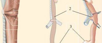
The organ is a hollow elastic tube. This tube consists of three shells:
- Internal . Mucous membrane with submucosal layer. Serves to moisturize and smoothly move the esophageal lump.
- Average . The muscular layer, in turn, is represented by two layers - the outer longitudinal, and the inner circular or circular. Contraction of smooth circular muscles leads to a narrowing of the esophageal lumen, and contraction of longitudinal muscles, on the contrary, leads to its expansion. The cooperative work of smooth and transverse muscles provides peristalsis, wave-like contractions of the esophagus to move food along. In addition, the esophagus has two sphincters, the upper pharyngeal and lower cardiac muscle valves. Both sphincters, closing, isolate the esophagus from the pharynx and stomach.
- Outdoor _ Adventitial membrane. It is represented by loose connective tissue, and externally covers the cervical and thoracic parts of the esophagus. The abdominal region is covered with peritoneum.
The esophagus originates in the neck at the level of the VI-VII vertebrae, extends through the chest cavity, and enters the abdominal cavity through an opening in the diaphragm. In this regard, there are 3 sections of the esophagus:
- Cervical . From VI-VII cervical to II thoracic vertebra. Length 5-8 cm.
- Chest . The main section is 15-18 cm long, extending to the entrance to the diaphragm.
- Abdominal . From the diaphragmatic opening to the entrance to the stomach. Length 1-3 cm.
The total length of the esophagus in an adult is 25-30 cm. The diameter of the internal lumen is 2-2.5 cm, but can expand to 4-4.5 cm. There are three anatomical narrowings in the esophagus. Their localization:
- Transition of the pharynx into the esophagus.
- Level V of the thoracic vertebra, corresponding to bifurcation or bifurcation of the trachea into two bronchi.
- Passage of the esophagus through the diaphragm.
Partial or complete obstruction of the esophagus for food, as a rule, forms in the area of these narrowings.
2. The most common diseases of the esophagus
- Heartburn
. Heartburn occurs when the lower esophageal sphincter does not close completely. As a result, the acidic contents of the stomach enter the esophagus. This is called reflux. This reflux may cause heartburn, coughing, or hoarseness, or cause no symptoms at all. - Gastroesophageal reflux disease (GERD)
. If reflux occurs frequently or is accompanied by unpleasant symptoms, it is called GERD. - Esophagitis
. Esophagitis is inflammation of the esophagus. Esophagitis can be associated with irritation of the esophagus, either as a result of reflux or infection. - Barrett's esophagus
. Regular reflux of stomach acid causes irritation of the esophagus, which can result in changes in the structure of its lower part. In very rare cases, Barrett's esophagus progresses to esophageal cancer. - Esophageal ulcer
. With an esophageal ulcer, erosions form in the mucous membrane of the esophagus. This is often the cause of chronic reflux. - Esophageal strictures, or narrowing of the esophagus
. Chronic irritation from reflux is a common cause of esophageal strictures. - Achalasia of the esophagus
. Achalasia is a rare condition in which the lower esophageal sphincter does not relax. Difficulty swallowing and regurgitation of food are the most common symptoms of the disease. - Esophageal carcinoma
. Esophageal cancer is a serious disease, which, however, is not so common. Risk factors for the development of esophageal cancer are smoking, alcoholism and chronic reflux. - Mallory-Weiss syndrome, or gastroesophageal rupture-hemorrhagic syndrome
, occurs when tears occur in the surface of the esophagus due to frequent vomiting. Such ruptures are accompanied by internal bleeding and subsequent vomiting of blood. - Varicose veins of the esophagus
. In people with cirrhosis, the veins in the esophagus may become enlarged and protruding. These veins can cause life-threatening bleeding. - Ring-shaped formations in the lower parts of the esophagus
. This is a benign collection of tissue in the form of a ring around the lower end of the esophagus. Typically, these rings do not cause any symptoms, but in some cases they can cause difficulty swallowing. - A collection of tissue at the top of the esophagus
. A disease that develops in a similar way to ring-shaped formations in the lower part of the esophagus, which also usually does not cause unpleasant symptoms. - Plummer-Vinson syndrome
. This is a disease of the esophagus accompanied by chronic iron deficiency anemia, ring-shaped formations in the upper part of the esophagus and difficulty swallowing. Iron therapy and expansion of esophageal tissue are the main methods of treating the disease.
Visit our Gastroenterology page
Treatment of Barrett's esophagus using minimally invasive technologies: laparoscopy and endoscopy.
My experience in treating patients with hiatal hernia and reflux esophagitis of varying severity is 24 years. During this time, I was able to successfully operate and treat more than 2,000 patients laparoscopically. In 10% of them, Barrett's esophagus was identified, which we successfully treated in 97% using our comprehensive approach of minimally invasive treatment.
It is worth noting that only as a result of a clear and complete examination can we obtain sufficient information about the patient. If Barrett's esophagus is detected on endoscopic examination and confirmed by histological tests, the question of mandatory treatment is raised, since the risk of degeneration into cancer is very high.
Puchkov K.V., Filimonov V.B. Hiatal hernia: monograph. - M.: MEDPRACTIKA - M., 2003. - 172 p.
At this stage, the results of cytological and histological studies help to accurately select the method of surgical treatment. We have already said that Barrett's esophagus is characterized by hyperkeratic, metaplastic and dysplastic changes in the esophageal mucosa. Consequently, when hyperkeratosis, gastric, small intestinal and colonic metaplasia, mild and moderate dysplasia are detected, we are talking only about a benign process. In the case of severe dysplasia or when squamous cell non-keratinizing cancer is detected, a diagnosis of cancer is made. This allows us to clearly distinguish between two types of treatment - in the first case, organ-preserving and in the second case, organ-sapping (Lewis operation).
Immunohistochemical examination, along with histological examination of biopsy specimens, allows us to identify early forms of esophageal adenocarcinoma. Esophageal cancer is characterized by an increase in the expression area of the Ki-67 marker and the anti-apoptotic factor bcl-2 compared to values in gastroesophageal reflux disease and Barrett's esophagus. Thus, the differential diagnosis of Barrett's esophagus and esophageal adenocarcinoma can be further achieved by studying the results of immunohistochemistry of esophageal cells producing nitric oxide synthase and endothelin-1.
If, as a result of endoscopic examination, histological and immunohistochemical studies, severe dysplasia and adenocarcinoma of the esophagus are not detected, we should only talk about organ-preserving treatment of Barrett's esophagus. The stages of this algorithm will be described by me below.
In the absence of a hiatal hernia, especially in young patients, we use radiofrequency ablation (RFA) of Barrett's esophagus. In the postoperative period, therapy with proton pump blockers (suppression of gastric secretion) and motilium (improvement of gastric motility) is prescribed for 6-8 weeks. Dynamic observation - gastroscopy - is carried out for several months. If a relapse of the disease occurs, this procedure can be repeated, since the effect is only within the mucous membrane, without causing damage to the entire thickness of the esophageal wall.
In the presence of a hiatal hernia, the first stage requires surgical intervention - laparoscopic Tope fundoplication (bilateral fundoplication at 270 degrees) and crurorrhaphy. This intervention eliminates the hernia and stops the pathological reflux of aggressive stomach contents into the esophagus. Without this stage, further treatment of Barrett's esophagus, as a rule, does not bring a positive result. In the postoperative period, for 3-4 months, therapy with proton pump blockers is prescribed.
This stage allows in 90% of cases to stop pathological reflux into the esophagus and thereby stop the pathological transformation of the mucous membrane and the reverse restructuring of the esophageal epithelium.
After surgery, after 2-3 months, it is necessary to perform radiofrequency ablation (RFA) of the affected mucosa, and then conduct follow-up examinations of the esophagus (FGS) after 3 and 6 months. In case of positive dynamics, further observation is carried out 1-2 times a year.
In a number of patients with long-term changes in the mucosa (long segment of Barrett's esophagus), an additional 1-2 sessions of RFA are necessary.
If, upon re-evaluation of the condition of the esophageal mucosa, according to FGS, the damage to the mucosa decreases, then we carry out further dynamic observation, performing FGS at intervals of 6 months. After 1 year, a final assessment is made for the presence or absence of Barrett's esophagus. In most patients, manifestations of Barrett's esophagus were reversed (especially in patients with hiatal hernia and acid reflux). If morphologically, during biopsy, we see persistence of changes in the mucosa (metaplasia), then we move on to the next stage - endoscopic (RFA) or argon plasma coagulation of the esophageal mucosa in the area of Barrett's esophagus.
Radiofrequency ablation and argon plasma coagulation are performed under endoscopic control, usually under general anesthesia (short-term intravenous sedation). For lesion segments up to 3 cm, one session is usually sufficient. If the lesion is more than 3 cm, a repeat procedure may be necessary. After radiofrequency ablation, the damaged tissue is replaced by squamous epithelium and heals without scarring. In our practice, we prefer to use RFA, which in principle is an improved version of the argon plasma coagulation technique, as it has fewer side effects.
It is worth noting that if areas of high-grade neoplasia were detected in the metaplasia zone, in which the likelihood of invasive growth increases significantly, radiofrequency ablation can also be performed. For deep lesions, endoscopic radical removal of a section of the esophageal mucosa is performed. If the area of changes is up to 2 cm2, endoscopic resection of the esophageal mucosa (EMR-C) is usually performed, and if the area of changes is more than 2 cm2, dissection of the neoplasm in the submucosal layer (ESD) is performed.
Next, lifelong clinical observation is mandatory - during the first year after 3 months, then FGS is performed once a year with a possible biopsy of the mucous membrane of suspicious areas.
It is extremely rare that after such complex and staged treatment, relapses of Barrett's esophagus occur (less than 5%), as a rule, this occurs with relapse of the hiatal hernia and remnants of altered mucosa after RFA.
A number of authors perform surgical treatment in the reverse order - at the beginning of RFA, and then laparoscopic fundoplication. It is worth noting here that the optimal period for performing laparoscopic surgery on the esophagus is 3-4 months after the first stage. Thus, according to ultrasound data of the esophagus, swelling of its wall persists for a very long time (against the background of RFA) and fundoplication performed at an earlier date can lead to the development of stenosis in the area of the esophagogastric junction. Naturally, the healing of the mucous membrane in the esophagus will take much longer, since the reflux of aggressive contents from the stomach remains uncorrected. That is why we use reasonable consistency in our work. First of all, the pathogenetic stage of stopping reflux and only after that is radiofrequency ablation of the altered mucosa. As I wrote earlier, in some patients the second stage is not required, since the mucous membrane returns to normal thanks to its own reparative processes.
Thus, laparoscopic fundoplication at the first stage of treatment makes it possible to reduce the size of the affected segment of the esophagus, which in the future may require only 1-2 sessions of RFA and, accordingly, a lower risk of developing esophageal stricture, and in some patients the second stage can be completely avoided.
3.Diagnosis of diseases
Of course, depending on the symptoms of which disease the doctor sees during the initial examination and consultation, methods for further diagnosing diseases of the esophagus will be selected individually. Let's talk about some of them:
- Upper endoscopy, FGDS (esophagogastroduodenoscopy)
. In this procedure, a flexible, thin tube with a camera on the end (endoscope) is inserted into the esophagus through the mouth. An endoscope allows you to examine the stomach and duodenum (small intestine). - pH monitoring of the esophagus
. A probe that monitors pH levels is inserted into the esophagus. This method is used to diagnose GERD and monitor the progress of GERD treatment. - X-ray examination
with preliminary administration of drugs containing barium. This method is usually used to determine the causes of difficulty swallowing. - Biopsy
. A method for diagnosing diseases of the esophagus, in which a sample of esophageal tissue is taken using endoscopy, which is then examined under a microscope. - Confocal laser endomicroscopy
. This is a new procedure in which a microscope is inserted into the patient's esophagus. Endomicroscopy can be a good alternative to biopsy.
About our clinic Chistye Prudy metro station Medintercom page!
Diagnostics
The simplest diagnostic method is radiography of the esophagus with barium contrast. The patient swallows a suspension of barium sulfate, and the doctor uses an X-ray machine to evaluate how it is progressing.
A more reliable method is FGDS (fibrogastroduodenoscopy). Using endoscopic equipment, the doctor assesses the condition of the mucous membranes of the esophagus, stomach, and duodenum.
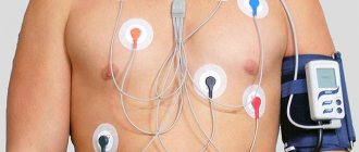
Modern methods of studying the esophagus are esophagomanometry and impedance pH-metry. These studies use probes equipped with sensors and measuring devices. After introducing these probes into the esophageal lumen, the contractility of the esophageal muscles, intraesophageal pressure, and acidity (pH) of the intraesophageal environment are determined. These methods are used to diagnose dyskinesia and gastroesophageal reflux.
4. Treatment of diseases of the esophagus
Just like the diagnosis of esophageal diseases, treatment depends on what specific disease is diagnosed. Among the treatment methods include:
- Use of H2 blockers
. The release of acid in the stomach is stimulated by histamine. Some antihistamines are called H2 blockers. They can reduce acid content and improve the condition of a patient with GERD and esophagitis. - Proton pump inhibitors
. These medications block many of the acid production processes in the stomach and also help with GERD. In addition, inhibitors help heal ulcers or esophagitis. - Esophagectomy, or removal of the esophagus
. This is a surgical procedure performed by thoracic surgeons. As a rule, it is performed for esophageal cancer. - Dilatation of the esophagus
. A special device is passed down the esophagus and then expands on its own, causing the esophageal ring, stricture, and other formations that prevent swallowing to widen. - Ringing of esophageal varices
. This procedure is performed endoscopically and involves wrapping rubber band-like devices around the esophageal varices. This reduces the chance of bleeding.