Esophageal cancer is rare, affecting just over 7.5 thousand Russians annually, or 8-9 people out of 100 thousand people, mostly elderly people. The highest rates, twenty times higher than in Russia, were recorded in China, Korea, Japan, Mongolia, Iran and Brazil.
In the structure of male cancer incidence, esophageal cancer accounted for 2.5%, while among female cancers it accounted for only 0.5%. This is not a female disease at all; men get sick almost four times more often and start getting sick earlier. In the male cohort, the average age of detection of cancer of the esophagus was 64 years, while in the female cohort it was after 70 years.
- Risk factors
- Clinical picture
- Diagnostics
- Treatment of esophageal cancer
- Palliative treatment of advanced esophageal cancer
Causes and risk factors of the disease
Malignant degeneration of cells of the esophageal mucosa can be provoked by many factors. Conventionally, they can be divided into several groups.
- Alimentary (food)
. This is the habit of eating too hot or too cold food, an excess of spicy dishes, pickles, marinades, foods with molds on the menu, a lack of fruits and vegetables, vitamin deficiency, especially a lack of vitamins A, B, E. - Bad habits
– smoking and alcohol. - Occupational hazards and exposure to chemicals
. This includes both accidental burns of the esophagus with caustic substances and constant exposure to harmful substances at work (working in poorly ventilated areas, inhaling toxic gases, industrial dust) or at home (living in areas with polluted air, frequent smoke due to fires). - Various diseases
. The development of neoplasms can be triggered by obesity, gastroesophageal reflux, hiatal hernia, esophageal achalasia, and Barrett's disease. The last two pathologies greatly increase the risk of tumor development. - Hereditary predisposition
.
Symptoms
At the latent stage of the disease, there are no symptoms.
The first symptoms appear due to tumor growth:
- Pain in the area where the tumor is located;
- Presence of blood clots;
- The appearance of constipation;
- Growth of lymph nodes;
- Weight loss;
- Decreased hemoglobin;
- Fatigue;
- Low ability to work;
- Bad dream.
At the stage of intensive growth of cancer cells and the appearance of metastases, the severity of symptoms increases.
Let's look at the symptoms of the formation of adenocarcinoma in various organs.
Intestines
- Painful stomach;
- Unpleasant sensations after eating;
- Poor intestinal permeability;
- Loose stools alternating with constipation;
- Blood and mucus in the stool.
Esophagus
- Pain when swallowing;
- Dysgaphia;
- Active salivation.
Nasal cavity
- Swelling of the tonsils;
- Persistent inflammation of the tonsils;
- Pain in the larynx, pharynx, nose;
- Unpleasant sensations when swallowing;
- Ear pain;
- Speech impairment;
- Enlarged lymph nodes.
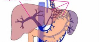
Liver
- Pain in the area of the right rib and hypochondrium;
- The eyes and skin are yellowish.
Symptoms of esophageal cancer
Patients often seek medical help when they have difficulty swallowing food. By this time, the tumor has already reached a significant size, which significantly worsens the survival prognosis. However, there are also early signs of the disease that are also characteristic of other pathologies. It is important to consult a doctor as soon as they appear for a timely diagnosis and initiation of treatment.
If the tumor is located in the upper or middle part of the esophagus, the first symptoms will be choking and discomfort (burning, rawness, slight pain) when swallowing solid food. When a tumor develops in the lower part, at the junction of the esophagus with the stomach, an early sign is constant regurgitation of air. Early signs of esophageal cancer, characteristic of tumors of all its parts, include dyspepsia - belching, heartburn, nausea.
Late signs of a malignant neoplasm of the esophagus are symptoms of dysphagia, or difficulty swallowing.
They are divided into degrees II to V (I refers to early signs).
- II – difficulty swallowing solid food; in order to swallow, food must be washed down with water. To facilitate the passage of food, profuse salivation occurs. Regurgitation of saliva and mucus and esophageal vomiting may occur due to food retention above the site of narrowing.
- III – inability to swallow solid food, regurgitation when trying to swallow. Patients can eat only liquid and semi-liquid foods.
- IV – only liquids can be swallowed.
- V – complete obstruction of the esophagus. Patients cannot swallow water or saliva.
As the symptoms of dysphagia develop, due to the stagnation of food in the area of constriction, its decomposition occurs, accompanied by local inflammatory changes, putrid odor from the mouth and pain appear, first periodic, then constant. Minor or heavy bleeding may occur. The accumulation of food can lead to its reflux into the respiratory tract (this mainly happens at night) and aspiration pneumonia. In the later stages, there is a risk of fistulas between the esophagus and trachea, mediastinum. Due to difficulty swallowing, patients limit themselves to food, lose weight, and even become exhausted.
Rehabilitation
In the first few days after surgery, the patient is usually prescribed intravenous nutrition, or nutritional mixtures are introduced into the stomach through a thin tube. Routine pain relief is performed to facilitate the recovery process. An important element of clinical recommendations after esophageal cancer is therapeutic and breathing exercises, which are first performed in bed, and then in a sitting and upright position. A bed with a raised headboard prevents the development of reflux. As the patient recovers, he is allowed to eat normally while following a special diet.
Types of esophageal cancer
Classification of pathology is carried out according to different criteria.
According to the location of the tumor and metastases:
- Cancer of the cervical (upper) esophagus
. It is diagnosed less frequently than other types, in no more than 10% of cases. Gives early metastases to the mediastinum, cervical tissue and supraclavicular space. Metastases also spread to the lymph nodes of the neck, subclavian, mediastinal and paratracheal. - Cancer of the thoracic (middle) region
. The most common, it accounts for 60% of all malignant neoplasms of the esophagus. Early metastases are detected in the surrounding tissue, lymph nodes of the mediastinum, and the lesser omentum. Late - in the bronchi, lungs, liver. - Abdominal (lower) cancer
. Accounts for 30% of all cases of esophageal cancer. Early metastases are found in the subphrenic, paraesophageal, pericardial lymph nodes, in nodes located along the left gastric artery and the lesser curvature of the stomach. Late metastases are detected in the bones and liver.
All types are characterized by Virchow's metastases
– late metastases to the lymph nodes of the left supraclavicular region.
By direction of growth and external structure:
- Endophytic (ulcerative) form
. Its share is about 30% of all cases of esophageal cancer. It grows deep into the organ, causing spasm and stenosis. The endophytic form is divided into: saucer-shaped cancer - a ring-shaped tumor formation with raised, well-defined edges and ulceration in the middle; - ulcerative cancer - the neoplasm is not delimited by a ridge from the adjacent tissue, spreading to it in the form of “tongues” or “daggers”.
. It accounts for about 60% of cases. The neoplasm grows into the lumen of the esophagus. It can cause stenosis, but it will be associated with the filling of the organ lumen with tumor tissue. Divided into:
- polypous cancer - the formation grows in the form of many irregularly shaped nodes, reminiscent of cauliflower, supported by a clearly visible stalk;
. Accounts for about 10% of esophageal cancer cases. It looks like a roughened area of the mucous membrane. It grows in a circle, narrowing the lumen of the esophageal tube.
Mixed esophageal cancer
– one of the forms passes into another. Thus, a polypous tumor can ulcerate and acquire a saucer-shaped shape.
The classification ulcerative, sclerosing and nodular is also used to macroscopically describe the structure of the neoplasm.
According to histological structure:
- Squamous cell carcinoma (carcinoma)
. It develops from stratified squamous epithelium in the upper layer of the esophageal mucosa. The most common - 98% of cases of malignant tumors of the esophagus. It can be keratinizing and non-keratinizing (more aggressive). - Cylindrical cell carcinoma (adenocarcinoma)
. It develops from cylindrical cells of the esophageal glands located in the submucosal layer. It is often secondary, spreading from the stomach. Often and quickly metastasizes. - Sarcoma of the esophagus
. Arises from stromal (connective or muscle) tissue. It can be mixed with other types, extremely malignant, and often recurs. - Rare tumors - mucoepidermoid, small cell carcinoma, melanoma
. Characterized by high malignancy. - Undifferentiated cancer
- the histological appearance of the tumor cannot be determined due to low cell differentiation.
By cell differentiation:
- Highly differentiated cancer
- cells are as similar as possible to healthy ones, but have signs of atypia. The course is slow, the likelihood of a successful outcome is greater. Gives single metastases and responds well to therapy. Moderately differentiated. The intermediate form is characterized by moderate malignancy. Relatively favorable prognosis with timely diagnosis. - Poorly differentiated
- abnormal, unequal structure of cells (polymorphism), which quickly divide and grow. It is highly malignant. - Undifferentiated cancer
is the most aggressive and is often secondary. In this situation, the severity of the condition is determined by the existing primary focus.
What's happened
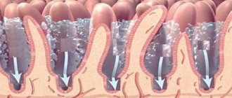
Adenocarcinoma is a malignant tumor developing from secretory glandular epithelial cells. It is localized on various internal organs, on the internal membranes of hollow organs, as well as human skin.
A distinctive feature of adenocarcinoma is the ability to produce secretions.
Such tumors can have a variety of sizes and shapes, which are directly dependent on the cellular and tissue functions, cellular and tissue structure of the organ that was affected.
Grades of esophageal cancer
Staging takes into account the depth of tumor penetration, the presence of regional and distant sites of metastasis.
| I | neoplasm in the mucous membrane and submucosal layer. The lumen of the esophagus is normal. There are no metastases. |
| II | The tumor, in addition to the mucous and submucosal layers, affects muscle tissue. The lumen of the esophagus is slightly reduced. Individual metastases to regional lymph nodes are possible. |
| III | the tumor spreads to the outer shell without affecting neighboring organs. The lumen of the esophagus is severely stenotic. There are numerous metastases in the regional lymph nodes. |
| IV | the neoplasm penetrates all layers of the esophagus and spreads to nearby organs. Regional and distant metastases are detected. |
The international classification uses the designation TNM, where T (x, 0, Is, 1, 1a, 1b, 2, 3, 4a, 4b) is the primary tumor, N (x, 0, 1, 2, 3) is regional lymph nodes, M (0.1) – distant metastases.
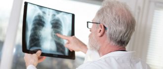
Diagnosis of esophageal cancer
During the initial visit, the doctor will collect an anamnesis of the patient’s life and illness (bad habits, occupational hazards, complaints, when the first symptoms appeared), conduct a visual examination, palpation of the lymph nodes, and give a referral for general blood tests, urine tests, and biochemical blood tests.
Additionally, the following studies are prescribed:
- endoscopic ultrasound examination
of the esophagus is usually combined with taking a biopsy; - radiography
with contrast - to identify narrowing of the esophagus; - computed tomography
with contrast enhancement - to detect metastases in regional lymph nodes, distant metastases; - positron emission tomography
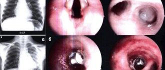
– to confirm the metastatic nature of enlarged lymph nodes; - bronchoscopy
– to exclude the penetration of the process into the bronchi; - thoracoscopy, ultrasound of the abdominal cavity
- to assess the involvement of the organs of the chest cavity and gastrointestinal tract in the process, detection of metastases; - blood test for tumor markers
.
Observation
Active surveillance is indicated for patients for early detection of relapse in the esophagus for the purpose of surgical intervention or relapse in the mediastinum for the purpose of chemoradiotherapy. The scope of the examination depends on the stage of the disease and previous treatment:
Stage I (after endoscopic mucosal/submucosal resections) and stages II–III (after chemoradiotherapy, with prospect of esophagectomy in case of recurrence):
- EGDS – every 3–4 months. during the first two years, every 6 months. – during the third year, then annually until the total observation period is 5 years.
- CT scan of the chest and abdominal organs – every 6 months. during the first 2 years, then annually until a total observation period of 5 years;
Stage I–III (after surgical treatment):
- CT scan of the chest and abdominal organs – every 6 months. during the first 2 years, then annually until a total duration of 5 years.
Other examination methods are recommended for other categories of patients if clinically indicated. Performing PET/CT and determination of serum markers for monitoring patients is not recommended.
Treatment of esophageal cancer
Therapeutic measures are selected by an oncologist, surgeon, radiologist and other specialists depending on the time of detection of the malignant neoplasm, the location of the tumor, its stage, the absence or presence of metastases, and the general condition of the patient.
If surgical treatment
it is usually combined with radiation therapy. During the operation, either complete excision of the tumor with adjacent tissues and nearby lymph nodes, or partial resection to free the lumen of the esophagus can be performed. Tissue from the small or large intestine is used to replace part of the removed esophagus.
If surgical treatment of esophageal cancer is not possible, radiation therapy
. Also, as a palliative treatment, photodynamic therapy can be used - a light-sensitive element is introduced into the tumor tissue, and it is destroyed by a laser. The procedure is carried out to facilitate swallowing; it cannot completely destroy the tumor.
Chemotherapy
in this type of cancer it is usually used as an auxiliary method to suppress the activity of malignant cells.
After treatment, all patients are registered with an oncologist and undergo regular examinations.
Surgical treatment in the department of thoracoabdominal surgery and oncology of the Russian Scientific Center for Surgery
Treatment in the department is carried out under the programs of compulsory medical insurance, voluntary medical insurance, VMP, as well as on a commercial basis. Read how to get treatment at the Department of Thoracoabdominal Surgery and Oncology of the Russian Scientific Center for Surgery.
To schedule a consultation, call:
+7 (499) 248 13 91 +7 (903) 728 24 52 +7 (499) 248 15 55
Submit a request for a consultation by filling out the form on our website and attaching the necessary documents.
Disease prognosis
As with other cancer pathologies, survival and the possibility of cure depend on the time of detection of the tumor. Esophageal cancer is one of the most difficult to treat. In general, with late diagnosis and lack of therapy, life expectancy from the moment of diagnosis is no more than 5-7 months. If detected early - up to 6-7 years.
The survival prognosis depends on the location of the tumor and the presence of metastases. When carrying out complex therapy (surgical treatment and radiation/chemotherapy), survival rate of more than 5 years is:
- when cancer is detected at stage I – 80-90%;
- on II – 40-50%;
- at III – 5-10%.
New treatment methods are helping to increase the life expectancy of patients. The use of stereotactic radiosurgery (cyberknife) in combination with radiation therapy at a linear accelerator increases survival by 25%. A radical radiotherapy program with differentiated irradiation of the primary tumor, zones of perifocal infiltration, as well as regional metastasis pathways, increases survival for all stages of esophageal cancer by 15%.
For inoperable tumors, radiation therapy is used, which can increase life expectancy to 12 months in 10% of patients.
Statistics
This is one of the most aggressive malignant diseases. Esophageal cancer is the 8th leading cause of death worldwide. According to the International Agency for Research on Cancer, in 2018 the incidence was 7.49 cases per 100,000 people per year, and the mortality rate was 6.62. Calculations by Rosstat of the Russian Ministry of Health say that the incidence is 5.6 cases per 100,000 people. Among men – 9.43 per 100,000, among women – 2.29 per 100,000. The disease is most often diagnosed in the so-called “Asian belt”, that is, from the northern part of Iran, through Central Asia and to the central regions of Japan and China, also capturing Siberia. This is largely due to the peculiarities of the diet of people living in these areas.
Most often (up to 80% of cases), the neoplasm is located in the lower and middle thoracic sections of the esophagus. With a frequency of 10-15% of cases, cancer of the cervical esophagus is diagnosed.
Prevention of esophageal cancer
There is no specific prevention of the disease. Quitting alcohol and smoking, normalizing diet and weight, regular examination of the gastrointestinal tract, and consulting a doctor at the first signs of the disease will help reduce the risk of developing cancer. In patients with gastroesophageal disease, annual endoscopic examination is recommended; in patients with Barrett's esophagus, a biopsy is recommended.
The information in this article is provided for reference purposes and does not replace advice from a qualified professional. Don't self-medicate! At the first signs of illness, you should consult a doctor.
Chemotherapy
Chemotherapy, that is, taking drugs that have an inhibitory and destructive effect on neoplasm cells, can be adjuvant, neoadjuvant, or independent.
Preoperative chemotherapy is especially effective for adenocarcinoma, for which 2-3 courses of treatment are prescribed. Postoperative chemotherapy is usually indicated in conjunction with neoadjuvant chemotherapy and is not recommended without it for adenocarcinoma and squamous cell carcinoma. Completely independent chemotherapy is recommended only for inoperable patients with contraindications to radiological treatment methods.