Cardiac hypertrophy is an increase in heart size. This is a heart condition that can lead to serious problems with the structure and functioning of the heart. The heart enlarges due to the fact that cells, cardiomyocytes, proliferate. They make up about 25% of all cells in this organ.
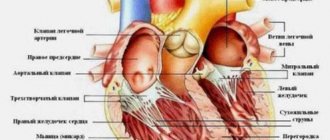
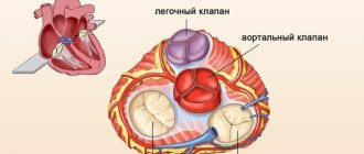
It is known that the heart is a hollow organ consisting of four chambers: the left atrium and ventricle, the right atrium and ventricle. Blood circulates in these chambers. Blood enters the atria and is expelled from the ventricles. In a normal process, the picture will be like this: the pressure in the left side of the heart is always normally higher than in the right chambers. This happens because the chambers of the left side of the heart participate in the systemic circulation, and the right ones participate in the pulmonary circulation.
With heart pathologies, this pattern can change to one degree or another. Doctors note hypertrophy of different parts of the heart chambers, although the incidence of hypertrophy of one or another part of the heart in children varies.
Geneticists are studying the topic of hereditary causes of hypertrophy in children and adults. Previously, it was believed that this disease in patients of different ages was associated with different genetic backgrounds. At this stage, it has been established that mutations can cause hypertrophy in both children and older patients.
of cardiac hypertrophy in children include:
- right ventricular hypertrophy
- left ventricular hypertrophy
- hypertrophy of both atria
- left atrial hypertrophy
- right atrial hypertrophy
Right ventricular hypertrophy
This pathology occurs more often in children than in adults and is associated with an increase and lengthening of muscle fibers as a result of increased functioning. Often the pathology is physiological in nature and is associated with the fact that in the first months of life the load on the parts of the stomach increases. However, more often there is a relationship between hypertrophy and congenital heart defects, which lead to the right ventricle receiving more blood than it can process (with a defect, blood flow may be disrupted). This leads to overload of the right ventricle.
The enlarged myocardium loses its physiological properties. Electrical conductivity is disrupted, which in turn causes arrhythmia, changes in contractility and other disorders.
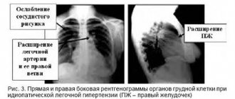
It should be noted that when hypertrophy of the right ventricle , the vessels associated with it are also negatively affected, which ultimately can lead to their sclerotization and disruption of the functioning of the pulmonary circulation.
The main methods for diagnosing the disease are electrocardiography and echocardiography (ultrasound). The first method may indicate a disturbance in the electrical conductivity of the heart and is considered an indirect method. The second method is more informative. It indicates the exact size of the heart, the presence of stenosis, the location of defects, allows you to estimate the pressure in the chambers, etc. These two methods complement each other, because the diagnosis of right ventricular hypertrophy cannot always be confirmed by ultrasound.
Cardiac hypertrophy
Cardiac hypertrophy
Cardiac hypertrophy is an enlargement of the heart muscle, which occurs mainly due to an increase in the number of cardiomyocytes - specialized muscle cells of the heart. This condition occurs in children, adolescents, young adults and the elderly.
Cardiac hypertrophy is a manifestation of a special state of the body: physiological or pathological. That is, it is not a disease, but a symptom.
Physiological
Physiological hypertrophy of the heart is observed in athletes and people who lead an active lifestyle. For regular physical activity, the body requires large amounts of oxygen. Oxygen is delivered through the blood. And to meet the increased oxygen needs, the heart increases the frequency and strength of contractions. And this requires greater metabolism in the heart muscle itself. This gradually increases the volume and mass of cells (cardiomyocytes). More often in athletes, cardiac hypertrophy begins from the left ventricle. Sports that can lead to cardiac hypertrophy are rowing, hockey, football, cross-country skiing, cycling, long-distance running, etc. When you stop training, this condition reverses. That is, the hypertrophied heart again becomes of normal size with normal wall thickness.
Pathological
Pathological hypertrophy of the heart occurs due to various diseases of the body. The human heart consists of four sections: two atria and two ventricles. The atria are reservoirs where blood enters from the body's circulation (blue vessels). The ventricles are a buoyant force that pushes blood through the vessels (red vessels). So each department has its own reasons for increasing.
Causes:
- Left ventricle – enlarges due to arterial hypertension, aortic valve stenosis, aortic atherosclerosis, general obesity, diabetes mellitus
- Right ventricle – due to congestive heart failure, chronic pulmonary failure
- Left atrium – with arterial hypertension, general obesity, aortic and mitral valve defects
- Right atrium - due to pulmonary diseases (when there is stagnation in the pulmonary circulation).
Development
The above reasons force us to maintain normal blood flow by increasing the mass of the heart. It must be taken into account that an increase in one part of the heart leads to hypertrophy of another. In addition to cardiomyocytes, the heart also contains connective tissue. With cardiac hypertrophy, it also grows, and this leads to a decrease in the elasticity of the walls and disruption of the heart.
If the load on the heart does not decrease, then the myocardium is gradually depleted because the blood flow cannot cope with the nutrition of the enlarged heart. This can lead to disruption of nerve impulses (arrhythmia), sclerosis and atrophy of the heart muscle.
Symptoms
- An asymptomatic course of cardiac hypertrophy is possible.
- If the left half of the heart is affected: pain in the heart area (increases after physical activity), arrhythmia, loss of consciousness, shortness of breath, dizziness.
- If the right half of the heart is affected: cough, shortness of breath, cyanosis (cyanosis) or pallor of the skin, swelling, arrhythmia.
Diagnostics
- Ultrasound examination of the heart
- ECG (electrocardiography)
- X-ray of the chest organs.
Treatment It is necessary to eliminate the cause of cardiac hypertrophy.
If it is arterial hypertension, it is necessary to take antihypertensive and diuretic drugs. Severe heart valve defects require surgical treatment and prosthetics. Respiratory diseases require anti-inflammatory and bronchodilator therapy. In any case, the approach is always individual. To monitor blood pressure and early detect arrhythmia, I recommend using automatic tonometers from the manufacturer Microlife, presented in our online store.
The author of the article is a practicing neurologist Maxim Nikolaevich Starshinin.
Left ventricular hypertrophy
With this pathology, the wall of the ventricle thickens. It happens that the septum between the right and left gastric chambers thickens. The thickening may be uneven, which determines the course of the disease.
This is a fairly common condition among childhood heart diseases with the following pathologies:
- arterial hypertension of the systemic blood circulation
- congenital heart defects
- acquired heart defects
- carditis
- myocardial dystrophies
The characteristic signs of left ventricular hypertrophy are heterogeneous. The patient may not know that he has this disease. Genetics note a high probability of transmission of the disease by inheritance, so a competent doctor studies the family history.
Diagnosis of left ventricular hypertrophy in newborns is quite difficult because they have a predominance of right ventricular mass. In modern pediatrics and cardiology, the diagnosis of left ventricular hypertrophy is highlighted as a separate area of study. The most informative are both electrocardiography (ECG) and echocardiography.
Treatment of this disease can be either surgical or medicinal. During the operation, the septum is brought to its normal physiological state.
Symptoms of right ventricular hypertrophy and its degree
In medical practice, right ventricular hypertrophy is quite rare. Moreover, the disease is difficult to diagnose. This is due to the fact that on the electrocardiogram, due to the small mass of the right ventricle compared to the left, the electrical activity of the left suppresses the parameters of the right ventricle. Normally, the left ventricle of the heart weighs three times more than the right.
Therefore, an increase in the size of the right ventricle is detected only when its mass increases many times.
As the right ventricle enlarges, the degrees of its damage are distinguished.
Moderate hypertrophy is observed when the size of the ventricle has a slight increase.
With a slower course of excitation processes compared to the left ventricle and a significant increase in the volume of the right ventricle, the average degree of hypertrophy is determined.
If the mass of the right ventricle significantly exceeds the mass of the left, pronounced hypertrophy of the right ventricle is diagnosed.
At the initial stage of the disease, the symptoms of right ventricular hypertrophy are not obvious and sometimes are completely absent.
As right ventricular growth progresses, symptoms of hypertrophy manifest as abnormal heart rhythms, defined by patients as fluttering in the chest or missed beats.
There is heavy breathing, pain and heaviness in the chest.
Patients often complain of sudden dizziness and loss of consciousness.
A characteristic symptom of right ventricular hypertrophy is swelling of the lower extremities.
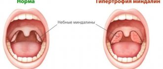
Atrial hypertrophy
A relatively common pathology in children. It is observed in the case of simultaneous violation of the mitral valve and tricuspid valve, with other heart defects that are congenital in nature, as well as with an atrial septal defect. Also, hypertrophy of both atria occurs as a concomitant disorder with carditis.
Doctors note that diagnosing biatrial enlargement (atrial hypertrophy) is more difficult than with isolated atrial defects. As with other types of hypertrophy, the main diagnostic methods are ECG and ultrasound of the heart.
Ventricular myocardial hypertrophy
the myocardium of both ventricles working without disturbances and the synchronous contractility of both chambers. But when the physiologically normal functioning of the cardiac system is disrupted, creating obstacles or changes in the direction of blood flow, it leads to intensive work. As a result of greater load, cardiac hypertrophy , which is combined with simultaneous changes in the myocardium itself. Conductivity and metabolism deteriorate.
The disease is quite common in young children. But diagnosing it, especially in early childhood, is quite difficult. Compared to diagnosing atrial hypertrophy, identifying ventricular myocardial disorders is considered a more difficult task, since it is more difficult to detect asynchrony in the work of the right and left sections.
Atrial hypertrophy
Left atrial hypertrophy occurs in children with the following diseases:
- congenital heart defects (mitral valve insufficiency, left ventricular hypoplasia, transposition of blood vessels, etc.)
- acquired heart defects (mitral valve stenosis and insufficiency, etc.)
- hypertension
- atrial tumors, myocarditis, cardiomyopathy
- mitral valve prolapse
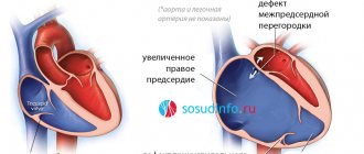
Right atrium hypertrophy , compared with left atrium pathology, is much less common in pediatric patients. The process of enlargement of the right atrium is given rise to diseases such as:
- congenital heart defects (tetralogy of Fallot, Eisenmenger disease, pathology of the interatrial septum and others)
- cor pulmonale
- tricuspid malformations
In conclusion, it should be noted that depending on the degree of pathology and its types, the doctor prescribes either conservative treatment or insists on the need for surgery.
leave a comment
Causes of hypertrophy of the right ventricle of the heart
The most common causes of right ventricular hypertrophy include mitral stenosis or congenital heart disease.
Such pathologies often develop in children with congenital heart defects of various types. For adults, its manifestation is typical for lung diseases complicated by changes in cardiac activity or with valvular heart defects.
Features of the development of hypertrophy and the severity of the course determine various forms of the disease.
The causes of right ventricular hypertrophy are often pulmonary hypertension, which causes increased pressure in the pulmonary artery. In this case, shortness of breath, fainting and frequent dizziness are observed.
Congenital heart disease in children - tetralogy of Fallot, is detected in infants at birth and accompanies it throughout the first year of life. It is caused by a violation of the outflow of blood from the right ventricle and is expressed by the “blue baby” syndrome.
With pulmonary valve stenosis, the movement of blood into the artery from the right ventricle is impaired.
A congenital defect of the interventricular septum leads to mixing of the blood of the two parts and causes hypoxia. As a result, the work of the entire organ is enhanced, incl. of the right ventricle and leads to an increase in its size.
Often the causes of right ventricular hypertrophy are lung diseases such as emphysema and pulmonary fibrosis, pneumonia, chronic bronchitis, bronchial asthma and pneumosclerosis, obesity, alcohol and nicotine.