Lacrimal organs - the system of the adnexal apparatus of the eye, which protects the eyes from drying out, produces tear fluid and drains it into the nasal cavity.
Lacrimal organs are divided into tear-producing ( tear-secretory ) and lacrimal-draining organs .
The tear duct consists of the main lacrimal gland and several additional ones located in the conjunctival sac.
The lacrimal ducts include: lacrimal openings, lacrimal lake, lacrimal stream and other formations along which tears move before entering the nasal cavity.
1
Diseases of the lacrimal organs. Diagnosis and treatment
2 Diseases of the lacrimal organs. Diagnosis and treatment
3 Diseases of the lacrimal organs. Diagnosis and treatment
Embryology
The orbital, or orbital, part of the lacrimal gland is formed in the embryo at the age of 8 weeks. By the time of birth, tear fluid is almost not secreted, because the lacrimal gland is not yet sufficiently developed (in 90% of children, active tear secretion appears only by the 2nd month of life). The formation of the lacrimal ducts begins at 6 weeks. embryonic life. From the orbital end of the nasolacrimal groove, an epithelial cord is immersed in the connective tissue, which is gradually detached from the original epithelial covering of the face and reaches the epithelium of the lower nasal passage by the 10th week. At the 11th week, the cord transforms into a canal lined with epithelium, which initially ends blindly. It opens into the nasal cavity at 5 months. OK. 35% of children are born with the outlet of the nasolacrimal duct closed by a membrane; the membrane usually ruptures in the first days after birth.
At what age does dry eye syndrome most often appear?
Dry eye syndrome can be detected in patients at almost any age. But most often it is diagnosed in patients over 40-50 years old. However, due to environmental changes, the widespread use of technology at work and at home, and the increasing use of contact lenses, the age of patients with detectable dry eye syndrome is becoming younger.
You need to understand! If everyday life is associated with the constant presence of risks or taking medications, the side effects of which disrupt the secretory function of the glands, then regular monitoring by an ophthalmologist is necessary in order to timely correct and take the necessary measures.
Anatomy, histology
Lacrimal gland
(glandula lacrimalis) consists of two parts: the upper, or orbital (orbital part, T.; pars orbitalis), and the lower, or palpebral (agelid part, T.; pars palpebralis). They are separated by a wide tendon of the muscle that lifts the upper eyelid (m. levator palpebrae sup.). The orbital part of the lacrimal gland is located in the fossa of the lacrimal gland of the frontal bone on the lateral superior wall of the orbit (fossa glandulae lacrimalis). The sagittal size of the gland is 10-12 mm, frontal - 20-25 mm, thickness - 5 mm. Normally, the orbital part of the gland is not accessible to external examination. It has from 3 to 5 excretory canaliculi passing between the lobules of the palpebral part of the lacrimal gland and opening in the upper fornix of the conjunctiva (see) on the side at a distance of 4-5 mm from the upper edge of the tarsal plate (upper cartilage of the eyelid, T.). The palpebral part of the lacrimal gland is much smaller than the orbital one and is located below it under the upper fornix of the conjunctiva. Its size is 9-11×7-8 mm, thickness 1-2 mm. A number of excretory canaliculi of the palpebral part of the lacrimal gland flow into the excretory canaliculi of the orbital part, and 3-9 canaliculi open independently. The multiple excretory canaliculi of the lacrimal gland resemble a shower, from the openings of which tears enter the conjunctival sac.
The lacrimal gland is held in place by its own ligaments (lig. suspensorium Soemmerringii, lig. retinens glandulae lacrimalis), attached to the periosteum of the upper wall of the orbit. The gland is also strengthened by the Lockwood ligament and the muscle that lifts the upper eyelid (see Eyelids).
The blood supply to the lacrimal gland is provided by the lacrimal artery (a. lacrimalis), a branch of the ophthalmic artery. The outflow of blood occurs through the lacrimal vein (v. lacrimalis).
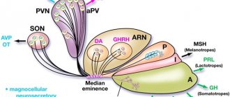
Rice. 2. Schematic representation of the innervation of the lacrimal gland (the path of secretory fibers is indicated in black): 1 - lacrimal gland; 2 - connecting branch of the zygomatic nerve with the lacrimal nerve; 3 - lacrimal nerve; 4 - zygomatic nerve; 5 - pterygopalatine node (switching point of secretory fibers); 6 - greater petrosal nerve; 7 - I branch of the trigeminal nerve (ophthalmic nerve); 8 - II branch of the trigeminal nerve (maxillary nerve); 9 - III branch of the trigeminal nerve (mandibular nerve); 10 - trigeminal node; 11 - facial nerve; 12 - elbow assembly.
The lacrimal gland is innervated by branches of the ophthalmic and maxillary nerve (see Trigeminal nerve), branches of the facial nerve (see) and sympathetic fibers from the superior cervical ganglion. The main role in regulating the secretion of the lacrimal gland belongs to the parasympathetic fibers that are part of the facial nerve (Fig. 2). The center of reflex lacrimation is located in the medulla oblongata (see). In addition, there are a number of vegetative centers, irritation of which increases lacrimation.
The lacrimal gland belongs to the complex tubular serous glands (see), in structure it is similar to the parotid gland (see). Excretory tubules of large caliber are lined with double-layer columnar epithelium, and smaller ones with single-layer cuboidal epithelium. In addition to the main lacrimal gland, there are small tubular accessory lacrimal glands (glandulae lacrimales accesso-riae), located in the fornix of the conjunctiva - Krause's glands (conjunctival glands, T.) and at the upper edge of the eyelid cartilage, in the orbital part of the conjunctiva - Waldeyer's glands. In the upper fornix of the conjunctiva there are 8-30 accessory glands, in the lower - 2-4.
Lacrimal ducts
begin with a tear stream (rivus lacrimalis). This is a capillary gap between the posterior edge of the lower eyelid and the eyeball. The tear flows along the lacrimal stream to the lacrimal lake (lacus lacrimalis), located at the medial corner of the eye. At the bottom of the lacrimal lake there is a small elevation - the lacrimal caruncle (caruncula lacrimalis). The lower and upper lacrimal openings (puncta lacrimalia) are immersed in the lacrimal lake. They are located at the tops of the lacrimal papillae (papillae lacrimales) and normally have a diameter of up to 0.5 mm. The lower and upper lacrimal canaliculi (canaliculi lacrimales) originate from the lacrimal openings, which go up and down, respectively, for 1.5 mm, and then, bending at a right angle, flow into the lacrimal sac, often through a common mouth. At the site of their confluence from above, a sinus is formed - Meyer's sinus (sinus Meyeri), there are folds of the mucous membrane: below - the Huschke valve (valvula Huschke), above - the Rosenmüller valve (valvula Rosenmiilleri). The length of the lacrimal canaliculi is 6-10 mm, the lumen is 0.6 mm. The lacrimal sac (saccus lacrimalis) is located behind the medial ligament of the eyelids (lig. palpebrale med.) in the fossa of the lacrimal sac (fossa sacci lacrimalis), formed by the frontal process of the upper jaw and the lacrimal bone. Surrounded by loose tissue and a fascial sheath, the arch of the sac on Vs rises above the medial ligament of the eyelids, and below the lacrimal sac passes into the nasolacrimal duct (ductus nasolacrimalis). The length of the lacrimal sac is 10-12 mm, width 2-3 mm. The walls of the sac consist of elastic and intertwined muscle fibers of the age-old part of the circular muscle of the eye (pars palpebra-lis m. orbicularis oculi), as well as the lacrimal part of the circular muscle of the eye (pars lacrimalis m. orbicularis oculi) or the muscle of Horner's lacrimal sac (m. sacci lacrimalis Horneri), the contraction of the cut promotes the suction of tears. The mucous membrane of the lacrimal sac and nasolacrimal duct has the character of adenoid tissue, lined with cylindrical, sometimes ciliated epithelium. In the lower parts of the nasolacrimal duct, the mucous membrane is surrounded by a dense venous network similar to cavernous tissue.
Nasolacrimal duct
, the upper part of which is enclosed in the bony nasolacrimal canal, passes in the lateral wall of the nose. The nasolacrimal duct is longer than the bony nasolacrimal duct, the length of the nasolacrimal duct is from 10 to 24 mm, the width is 3-4 mm. At the exit to the nose there is a fold of the mucous membrane - the lacrimal valve of Hasner (valvula lacrimalis Hasneri). The nasolacrimal duct opens under the anterior end of the inferior turbinate at a distance of 30-35 mm from the entrance to the nasal cavity (from the nostrils) in the form of a wide or slit-like opening. Sometimes the nasolacrimal duct runs as a narrow tubule in the nasal mucosa and opens away from the opening of the bony nasolacrimal duct. The last two anatomical variants of the structure of the outlet of the nasolacrimal duct, as well as pronounced anatomical variants of numerous valves, ridges and sinuses located along the lacrimal canaliculi, lacrimal sac and nasolacrimal duct (valves of Huschke, Rosenmüller, Gasner, Meyer’s sinus, etc.) , can disrupt the mechanism of active lacrimal drainage into the nasal cavity and contribute to the development of inflammatory processes in the lacrimal ducts.
Symptoms
The symptoms of pathologies associated with disorders of the anatomical structure in question are quite diverse and may include the following signs:
- decreased vision;
- redness;
- soreness;
- blockage of tear ducts;
- swelling of the eyelid;
- increased lacrimation;
- dry eyes, etc.
A similar clinical picture can manifest itself both in the case of the development of acquired pathological processes and in congenital diseases of the visual organs.
Physiology
The tear produced by the lacrimal glands is a transparent liquid with a slightly alkaline reaction, medium beat. its weight is 1.008. It contains 98.2% water, the rest is protein, urea, sugar, sodium, potassium, chlorine, epithelial cells, mucus, fat, and the bacteriostatic enzyme lysozyme (see).
Tear is of great importance for the normal function of the eye. A thin layer of liquid covering the anterior surface of the cornea, along with other factors, ensures the ideal smoothness and transparency of the cornea, and therefore the correct refraction of light rays by its anterior surface. Tears also help cleanse the conjunctival sac of germs and foreign bodies.
The accessory lacrimal glands secrete 0.5-1.0 ml of tears per day, i.e., as much as is required to moisturize and clean the surface of the eye; the orbital and palpebral parts of the lacrimal gland are activated only when the eyeball, nasal cavity is irritated, when crying, etc. When crying, up to 2 teaspoons of tear fluid can be released. Lacrimal drainage is ensured by the following factors: capillary absorption of fluid into the lacrimal openings and lacrimal canaliculi; contraction and relaxation of the orbicularis oculi muscle, especially its lacrimal part (Horner’s muscle), which creates negative pressure in the lacrimal ducts; the presence of folds of the mucous membrane of the lacrimal ducts, which play the role of hydraulic valves.
Treatment
Treatment always depends on the causes of the disease.
To compensate for impaired tear production, replacement therapy is most often recommended in the form of regular administration of drugs with a composition identical to the tear fluid. For a longer presence of tears in the eye, the paths of its outflow - lacrimal puncta - are sometimes specially blocked with some kind of “plugs”.
Inflammatory diseases of the lacrimal ducts with disruption of their patency require the prescription of anti-inflammatory therapy or surgical restoration of the outflow tract - bougienage or surgical intervention.
Persistent obstruction of the patency of the nasolacrimal duct is an indication for dacryocystorhinostomy surgery, with the performance of an anastomosis - a direct connection between the lacrimal sac and the nasal cavity through the bone septum separating them.
Dagaev Adam Huseinovich
Examination methods
The examination of the patient begins with familiarization with the medical history. Only the palpebral part of the lacrimal gland is accessible to inspection; it is examined by turning the eye being examined inward and downward and by everting the upper eyelid. The orbital part of the lacrimal gland is examined by palpation. During an external examination of the lacrimal ducts, attention is paid to the presence of lacrimation, the position and severity of the lacrimal openings, especially the lower one, the condition of the conjunctiva, the skin of the eyelids, and the area of the lacrimal sac; for the presence and nature of discharge from the conjunctival sac, lacrimal openings and lacrimal sac.
Slit lamp examination (see) is used to diagnose the pathology of lacrimal openings (dislocation, eversion, etc.) after preliminary instillation of 3% collargol solution into the conjunctival sac.
Functional studies include tubular test and nasal test. Tubular test (see) is performed to check the suction function of the lacrimal openings, tubules and lacrimal sac. The nasal test is performed to determine the degree of patency of the lacrimal ducts. After instillation of 2 drops of 3% collargol solution into the conjunctival sac, a probe with moistened cotton wool is inserted into the nose under the inferior turbinate. The test is positive when paint appears on the cotton wool in the first 5 minutes, delayed - when it is detected after 6-20 minutes. and negative if the paint appears after 20 minutes. or not detected at all.
Probing and washing of the lacrimal ducts are carried out for diagnostic purposes, after anesthesia with 0.25% dicaine solution and expansion of the lacrimal punctum with a conical probe. Normally, the cylindrical Bowman probe No. 1 passes freely along the lacrimal canaliculus to the inner wall of the lacrimal sac. Probing of the nasolacrimal duct is not performed for diagnostic purposes. By washing the lacrimal ducts, their passive patency for fluid is established. A blunt cannula attached to a syringe is carefully inserted along the lacrimal canaliculus into the lacrimal sac. Normally, liquid (0.02% furatsilin solution, isotonic sodium chloride solution, etc.) flows from the corresponding nostril in a stream into the tray. When the lacrimal ducts are obliterated, fluid flows from the opposite or the same lacrimal punctum into the conjunctival sac.
X-ray of the lacrimal ducts with contrast allows you to obtain the most valuable information about the level and degree of obstruction of the patency of the lacrimal ducts (see Dacryocystography).
Rhinological examination allows us to identify various pathological changes and anatomical features of the structure of the nasal cavity and its paranasal sinuses (paranasal sinuses, T.), as well as select the optimal option for subsequent treatment.
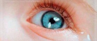
Dry eye syndrome
Dry eye syndrome is a complex of symptoms that appear when the stability of the tear film is disrupted for a long time. This syndrome occurs in almost 50% of patients visiting an ophthalmologist with various complaints. The frequency of diagnosing the syndrome only increases with age.
The pericorneal tear film consists of three layers: the mucin layer, the tear fluid layer, and the superficial lipid layer. In case of disruption of the secretion of one or many components of the tear film, as well as its destabilization due to the influence of external factors, dry eye syndrome develops.
Dry eye syndrome can be a consequence of inflammatory eye diseases, incomplete closure of the palpebral fissure, burns of the conjunctiva, dacryoadenitis, as well as extirpation of the lacrimal gland. In addition, dry eye syndrome can occur with systemic diseases (Mikulich's disease, Sjögren's syndrome, abnormal menopause, rheumatoid arthritis, hypovitaminosis A, etc.). Also, the secretion of tears is influenced by many exogenous factors, both physical and chemical.
The main signs of the disease are the sensation of a foreign body, burning, dryness and itching of the eyes. At the beginning of the pathological process, lacrimation may occur, which intensifies under the influence of external factors. Patients note fluctuations in visual acuity during the day and deterioration in eye performance. In addition, pain may be observed when instilling indifferent eye drops. An ophthalmological examination reveals conjunctival hyperemia and swelling; in severe cases, the shine of the conjunctiva and cornea often disappears (diffuse epitheliopathy, corneal erosion). Biomicroscopy allows you to identify mucin threads and certain inclusions that “contaminate” the tear film; tear film break-up time is reduced. The Schirmer test determines a decrease in the secretion of tear fluid.
Treatment includes eliminating the etiological factor. If this is not possible, symptomatic therapy is prescribed, with compensation for the missing tear fluid. Solutions of “artificial tear” drops are instilled conjunctivally, and the frequency of instillation is determined for each patient individually. Moisturizing gels are placed behind the lower eyelid at night. If the desired effect is not achieved, it is possible to perform a surgical operation - obturation of the lacrimal openings.
Pathology
There are pathologies of the lacrimal gland and pathologies of the lacrimal ducts.
Pathology of the lacrimal gland
Developmental defects
. The absence or insufficient development of the lacrimal gland is characterized by the absence of tears, which is especially pronounced when crying. No treatment is required because the accessory lacrimal glands produce enough tears to moisturize and clean the surface of the eye. It is necessary to protect the eye from infection.
Displacement of the lacrimal gland occurs when the ligaments supporting the gland are weak. This gland, in the form of a painless formation, is felt under the skin of the upper eyelid and lateral corner of the eye. It can be easily adjusted with a finger and falls out again. Treatment is surgical, aimed at strengthening the lacrimal gland in its bed. The prognosis is favorable.
Damage
lacrimal gland are rare, usually observed with damage to the orbit (see), the upper eyelid (see Eyelids). Surgery (removal of the gland) is required only in cases where there is significant destruction of the gland and its prolapse into the wound.
Diseases
. Functional disorders of the lacrimal gland manifest themselves in the form of hyperfunction - increased lacrimation (lacrimation, lacrimalization) in the normal state of the lacrimal ducts. The cause of hyperfunction of the lacrimal glands can be various reflex irritations and disorders of its innervation. In some cases, the cause cannot be determined. Reflex lacrimation can be caused by bright light, wind, cold, patol. process in the nasal cavity, etc. If lacrimation is persistent, then injections of 96% ethyl alcohol into the lacrimal gland, blockade of the pterygopalatine ganglion, electrocoagulation (see Diathermocoagulation) or partial adenectomy are performed. The prognosis is favorable once the cause of the disease is eliminated.
Hypofunction of the lacrimal gland is one of the manifestations of Sjögren's syndrome (see Sjögren's syndrome).
Inflammatory diseases of the lacrimal gland in isolated form are rare; more often inflammation develops as a complication of various infections. diseases, for example, influenza, scarlet fever (see Dacryoadenitis).
Tumors
lacrimal glands are rare. There are benign and malignant tumors.
Of the benign ones, mixed tumors are more common (see). The tumor process is manifested by a unilateral gradual painless enlargement of the gland, a slight displacement of the eye inward and downward. Visual disturbances are rare. Mixed tumors become malignant in 4–10% of cases.
In addition, benign tumors include a cyst (dacryops), which develops either inside the gland or from its excretory ducts after injury or inflammation. It is a translucent, painless tumor protruding at a considerable size from under the outer edge of the orbit.
Treatment of a lacrimal gland cyst consists of excision from the side of the conjunctival sac or diathermocoagulation. The prognosis is favorable. Other tumors of the lacrimal gland must be removed along with the gland.
Malignant tumors of the lacrimal gland (mainly adenocarcinomas), according to A.I. Paches et al. (1980), account for 68 - 76% of all neoplasms in this localization. Growing into surrounding tissues, malignant tumors fix the eyeball, cause severe pain, impair vision, and metastasize to distant organs.
In case of malignancy of a benign tumor or a primary malignant tumor, exenteration of the orbit (see) is performed, followed by radiation therapy (see).
Pathology of the lacrimal ducts
The most common wedge, a manifestation of congenital or acquired pathology of the lacrimal ducts, is constant lacrimation.
Developmental defects.
More common is congenital closure of the mouth of the nasolacrimal duct, leading to the development of dacryocystitis in newborns (see). In rare cases, congenital absence, displacement, narrowing or obliteration of the lacrimal punctum, absence of the lacrimal canaliculus, and fistula of the lacrimal sac are noted. If there is a violation of the tear flow, surgical treatment is indicated, aimed at creating a path for the drainage of tears into the nasal cavity.
Damage
. Ruptures of the lacrimal canaliculi are observed with trauma to the medial part of the eyelids; timely surgical treatment of the wound by an ophthalmologist is necessary. Damage to the lacrimal sac and nasolacrimal duct is more common in injuries with tissue damage to the medial corner of the eye and in fractures of the medial wall of the orbit, nasal bones and frontal process of the maxilla. As a rule, these injuries are recognized late, when complicated by purulent dacryocystitis. Treatment is surgical.
Diseases
. Pathological changes in the lacrimal openings in the form of displacement, eversion, narrowing, obliteration usually occur as a result of damage or inflammatory diseases of the conjunctiva of the eyelids. The most common occurrence is inversion of the inferior lacrimal punctum. Surgical treatment (see Blepharoplasty).
Inflammation of the lacrimal canaliculus (dacryocanaliculitis) often occurs secondary to inflammatory processes of the conjunctiva. The skin in the area of the tubules becomes inflamed. There is lacrimation and mucopurulent discharge from the lacrimal openings. Fungal dacryocanaliculitis is characterized by a strong expansion of the lacrimal canaliculus due to its filling with pus and fungal concretions. Treatment of dacryocanal iculitis is conservative, depending on the causes that caused it. For fungal dacryocanaliculitis, the lacrimal canaliculus is split and stones are removed, followed by lubricating the lacrimal canaliculus cavity with 5% alcohol iodine solution.
Sometimes there is atony of the lacrimal canaliculi, characterized by a negative canalicular test with a normal state of the lacrimal opening and a free lumen of the lacrimal canaliculus. Treatment is darsonvalization of the lacrimal canaliculus area, iontophoresis with calcium chloride and novocaine.
Stenosis and obliteration of the lacrimal canaliculus can result from inflammation or damage to the canaliculi. Treatment is plastic restoration of the tubule lumen.
Inflammation of the lacrimal sac - see Dacryocystitis.
Stenosis and obliteration of the nasolacrimal duct, resulting from its inflammation or damage, are characterized by a slow or negative nasal test with a positive canalicular test; often lead to the development of dacryocystitis. For stenosis, treatment begins with trial probing of the nasolacrimal duct and washing it with solutions containing proteolytic enzymes. In case of obliteration of the nasolacrimal duct, as well as in case of failure of conservative treatment of stenosis, an operation is performed to create an anastomosis between the lacrimal sac and the nasal cavity, which ensures the restoration of the outflow of tears into the nasal cavity (see Dacryocystorhinostomy).
Tumors
lacrimal ducts are rare. For benign tumors (fibromas, papillomas, polyps), the wedge, the picture, resembles hron. dacryocystitis. Surgical treatment: tumor removal with simultaneous dacriocystorhinostomy. The prognosis is favorable. A malignant tumor (carcinoma, sarcoma) can grow into the skin, nasal cavity, and paranasal sinuses. Treatment is surgical in combination with radiation therapy. If the tumor is detected late, the prognosis is unfavorable.
Operations on the lacrimal ducts
The goal of surgical interventions for lacrimal drainage disorders is to create a stable connection between the lacrimal sac and the nasal cavity. Dacryocystorhinostomy (see) is used in various modifications using external and intranasal approaches. During these operations, one of the difficult and crucial moments is the formation of a bone window in the lacrimal fossa. An improved method of endonasal operations on the lacrimal ducts using ultrasound equipment has greatly facilitated this stage of the operation. Thus, operations performed with the help of a raspatory knife and saw powered by an ultrasonic generator are much faster, with less surgical trauma and a significant reduction in postoperative treatment time. Modern endonasal operations on the lacrimal ducts provide good cosmetic and functional results, especially with recurrent dacryocystitis, combined injuries of the nose, orbit and lacrimal ducts, with bilateral pathology of the lacrimal ducts, etc.
Bibliography: Averbakh
M.I. Ophthalmological essays, M.-L., 1940; Beloglazov V. G. Intranasal method of operation of the lacrimal ducts with ultrasonic instruments, Vestn. otorhinolar., No. 5, p. 60, 1978, bibliogr.; Krasnov M. L. Elements of anatomy in the clinical practice of an ophthalmologist, M., 1952; Margolis M. G. Operations on the lacrimal organs, Guide to the eye. hir., ed. M. L. Krasnova, p. 52, M., 1976; Multi-volume guide to eye diseases, ed. V. N. Arkhangelsky, vol. 1, book. 1, p. 137, 206, M., 1962, vol. 2, book. 1, p. 187, M., 1960; Patten B. M. Human embryology, trans. from English, M., 1959; Der Augenarzt, hrsg. v. K. Velhagen, Bd 3, S. 7, Lpz., 1975, Bibliogr.; Mann J. Developmental abnormalities of the eye, L., 1937; Schirmer O. Mikroskopische Anatomie und Physiologie der Thraneorgane, Handb. ges. Augenheilk., begriind. v. A. Graefe u. Th. Saemisch, Bd 1, Abt. 2, Kar. 7, S. 1, B., 1931; System of ophthalmology, ed. by S. Duke-Elder, v. 13, pt 2, L., 1974.
V. G. Beloglazov.
Causes of disease development
Functional disorders of the lacrimal glands are the result of a disorder of innervation or reflex irritations. They can occur with autoimmune and endocrine diseases, when undergoing hormone therapy, when exposed to external irritants (frost, strong wind, smoke, bright light), prolonged work at the computer, and in heavy smokers.
The main causes of inflammation of the lacrimal glands are infectious diseases (influenza, tonsillitis, scarlet fever, mumps, pneumonia, acute respiratory viral infections, measles, typhoid fever). Other factors can also provoke the inflammatory process - narrowing of the tear ducts, trauma, a foreign body in the eye, overheating or hypothermia of the body, prolonged exposure to a dusty room, conjunctivitis, diabetes mellitus.
Forecast
Most often, acute dacryoadenitis has a benign course - the inflammatory infiltrate resolves and the disease resolves within 10-15 days. If an abscess of the gland forms, the disease becomes more severe. Treatment is lengthened if the abscess resolves on its own with pus draining into the upper conjunctival fornix or out through the skin of the eyelid, but the prognosis for recovery is favorable. With adequate treatment of canaliculitis, complete recovery occurs. But if the inflammatory process in the lacrimal canaliculi takes a chronic course, then a disturbance in the outflow of tears is observed.
List of sources
- Egorov E.A. Red eye: clinical picture and treatment // Russian Medical Journal. 1999;7,1(85):13-16.
- Therapeutic ophthalmology (ed. Krasnova M.L., Shulpina N.B.) // M., Medicine, 1985, pp. 63-87, 96-146.
- Brzhesky V.V., Astakhov Yu.S., Kuznetsova N.Yu. Diseases of the lacrimal apparatus: A manual for medical practitioners. — 2nd ed. corrected and supplemented - St. Petersburg: Publishing house N-L, 2009. - 108 p.
- Cherkunov B.F. Diseases of the lacrimal organs: Monograph. Samara. State Enterprise Perspective, 2001. -296 p. (140-152).
- Rational pharmacotherapy in ophthalmology: A guide for practitioners / Ed. ed. E.A. Egorova. - M.: Litterra, 2004. - 954 p.
Pathogenesis
With a wide variety of causes, inflammation proceeds in the same way, no matter what pathogens it is caused by and does not depend on the location. The pathogenesis of any inflammation includes the main components:
- Tissue damage, which plays the role of a trigger factor.
- Release and activation of biologically active substances (inflammatory mediators).
- Vascular reactions.
- Exudation is inflammatory edema (fluid accumulation in tissue).
- Emigration of blood cells to the site of inflammation ( neutrophils , eosinophils , lymphocytes , monocytes ).
The pathogenesis of chronic inflammation has not yet been fully disclosed, but its development is caused by sensitization of the body to bacterial flora and its metabolites. Against the background of sensitization, infectious-allergic inflammation develops. An important factor that leads to chronicity of inflammatory eye diseases is the similarity of the microflora of the mucous membranes of the eyes, nose and pharynx.