March 28, 2014
Tumors are defined as new growths that are composed of cells dividing uncontrollably, and these tumors can also be found in the bones. Constant bone pain is considered the most important symptom for identifying bone tumors. Professor Kaan Erler, a specialist at Anadolu Medical Center in the field of orthopedics and traumatology, says that “in most cases of bone tumors, the cause is unknown and sometimes has a genetic nature. Healthy tissues are replaced by abnormal tissues.
The tumor weakens the bones and leads to pathological fractures. If the necessary measures are not taken in time, rapidly developing (aggressive) tumors begin to move towards other tissues and cause metastases.”
Professor Dr. Erler says that most bone tumors are benign. And he adds: “Cancer that starts in the bone (primary) is different from cancer that starts in other parts of the body and moves to the bone (secondary). The development of tumors and the damage they cause to tissue depends on their biological behavior. Some of them are discovered by chance, some cause serious problems.” Prof. Dr. Erler answered our questions about the diagnosis and treatment of bone tumors.
What are the most common bone tumors?
The most common primary bone tumors are:
- Multiple myeloma: This is the most common type of primary bone tumor. This is a malignant bone marrow tumor. Every year it is detected in 20 people per million. It is most common between the ages of 50 and 70 and can affect any bone.
- Osteosarcoma: This is the second most common type of primary tumor of bone origin. Often found in teenagers and located in the knee area, it is fatal in 2-3 cases per million each year. Less commonly, this tumor is localized in the hip and shoulder area.
- Ewing's Sarcoma: Mostly occurs between the ages of 5-20 years. It is characterized by extensive neoplasm in soft tissues and destroys bone. Most often localized in the area of the upper and lower extremities, pelvic bone and chest.
- Chondrosarcoma: Most often occurs between the ages of 40-70 years. It provokes neoplasms in the hip, pelvic bone and shoulder.
Stages
Based on the stage of bone cancer, doctors determine an effective treatment regimen. The prognosis for recovery depends on the severity of the disease. There are the following stages of bone cancer:
- first. The cancer has not yet spread beyond the bone tissue. At the initial stage, the size of the tumor is less than 8 cm, and later it either increases or it appears in different parts of the bone. The degree of malignancy is still low;
- second. At this stage of bone cancer, the tumor becomes more malignant, the cells lose their differentiation;
- third. The tumor appears in several areas of the bone, the cells are already dedifferentiated;
- fourth. Stage 4 bone cancer is characterized by tumor spread beyond the bone tissue. The lungs, regional lymph nodes and more distant organs are affected. Metastases in the bones of cancer appear precisely at the 4th stage.
What are the symptoms of bone tumors?
Most patients with bone tumors complain of pain. The pain, as a rule, is long-lasting, causing discomfort with low intensity, the so-called “dull” pain. The pain persists even when the patient is resting, and it intensifies at night. Trauma is not the cause, but pain increases after injury. In weakened bones, a pathological fracture occurs, which increases pain. Some tumors cause fever and night sweats. Sometimes neoplasms are painless. Some tumors are discovered by chance during x-rays after ankle injuries.
What should be done in case of this kind of painful situation?
If a person thinks they may have a bone tumor, they should see a doctor right away.
What measures are taken at the diagnosis stage?
The doctor takes a detailed history of the patient in order to know the patient's medical history. The medical history includes all the details - from the drugs used to all previous diseases. The size and mobility of the tumor, its relationship to the joints and whether it has invaded are examined, and other systems are examined if necessary. First, the patient is given an x-ray. Different bone tumors produce different X-ray images. Some show excess calcification, some show bone resorption. Sometimes we see a combination of the two.
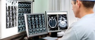
Is x-ray enough to identify a tumor, or is some other method necessary? Some tumors can be seen on X-rays, but we use detailed imaging techniques such as tomography, MRI, scintigraphy, PET scans and pulmonary tomography to determine the type of tumor. We use CT scans to see details of the bone, and MRIs to see how the tumor is growing in the bone or to see whether the tumor has spread to other distant sites. Bone scintigraphy provides information about the biological activation of the tumor and whether there are metastases.
Diagnostic methods
Typically, a patient who comes to the clinic with a complaint of bone pain is first prescribed an x-ray. This is the fastest and most accessible diagnostic method; it allows you to quickly assess the condition of bone tissue and detect pathological formations. The affected bone looks “eaten away” on the pictures, or a defect or “hole” is found in it. You can see how the tumor spreads into neighboring tissues. Some signs on radiographs suggest the presence of a malignant tumor with a high degree of probability, but only a biopsy can make an accurate diagnosis.
Computed tomography helps to clarify the size, location, number of tumor foci, the degree of germination into surrounding tissues, and detect metastases in the lymph nodes and internal organs. MRI is prescribed according to indications: it helps to better visualize the tumor and assess the condition of the soft tissues, brain and spinal cord.
Positron emission tomography helps in searching for small metastases. During the study, sugar molecules with a radioactive label are introduced into the body. Tumor cells absorb the radiopharmaceutical and become visible in pictures taken with a special device.
The most accurate method for diagnosing cancer is a biopsy. During the study, a fragment of presumably tumor tissue is obtained and sent to the laboratory to study the appearance of cells, tissue structure, and molecular genetic characteristics. A biopsy can be performed in different ways:
- For a fine-needle biopsy, a syringe with a needle is used. If the bone is deep and hidden by muscles, the procedure is carried out under the control of computed tomography.
- For trephine biopsy, a thick needle with a diameter of approximately one and a half millimeters is used. This method is considered better than fine-needle biopsy and allows for a more accurate diagnosis.
- Sometimes a large fragment of bone is needed for examination. In such cases, an excisional or incisional biopsy is performed: surgery is performed to remove all or part of the tumor.
Book a consultation 24 hours a day
+7+7+78
How is bone cancer treated?
Treatment of bone tumors is always the result of teamwork. The main members of this team are an orthopedic oncologist, an oncologist, a radiologist, a radiation oncologist, and a pathologist. The goal of treatment is to overcome the cancer and protect the affected limbs. Are there improvements in the treatment of bone tumors as medicine advances? Are there changes in surgical techniques? Previously, in order to remove a tumor from the body, there was mainly a method of amputation of limbs. But a surgical approach is now available that can both remove the tumor and protect the limbs. Surgery can be performed by removing the center of the tumor or removing it along with a small area of healthy skin. The goal of treatment is to ensure the functioning of the limb after cancer therapy. This is facilitated, in particular, by the development of reconstructive surgery. Also often used are prosthetics, bone grafts, which are pieces of bone taken from other parts of the skeleton, used to heal the affected area, and biological reconstruction techniques.
Causes
There are two theories for the origin of osteomas: from the remnants of embryonic cartilage or from the periosteum of mature bone. In some cases, the occurrence of osteomas is associated with an inflammatory process or injury Source: Toropova I.A. Features of the clinical course of osteoma of the nose and paranasal sinuses / I.A. Toropova // Bulletin of RUDN University. — Medicine Series. - 2005. - No. 1(29). — P. 95-97. .
It is believed that the development of osteomas is also facilitated by:
- injuries;
- hypothermia;
- inflammation and previous infections;
- some diseases (rheumatism, gout, syphilis);
- genetic predisposition.
What are the other methods for bone tumors?
Some bone tumors are sensitive to radiation therapy. Radiation therapy can be used as a stand-alone treatment or in combination with other treatments. Chemotherapy can be used for treatment depending on the biological behavior of the disease. It can be used before or after surgery.
Is there a difference between the sexes regarding the risk of developing this disease?
We can provide information on this issue in the USA. Malignant bone tumors are detected in 2500 cases per year, benign ones - in 200,000-300,000. Bone metastases occur in 250,000 - 300,000 cases per year. There were no differences between the sexes in terms of the risk of bone tumors.
What would you like to say about the follow-up of patients after treatment?
Follow-up of the patient is done initially at short intervals, and the follow-up itself will continue for many years. With this method, recurrence of the disease or its spread to other organs is detected at an early stage and a treatment plan is drawn up. At this stage, the patient needs psychological support and a return to a full social life. Lung, breast, thyroid and prostate cancers are the main causes of metastases, in addition to primary malignant bone tumors. In addition, it is necessary to carefully investigate the causes of bone pain that occurs in old age.
What is amputation?
Amputation is the removal of an affected part of the body in order to save the patient's life if it is impossible to save the organ. For example, if we are talking about the development of a disease in the leg, the leg must be amputated. But we are now far from such a surgical approach thanks to developments in diagnostics and treatment. If in the 80s 90% of patients diagnosed with cancer would have been treated with amputation, now this percentage is less than 10%.
Diagnosis of tumors of bone and cartilage tissue
SIGNS AND SYMPTOMS OF THE DISEASE
Pain in the affected bone is the most common symptom of bone and cartilage tumors. At first, the pain is not constant and worries more at night or when walking in case of damage to the lower limb. As the tumor grows, the pain becomes constant.
The pain increases with movement and can lead to lameness if there is a tumor in the lower extremity.
Swelling in the area of pain may take several weeks to appear. Sometimes the tumor can be felt with your hands.
Fractures are uncommon and can occur both in the area of the tumor itself and in the immediate vicinity of it.
General symptoms occur during a widespread process and are expressed in the form of weight loss and increased fatigue.
A standard X-ray will most often detect a bone tumor, which may appear as a cavity or additional growth of bone tissue.
Computed tomography (CT) (sometimes with the additional injection of a contrast agent) makes it possible to identify tumors of the shoulder girdle, pelvic bones and spine.
Magnetic resonance imaging (MRI) is especially useful for lesions of the spine and spinal cord.
Radioisotope bone scanning using technetium can detect both the extent of local tumor spread and damage to other bones. This method is more effective compared to standard x-ray examination of bones.
A biopsy (taking a piece of tumor for examination) is mandatory, as it gives the right to confirm or exclude tumor damage to bone or cartilage tissue. In this case, a biopsy can be performed with a needle or during surgery.
After a detailed examination, the stage of the disease is determined.
Depending on the prevalence of the process, stages can be established - from I to IV.
In addition, examination under a microscope makes it possible to determine the degree of malignancy of the tumor.
Mature tumors have a low degree of malignancy, moderately differentiated tumors have an intermediate degree of malignancy, and poorly differentiated (immature) tumors have a high degree of malignancy.
Undifferentiated (most immature) tumors are the most aggressive.
What can you say about benign bone tumors and their treatment?
The most common benign bone tumors are non-osteogenic fibroma, simple bone cyst, osteochondroma, giant cell tumor, cartilaginous tumor, and fibrous dysplasia. Since treatment for benign bone tumors depends on the type of tumor and the age of the patient, monitoring the patient is sufficient in most cases. In some cases, drug treatment relieves pain. In some cases, especially in pediatric patients, tumors may disappear spontaneously over time. Some benign tumors can develop into malignant tumors and then metastases may begin to develop. Sometimes the doctor recommends removing the tumor. This approach prevents possible pathological fractures. Some tumors may recur even though they have been excised. Giant cell tumor is the most common benign tumor, which can have an aggressive course of development. In some cases, osteochondroma, which is localized in several areas, can develop into cancer.
BE ATTENTION TO THESE SYMPTOMS!
Specialist in orthopedics and traumatology, Professor Kaan Erler says: “If you experience pain despite being rested or if you suspect a bone tumor, please consult a doctor immediately. Orthopedic oncologists will help you in any situation.”
Prevention
There are no special preventive measures to prevent the occurrence of osteoma. Doctors recommend taking an x-ray every year to detect the tumor in a timely manner and, if necessary, remove it.
Specialists of the medical surgical department successfully perform operations to remove various types of osteomas. If you notice a lump on any bone, contact a specialist who will make a diagnosis and promptly prescribe treatment.
There is no special prevention for this disease. The main cause of osteomas is considered to be genetic predisposition.
Some recommendations:
- avoid injury;
- timely cure diseases of the musculoskeletal system;
- undergo examination if any neoplasms of unknown origin are detected.