Disseminated processes in the lungs mean a fairly large group of diseases and pathological conditions (more than 100 known to modern medicine), in which multiple inflammatory foci and (or) fibrotic changes are diagnosed in the respiratory organ, usually located chaotically and on both sides. Pathological changes can affect almost all pulmonary segments, or can be concentrated only in certain places, for example, around the bronchi, in the peripheral parts of the lungs, etc.
Primary disseminated lung diseases are identified based on the results of hardware medical examinations (CT, X-ray). On high-resolution cross-sectional computed tomography of the lungs (MSCT), foci of dissemination and fibrosis are best visualized. A radiologist can assess the volume of damaged lung tissue, identify fibrous cords (connective tissue adhesions and scars), accumulation of fluid and (or) pus in the alveolar vesicles, and concomitant pathologies of the respiratory tract, arteries, and lymphatic system.
In this article we will talk in more detail about pulmonary dissemination and the diseases in which this symptom is observed.
What is dissemination in the lungs?
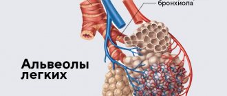
Dissemination consists of multiple pathological foci (compressions) with a diameter of 1-10 mm. On CT scans they appear as light spots, while normally the lung tissue is visualized as an almost uniform dark color. The lesions can be completely different in size, shape (ellipsoid, with uneven edges) and morphology. Perifocal inflammation is often found around the lesions. They can merge and in this case resemble infiltrative processes in pneumonia. Dissemination also manifests itself in the form of focal micro lesions with blood and swelling.
During disseminating processes in the lungs, the respiratory organ partially (depending on the volume of damage) ceases to perform its main function - breathing and transporting oxygen to other organs, in particular to the heart and brain. With total disseminated lung damage, the patient may die.
Diet for pneumoconiosis
Diet for bronchitis
- Efficacy: no data
- Terms: 7-14 days
- Cost of products: 1600-1800 rubles. in Week
The priorities of therapeutic nutrition are the principles of rationality, detail and balance. When choosing menu products, preference is given to lipotropic products, as well as those containing large quantities of vitamins and antioxidants, so the menu consists of:
- liquid porridges and vegetable soups and salads, herbs, all this with the addition of milk and vegetable fats has a qualitative effect on the human diet;
- eggs, dietary fish and meat;
- to improve the functioning of the gastrointestinal tract - fermented milk products and drinks;
- for sweets – honey, various fruits and berries, fruit drinks, compotes and jelly made from them;
- dried fruits and nuts for snacks.
If a person experiences weight loss and loss of appetite, then he should diversify his diet and add spices, lightly salted snacks and delicacies, for example, red caviar, pickles, lightly salted herring or salmon fillets.
Symptoms of pulmonary dissemination
Like most lung diseases, disseminated lung disease does not have specific symptoms that can be used to accurately diagnose it. With dissemination, patients may experience standard respiratory symptoms:
- Dyspnea;
- Cough;
- Difficulty breathing (it becomes more frequent, it becomes difficult to hold it);
- Unpleasant sensations in the chest;
- Cough;
- Loss of appetite;
- Temperature increase;
- Weakness (due to impaired respiratory function and lack of oxygen);
- Decrease in blood oxygen saturation;
- Change in blood pressure.
Prevention
To prevent pneumoconiosis, a set of measures is used, which are based on improving working conditions, compliance with all production safety requirements, and improvement of existing technological processes. In order to prevent the occurrence of pneumoconiosis, it is necessary to use personal (special dust respirators, as well as safety glasses and dust-proof clothing) and collective protective equipment (local supply and exhaust local ventilation, ventilation and humidification of all production premises).
Causes of pulmonary dissemination
When dissemination is detected in the lungs, it is important to accurately determine the cause and identify the specifics of the pathological process. The causes of pathological processes, depending on the shape, density and nature of the distribution of lesions, can be:
- Tuberculosis;
- Pneumonia;
- Poisoning by toxic gases and drugs;
- Prolonged inhalation of industrial dust (pneumoconiosis);
- Hamman-Rich disease;
- Other types of alveolitis (including allergic);
- Goodpasture's syndrome;
- Idiopathic pulmonary fibrosis;
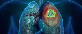
- Oncological diseases of the lungs or other organs (sarcoidosis, metastases, carcinomatosis, bronchoalveolar cancer);
- Acute respiratory distress syndrome;
- Vasculitis (including rheumatoid);
- Some diseases of other internal organs (heart, liver).
Note that the causative agents of infectious and inflammatory lung diseases that can cause disseminated lung damage include a large group of viruses, bacteria and fungi (mycobacterium tuberculosis, SARS-CoV-2, coccidioides immitis), etc. Sometimes it is possible to identify the exact cause of diffuse inflammatory foci and fibrosis is not possible. If the disease has symptoms similar to tuberculosis or pneumonia, or there is severe fibrosis, but even after medical examinations the cause remains unclear, then such pathological changes are called “idiopathic”, and treatment tactics are selected individually based on the data obtained and the patient’s medical history.
Therefore, disseminated lung diseases are considered quite difficult to diagnose. CT scan of the lungs is much more informative than conventional radiography, however, even this method, isolated from laboratory tests, does not provide enough information to prescribe therapy. If a pulmonologist or radiologist suspects a malignant process, a biopsy may be recommended for the patient.
Literature and sources
- Vorontsova E.I., Senkevich N.A., Bogolyubov V.M., Grigoryan E.A., Likhachev Yu. Pneumoconiosis // Great Medical Encyclopedia: in 30 volumes / ch. ed. B.V. Petrovsky. — 3rd ed. - Moscow: Soviet Encyclopedia, 1982.
- L.V. Artemova et al. Federal clinical recommendations for the diagnosis, treatment and prevention of pneumoconiosis (Russian) // Federal State Budgetary Institution "Research Institute of Occupational Medicine" and Rospotrebnadzor Occupational Medicine and Industrial Ecology. — Moscow, 2021.
- Kovnatsky M. A. Clinic of pneumoconiosis / Ed. N. A. Grodzenchik. — L.: Medgiz. Leningr. department, 1963.
Video on the topic:
What diseases are characterized by the symptom of pulmonary dissemination?
Let's consider the most common diseases that can manifest as a disseminated pathological process in the lungs.
Tuberculosis
Mild tuberculosis is a fairly common, dangerous and serious disease. Its causative agent is bacteria - Koch bacilli, which are easily transmitted by contact and airborne droplets, can live for years in street dust and even in human lungs without causing any symptoms.
The disease manifests itself when the human immune system cannot independently restrain the active phase and growth of mycobacterium tuberculosis. Bacterial damage to the lungs in tuberculosis is usually visualized as multiple inflammatory foci - granulomas.
In the center of tuberculous granulomas there are foci of necrosis. Seals form around them - the pulmonary alveoli are filled with a liquid substrate, which contains the bacteria themselves, epithelial and plasma cells, dead lymphocytes, large Langhans cells, and macrophages.
In the initial stages, tuberculosis progresses almost asymptomatically; over time, the patient begins to notice weakness, deterioration in general health, cough and changes in breathing.
At the same time, something suspicious is rarely noted on auscultation. Sputum analysis does not show mycobacteria. Diagnosis of tuberculosis is possible based on the results of an X-ray examination and a tuberculin skin test.
Granulomatosis and diffusely located “ground glass” on CT are not a specific sign of tuberculosis. The former are also found in sarcoidosis, and the latter in pneumonia and other diseases.
To treat tuberculosis, the patient is prescribed a course of antibacterial therapy. It is important to prevent fibrosis (scarring of the lungs) as such changes can be irreversible.
Pneumoconiosis
Pneumoconiosis is a disseminated lung disease caused by inhalation of construction or industrial dust. Most often, pathological processes occur with a pronounced development of primary diffuse fibrosis, in which the pulmonary alveoli “stick together” as connective tissue grows in them.
Pneumoconiosis is also granulomatous in nature. CT scans clearly show multiple nodular densities of varying densities. Such compactions should be checked to determine whether the process is benign and whether there is an oncological threat.
Pneumoconiosis is a so-called “occupational” disease. Most often it affects:
- Electric welders;
- Builders and workers (the disease is caused by inhalation of dust from silicon dioxide, silicates, metals, carbons);
- Industrial workers (the disease is also caused by inhalation of dust from cotton, flax, grain, and some chemicals).
Silicosis is a common and severe lung disease, in which multiple diffuse fibrosis and nodular compactions are diagnosed. As a result, the patient's functional lung volume is significantly reduced. Occurs due to prolonged inhalation of dust with free silicon dioxide (contained in quartz sand and the surrounding air). Therefore, sandblasters, employees of relevant (abrasive blasting) enterprises, and residents of “sandy” regions are at risk.
Asbestosis is a chronic lung disease that also causes multiple pulmonary fibrosis. Occurs due to inhalation of asbestos dust or asbestos fibers. Employees of asbestos mining and asbestos processing enterprises are at risk.
Pneumonia and complications of pneumonia
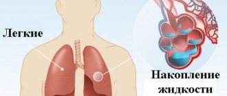
Pneumonia is a group of inflammatory lung diseases. Against the background of this disease, the patient may experience more dangerous complications, which are also characterized by dissemination: acute respiratory syndrome (ARDS), interstitial lung disease, cobblestone symptom, honeycomb lung, pulmonary edema, fibrosis, etc.
In pneumonia with multiple diffusely located loci of damage, the vital capacity of the lungs is significantly reduced, since the inflammatory foci and infiltrates are large, the alveoli are filled with liquid exudate, not air.
Pneumonia with dissemination can be a consequence of pulmonary candidiasis, pneumoconiosis and other diseases. It is necessary to accurately establish the cause of pathological changes. Sometimes it is also necessary to exclude a malignant process (based on laboratory diagnostic results).
Sarcoidosis
Pulmonary sarcoidosis is an oncological disease. Its main features on CT are dissemination and mediastinal lymphadenopathy. Dissemination on scans is not as pronounced as with progressive tuberculosis, but there is a certain similarity. Diagnosis is complicated by pulmonary fibrosis. Along with disseminated damage to the respiratory organ, vasculitis, perivasculitis, and peribronchitis are present.
Multiple foci (from 2 mm to 1 cm) are often located along the bronchovascular bundles, interlobular fissures, costal pleura, and in the interlobular septa (perilymphatic type of dissemination). In sarcoidosis, granulomatosis is often (about 35% of cases) found not only in the lungs, but also in the bronchi. At the same time, their mucous membrane may not be changed - swelling, hyperemia, and areas of thickening of the epithelium indicate damage to the bronchi in sarcoidosis.
In the early stages, sarcoidosis develops asymptomatically; in later stages, the patient begins to be bothered by a nonproductive cough, breathing problems (for no obvious reason), discomfort and burning in the back, and heaviness in the chest.
Pathogenesis
Occupational diseases associated with work in dusty rooms, workshops and mines are dangerous due to their hidden, almost asymptomatic course against the background of diffuse growth of connective tissue in the lungs, as well as, for example, as in silicosis, the formation of characteristic nodules. Foreign tissue reduces the lungs' ability to process oxygen.
External view of the lung of a healthy 78-year-old man suffering from pneumoconiosis
The pathogenesis is considered not to be thoroughly studied; scientists put forward various theories. The most common is phagocytic: after the penetration of foreign small dust particles into the lungs, they settle in the alveoli, through absorption by macrophages or white blood cells, the body tries to remove them, but macrophages are not able to cope with this task, so they rupture, which leads to the release of particles into the lung tissue. The fibrotic process with scar formation is caused by the accumulation of lactic acid, subsequent activation of the enzyme - ketoglutorate , which directly triggers the process of collagen . As a result, the respiratory surface of the lungs decreases.