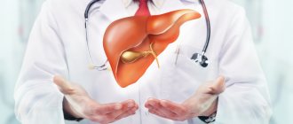
There is an organ in the human body that plays an outstanding role in ensuring life. This organ is the liver, and its functions are so diverse that even a brief listing of them would take up a lot of space.
The liver (hepar) is the largest digestive gland. It produces bile, which enters the duodenum and is necessary for digesting food. The liver performs a “barrier” function and neutralizes harmful compounds that enter it from the intestines and other organs. It is involved in all types of metabolism: it synthesizes proteins (albumin and globulins in blood plasma, factors of the blood coagulation system and the main components of protective antibodies), converts carbohydrates (glucose is deposited in the liver in the form of glycogen) and fats (cholesterol is formed, and excess fat acids are destroyed), most vitamins are deposited (carotene is converted into vitamin A in the liver), some hormones are synthesized and destroyed. The liver serves as a blood depot, up to 20% of which it can retain. In the fetus, the liver performs a hematopoietic function: it produces red blood cells.
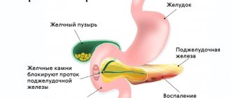
Location of the liver and gallbladder
Liver and gallbladder
The liver is located in the abdominal cavity, in the right hypochondrium, directly under the diaphragm (Fig. 1). The variety of functions leads to the fact that the weight of the liver in an adult reaches 1.5–2 kg (approximately 1/36 of body weight). In the fetus, the relative weight of the liver is twice as large (1/18 of body weight), and it occupies half of the abdominal cavity. The shape of the liver corresponds to the formations surrounding it: the upper surface is convex, like the dome of the diaphragm, and on the lower surface there are grooves and depressions from adjacent organs (right kidney, duodenum and colon). The surface of the liver is smooth and shiny from the peritoneum covering it, the color is red-brown (accumulations of fat give a yellowish tint). The ligaments of the liver fix it in a certain position and are folds of the peritoneum that passes to the liver from the diaphragm and neighboring organs. The ligaments divide the liver into lobes: the larger right one and the smaller left one.
On the lower surface of the right lobe of the liver, the gallbladder is located in a small depression (see Fig. 1). Nearby, in the transverse groove, there is the portal of the liver - the place where blood vessels, nerves enter the liver and where the bile ducts exit. The peculiarity of the liver is that it receives blood from two sources: like all organs, it is supplied with arterial blood (from the hepatic artery), and venous blood, flowing from the stomach, intestines, pancreas and spleen, comes from the portal vein. This blood contains nutrients from the gastrointestinal tract, which are neutralized in the liver and partially stored (as glycogen), insulin from the pancreas, which regulates sugar metabolism, and breakdown products of blood cells from the spleen, which are used to produce bile. Within 1 hour, all the blood passes through the vessels of the liver several times, “unloading” some substances here and being saturated with others. “The busiest harbor in the entire river of life” is called the liver.
Causes of vomiting bile
In most cases, the discharge of vomit containing bile components is caused by non-infectious and infectious diseases of the digestive tract, however, there are a number of situations in which the condition is noted once or occasionally due to dietary errors. Disruption of the normal digestion of products in the lumen of the duodenum with the occurrence of acute duodenostasis, reflux of chyme into the stomach and then vomiting of bile contents is possible when overeating fatty foods, especially in combination with alcoholic beverages.
Cholelithiasis
With lesions of the hepatobiliary system, vomiting of bile is visceral in nature and develops due to overstretching of the walls of the gallbladder and bile ducts. Vomiting often occurs at a height of pain, is accompanied by the release of a small amount of mucus and bile acids, and does not alleviate the patient’s condition. The symptom can be observed when the mechanisms of bile formation and bile excretion are dysregulated, which is caused by organic disorders in the biliary tract. The main causes of vomiting of duodenal contents with bile in pathologies associated with cholelithiasis:
- Cholangitis
. In acute inflammation of the bile ducts, vomiting is associated with symptoms of general intoxication of the body and does not bring relief to the patient. Dyspeptic disorders are secondary signs of the disease; a triad of symptoms comes to the fore: pain in the projection of the gallbladder, high fever, jaundice. In severe cases, disturbances of consciousness and a drop in blood pressure may occur. - Choledocholithiasis
. Vomiting when the bile duct is blocked by a stone is usually of a reflex nature and is caused by viscero-visceral interactions of the autonomic nerves. The symptom manifests itself against the background of severe pain in the right hypochondrium, which occurs due to stretching of the gallbladder wall. Also, choledocholithiasis is characterized by yellowing of the skin and mucous membranes, discoloration of feces, and the appearance of dark urine. - Postcholecystectomy syndrome
. After surgical removal of the gallbladder, pathological circulation of bile develops, which disrupts the processes of digestion and absorption of nutrients. Dyspeptic disorders (bitter belching, bloating, vomiting) are combined with intense pain, observed in 70% of patients. When the process worsens, fever and jaundice are observed. - Biliary dyskinesia
. Discoordination of the contraction of the sphincters of the hepatic and bile ducts leads to a change in the normal rhythm of bile secretion into the intestines, which is accompanied by the appearance of pain and dyspeptic symptoms - nausea, vomiting of bile, flatulence, and loss of appetite. The hypermotor variant is characterized by a predominance of pain; with hypomotor dysfunction, dyspeptic manifestations prevail.
Duodeno-gastric reflux
The reflux of contents digested in the duodenum develops with various organic and functional pathologies of the gastrointestinal tract. The release of vomit with bile is caused by disturbances in the pyloroduodenal zone, which is manifested by periodic gaping of the pyloric sphincter and increased pressure in the upper part of the duodenum. When aggressive contents enter the stomach, an involuntary contraction of smooth muscles and abdominal muscles occurs, which causes vomiting. The appearance of bile in vomit is most often associated with the following diseases:
- Hyperacid conditions
. An increase in the production of hydrochloric acid leads to a pronounced decrease in pH and frequent opening of the pyloric sphincter, accompanied by regurgitation of bile into the stomach. The development of chronic gastritis or peptic ulcer worsens the patient’s condition, causing nausea and repeated vomiting. There is a pronounced pain syndrome, which has a characteristic rhythm associated with food intake. - Acute duodenitis
. Dyspeptic disorders, along with intense night and morning epigastric pain, always occur during inflammatory processes in the duodenum. Overfilling and distension of the upper intestines potentiates relaxation of the pylorus and reflux of contents into the gastric cavity. In the initial stages of duodenitis, the patient complains of bitter belching and heartburn, which are aggravated by vomiting with bile. - Acute gastric dilatation
. Stretching of the walls of the organ, caused by an acute disruption of innervation, leads to the lowering of the stomach and compression of the intestinal loops. As a result, a retrograde flow of intestinal contents is formed, vomiting occurs with the presence of bile acids, which abundantly enter the enlarged stomach. The disease is characterized by the release of a large amount of vomit (up to 7-8 liters per day). - Functional stomach disorders
. The appearance of bile vomiting in such patients is associated with primary disorders of motility of the upper gastrointestinal tract, incoordination of smooth muscles and sphincters. The severity of the symptom increases after eating fatty foods that stimulate bile secretion. Functional dyspepsia is characterized by variability and inconstancy of complaints, combination with psychasthenia. - Duodenal stenosis
. The presence of bile in the secreted vomit is characteristic of a narrowing of the descending intestine. In this case, long-term stagnation of partially digested chyme occurs, which causes intestinal overdistension and retrograde duodenal-gastric flow. In the later stages of the disease, the vomit acquires a foul odor due to prolonged fermentation and rotting of food.
Pancreatic diseases
Dyspeptic symptoms in combination with severe pain are the leading ones in the clinical picture of pancreatic lesions. Vomiting of bile more often occurs in chronic pancreatitis and is caused by relaxation of the pyloric sphincter muscles and an increase in the amount of bile acids in the intestinal lumen. Symptoms may occur when intestinal patency is impaired due to compression of the pancreas by neoplasia with stagnation of intestinal contents and reverse peristalsis. The most common pancreatic causes of bile entering the vomit are:
- Chronic pancreatitis
. Inflammation of the pancreas is characterized by repeated debilitating vomiting, first of gastric contents and eaten food, and then of bile and mucus. The symptom is noted against the background of intense girdling pain radiating to the heart, left arm and shoulder blade. Various stool disorders are also observed: alternation of constipation with diarrhea, the appearance of fat and food residues in the stool. - Pancreatic tumors
. New growths of the head of the pancreas, with an increase in volume, compress the duodenum, leading to duodenostasis and high intestinal obstruction. Vomiting of bile can be observed in the initial stages of the disease, until the sphincter of Oddi is completely obstructed, then the vomit acquires a putrid odor and contains partially digested food.
Acute surgical pathology
The development of bile vomiting is caused by a reflex reaction of the autonomic ganglia and higher centers of the medulla oblongata to intense inflammation in the abdominal cavity. This symptom is often observed with appendicitis, peritonitis, acute cholecystitis, when, against the background of forced restriction of food intake, repeated vomiting occurs with the release of duodenal contents. In case of acute intestinal obstruction and impaired passage of feces, vomiting occurs as a reaction of the smooth muscle layer of the gastric wall to an increase in pressure in the intestines.
Acute intoxication
Vomiting in case of poisoning is caused by stimulation of the vomiting center by impulses from the autonomic nerves and irritation of the trigger zone in the medulla oblongata by toxic substances. Often, an admixture of bile in vomit is observed during acute alcohol intoxication. This is due to the irritating effect of ethanol and its metabolites on the pancreas and liver, activation of proteolytic enzymes, and increased contractions of the gallbladder muscles. Vomiting due to alcohol poisoning is combined with facial redness, disturbances of consciousness, and psychomotor agitation.
Pregnancy
Vomiting of bile during pregnancy is most often observed in the first trimester and is caused by toxicosis. The symptom usually occurs in the morning, on an empty stomach, so when vomiting, the contents of the upper part of the duodenum are released. Repeated vomiting, making it difficult to eat, is considered a pathological condition. Bile in the vomit sometimes appears due to exacerbation of chronic diseases of the biliary tract, which in pregnant women is caused by hormonal changes and increased stress on all body systems.
Worm infestations
The occurrence of vomiting with bile impurities is usually observed during infection with protozoa and intestinal parasites that multiply in the gallbladder and ducts - opisthorchiasis, giardiasis. Microorganisms have both a mechanical irritant effect on the digestive organs, leading to inflammatory processes in the biliary system, and a general sensitizing effect with the development of allergic reactions. In addition to bilious vomiting, the patient is usually bothered by bitter or rotten belching, nausea, flatulence, pain in the navel and right hypochondrium.
Structure of the liver
Liver and gallbladder
The performance of numerous functions is associated with the peculiarities of the internal structure of the liver. The dense membrane covering the liver under the peritoneum goes deep into the organ and divides it into prismatic lobules with a diameter of about 1.5 mm. The number of such hepatic lobules in humans reaches 500 thousand; they are the structural and functional unit of the liver (Fig. 2). In the lobule, liver cells (hepatocytes) are grouped in the form of radial plates, between which there are wide blood capillaries (sinusoidal), converging to the liver). Inside the radial plates, between two adjacent rows of hepatocytes, slits are formed, called bile ducts: bile produced by hepatocytes enters them.
Each liver cell is in contact with the wall of the blood capillary on one side, and with the lumen of the bile duct on the other. This structure allows hepatocytes to work in two directions: secrete bile into the bile ducts and send glucose, proteins, fats, vitamins, urea, etc. into the blood. Raw materials for the production of bile and numerous substances also enter through the capillaries with arterial and venous blood. As already mentioned, arterial blood arrives in the liver through the branches of the hepatic artery, and venous blood through the branches of the portal vein. In the wide capillaries of the hepatic lobules, arterial blood mixes with venous blood and flows very slowly, which promotes the exchange of substances between blood and hepatocytes. The capillary wall also contains special cells - stellate macrophages, which perform a protective function. They can capture from the blood and destroy various foreign particles, microorganisms, and damaged cells. Blood saturated with waste products of hepatocytes flows from the capillaries into the central vein of the lobule, and from there into larger veins, through which it is carried out of the liver and enters the inferior vena cava, i.e. returns to the general bloodstream.
Bile acids
Bile acids are tetracyclic monocarboxylic hydroxy acids from the class of steroids. By chemical nature they are derivatives of cholanic acid. They are the main end product of cholesterol metabolism. Bile acids are formed in the liver and excreted in bile, both in free form and as paired compounds (paired or conjugated bile acids) with glycine and taurine. Glycine and taurine are linked to bile acids by peptide bonds. Human bile mainly contains cholic, deoxycholic and chenodeoxycholic. In addition, lithocholic, allocholic and ureodeoxycholic acids are present in small quantities. In hepatocytes, primary bile acids are synthesized directly from cholesterol: chenodeoxycholic and cholic acids. After the release of bile into the intestine, under the action of enzymes of the intestinal microflora, secondary bile acids are formed from primary bile acids: lithocholic and deoxycholic acids. They are absorbed from the intestine, enter the liver with the blood of the portal vein, and then into the bile. It should be noted that intestinal microorganisms form about 20 different secondary bile acids, but only deoxycholic acid and, to a lesser extent, lithocholic acid are absorbed in noticeable quantities; the rest are excreted from the body. Due to the presence of α-hydroxyl groups in the structure, bile acids and their salts are amphiphilic compounds and have detergent properties. The main functions of acids are to form micelles, emulsify fats and solubilize lipids in the intestine, which increases the effectiveness of pancreatic lipase and promotes lipid absorption. Bile acids are also involved in the regulation of cholesterol synthesis, minimizing the increase or deficiency of cholesterol in the body, and the regulation of the formation and secretion of bile. Their pronounced influence on the functional state of various parts of the nervous system was discovered. It has been determined that they act as surfactants in the internal environment of the body: they do not pass through membranes, but regulate membrane processes of cells and intracellular structures.
If bile formation or bile secretion is impaired (for example, due to blockage of the bile duct with a gallstone), the conditions for the digestion of fats and the absorption of hydrolysis products worsen, and a significant part of them is excreted from the body. Fat-soluble vitamins are also not absorbed, which leads to the development of hypovitaminosis. A blood test is indicated for patients with impaired liver excretory function. An increase in the amount of bile acids is possible even with mild deviations. The level increases with cholestasis (primarily with a long course), which develops against the background of primary biliary cirrhosis. Another reason for an increase in titer is insufficient bile secretion during treatment with pharmaceuticals; tests can also confirm or exclude:
- subhepatic jaundice,
- alcoholic liver damage,
- hepatitis-like syndrome in infants,
- viral or toxic hepatitis,
- cystic fibrosis,
- congenital obstruction of the bile ducts,
- acute cholecystitis.
Liver bile
Hepatocytes produce bile constantly, 0.5–1 liters per day. 95–98% of liver bile consists of water and has an alkaline reaction. It contains bile salts, bilirubin, cholesterol, fatty acids, lecithin, ions Na+, K+, Ca2+, Cl-, HCO3-, etc. The color of bile is due to bile pigments (bilirubin, etc.), which are formed from the breakdown products of red blood cells. It is bile pigments that color the intestinal contents brown. The role of bile in digestion is reduced to emulsification and breakdown of fats, which facilitates their digestion and absorption. Bile enhances intestinal motility.
Possibilities of enzyme therapy in the treatment of biliary and pancreatic insufficiency
Professor Drapkina O.M.: : - We are moving on to the next section, gastroenterology, and once again I ask, dear colleagues, to comply with the regulations. Professor Shatikhin Andrey Ionovich, “Possibilities of enzyme therapy in the treatment of biliary and pancreatic insufficiency.”
Professor Shatikhin A.I.: — Gastroenterologists know well that, according to modern concepts, chronic pancreatitis is a group of diseases of the pancreas of various etiologies, characterized by pain and progressive functional failure of the organ.
What will we talk about today? We will talk about the treatment of functional pancreas insufficiency. Gradual fibrosis, exogenous pancreatic insufficiency - all this can lead to diabetes mellitus.
Pancreatic fibrosis with the development of functional pancreatic insufficiency in chronic pancreatitis is the outcome of a wide range of exogenous influences and endogenous factors. In chronic pancreatitis, both the secretory and endocrine parts of the pancreas are affected, which in the later stages leads to the development of diabetes mellitus.
What etiological factors should attract the attention of practicing gastroenterologists when diagnosing chronic pancreatitis? The most common causes are alcohol abuse up to 90%, the presence of diseases of the biliary system, and aging. In connection with the above, biliary pancreatitis and alcoholic pancreatitis are often distinguished. Hereditary or involutive pancreatitis is also possible.
Clinical symptoms of chronic pancreatitis. Its main manifestations are:
- constant pain in the upper abdomen after eating;
- constant decrease in exocrine pancreatic function; dyspeptic disorders;
- jaundice up to 33% in patients of a transient nature, associated with edema, enlargement of the head of the pancreas and compression of the common bile duct;
- formation of pseudocysts due to ruptures of the pancreatic ducts.
A rare manifestation of chronic pancreatitis may be portal hypertension due to inflammatory compression of the vena portae (portal vein) and v. lienalis (splenic vein).
Pancreas research methods:
- these are, of course, complaints, an absolutely deep, clear, meticulous history of the disease;
- laboratory tests of pancreatic enzymes in blood and urine;
- plain radiography to detect calcifications in an oblique projection, which often happens in people who abuse alcohol;
- Ultrasound examination: size, contour irregularities, ducts, pseudocysts, lesions, parenchyma compactions on the gray scale of the screen. I must warn you that now a diagnosis is often made only on the basis of an ultrasound examination, as if exaggerating the significance of an ultrasound examination. This is not true;
- ERCP (endoscopic retrograde cholangiopancreatography) is a change in the pancreatic ducts and its branches, identifying the so-called “chain of lakes” symptom;
- computed tomography (CT) with contrast, signs of chronic pancreatitis in this case: the absence of accumulation of the contrast agent is revealed - this is a zone of necrosis;
- angiography;
- and what is often carried out in practice by doctors and is of great importance for determining exocrine function is stool macroscopy: stool has a gray tint, has a shiny color, is greasy, sticky due to steatorrhea, and is poorly washed off the toilet; and stool microscopy - amilorrhea, creatorrhoea and steatorrhea due to the large amount of neutral fat in the stool of large droplets with a diameter of up to 8 microns. Study of elastase in feces.
General principles of treatment of chronic pancreatitis:
- refusal to drink alcohol, including beer;
- diet: fifth, 5p table (pancreatic) low in fat and with a lot of protein;
- pain relief: these are antispasmodics and non-narcotic analgesics: papaverine, no-spa, metamizole, paracetamol;
- Narcotic analgesics may be prescribed for severe pain, in particular promedol;
- tramadol – orally;
- antacids - very good for inactivating HCl, which stimulate pancreatic secretion;
- aluminum hydroxide 15 minutes before meals and an hour after meals;
- anticholinergics (m-anticholinergics);
- antidepressants;
- octreotide, in particular sandostatin, as an inhibitor of pancreatic secretion, reduces the number of pseudocysts;
- Proton pump blockers are a big thing now. This is a whole group of drugs, which are prescribed to these patients 1-2 times a day, 20 mg;
- beta blockers (anaprilin);
- lateral pancreatojejunostomy and distal pancreatectomy are surgical interventions;
- percutaneous denervation of the plexus solaris by administration of alcohol.
Now we come to the drugs that are the topic of today’s message in the treatment of exocrine pancreas function:
- these are drugs containing lipase, amylase, protease in different concentrations;
- tablet preparations: mezim, panzinorm, pancreoflat, pancitrate (capsules with microtablets);
- encapsulated preparations: micrazim, enteric capsules with microgranules, the now famous Creon - enteric capsules with minimicrospheres with various doses of lipase from 10 thousand ED to 40 thousand ED; preparations containing, in addition to lipase, amylase, protease, components of bile from cattle, often from pigs, and in enteric tablets.
Enzyme preparations:
- facilitate the digestion of proteins, fats and carbohydrates;
- stimulate the secretion of their own enzymes of the pancreas, stomach, small intestine and bile secretion;
- stimulate choleretic action;
- stimulate emulsification of fats;
- stimulates lipase activity;
- stimulates the absorption of fats and fat-soluble vitamins.
Creon and chronic pancreatitis. About Creon, it must be said that this is a relatively new drug, and here the doctor’s curiosity and passion for a new, perhaps better drug are triggered. I have absolutely nothing against Creon, but Creon also does not make a difference in the treatment of exocrine function. Why Creon? Why is it preferred? Since it has a high lipase content? This tendency is generally accepted among all gastroenterologists: if there is a lot, it means better.
In recent years, due to the appearance of Creon, the problem of chronic pancreatitis with pancreatic insufficiency has been very actively discussed, and, consequently, other enzymes and indications for them are forgotten.
There is overdiagnosis of chronic pancreatitis. I already stopped at this when I talked about ultrasound examination. But overeating is where the search for enzyme preparations takes place. In practice, there is an overdiagnosis of chronic pancreatitis with true pancreatic insufficiency. This is deep pancreatic insufficiency, which is quite rare. In this case, chronic pancreatitis is diagnosed, I emphasize once again, using only echography. It should be noted that true pancreatic insufficiency is rare and requires, first of all, clinical and laboratory confirmation (scatology and elastase test).
The most common cause of digestive disorders is overeating due to diseases of the biliary tract and liver or relative deficiency of pancreatin due to hyperacidity.
Why should doctors be interested in bile enzymes? Today the situation is changing due to the non-infectious epidemic of diabetes mellitus, which is accompanied by diseases of the biliary tract and biliary insufficiency.
The problem of biliary insufficiency is currently not relevant in clinical practice, while in patients with biliary tract diseases with diabetes mellitus, metabolic syndrome and alcoholism, it is very relevant. This leads to renewed interest in choleretics, in particular festal, which can correct the symptoms of biliary insufficiency.
Let's remember the mechanisms of action of bile and bile acids. There is an opinion among doctors that tableted enzymes are ineffective, since they are destroyed by the action of HCl, while they forget that although there are tablets, they are enteric drugs, please, your festal.
Doctors do not sufficiently understand, or have forgotten, the mechanisms of action of bile, bile acids and lipase, which does not allow them to deeply understand the mechanisms of action of some enzymes.
Bile is a secretion secreted by hepatocytes; in the human intestine plays an important role in the physicochemical processing, digestion and absorption of fat.
From a physiological point of view, bile acids are the main component of bile, associated with the processes of digestion and absorption of fat.
Bile acids activate pancreatic lipase; in the presence of a bile component, the pH optimum for lipase action shifts from 8 to 6, that is, the level that corresponds to the duodenum.
Bile acids (cholic and chenodeoxycholic) transport lipids in an aqueous environment and are capable of dissolving lipids by forming a micellar solution - a lipid complex of bile, which is transferred to the intestine.
Bile acids are involved in the emulsification of fat as part of the emulsifying system, where saturated monoglyceride, unsaturated fatty acid and bile acid are fat emulsion stabilizers.
After fat is broken down by lipase, the breakdown products – monoglycerides and fatty acids – form a micellar solution. Thanks to bile acids, micelles that are stable in an aqueous environment are formed containing fat breakdown products, cholesterol and phospholipids, which are transferred from emulsified particles to the absorption surface of the intestinal epithelium.
This is where fat absorption begins. Lipases catalyze the hydrolytic breakdown of triglycerides into fatty acids and glycerol, which are absorbed in the digestive tract as energy, and I emphasize, plastic material. This is something that is difficult to digest without, so lipase, by catalyzing hydrolytic cleavage, provides us with energy and plastic materials.
About the results of the positive use of festal containing bile components. Festal is especially popular among doctors and consumers as an enzyme containing bile components, in particular bile acids. In case of pancreatic disease, festal compensates for the insufficiency of its exocrine function. Festal ensures success in treating biliary dyskinesia, hypomotor hypokinesia of the gallbladder, and enhances intestinal motility. Has a beneficial effect on the function of the entire gastrointestinal tract; good tolerance is noted; relieves dyspeptic symptoms. This is the data of Okhlobystin A.V. and academician Ivashkin V.T. from their book “Rational Pharmacotherapy of Digestive Diseases.”
It also postulates that drugs containing bile and bile acids, in particular festal, can serve as a means of replacement therapy for endogenous bile acid deficiency.
About the results of the positive use of festal containing bile components. Thus, in patients with biliary pancreatitis, involuting chronic pancreatitis, especially in those with alcoholic pancreatitis, a significant progressive decrease in the total content of bile conjugates was noted in the duodenal bile, which can lead to a decrease in the absorption of cholesterol in the intestine and affect the exocrine function of the pancreas. In patients with no severe exogenous pancreatic insufficiency and only dyspeptic complaints, the use of drugs such as Festal is justified, as they contain a bile extract that promotes the emulsification of fats and increases lipase activity.
So, about the results of the positive use of festal containing bile components. This is where I cited the data of L.V. Vinokurova. from the book “Effective Pharmacotherapy-Gastroenterology” for this year.
Yakovenko E.P. et al. says that it is advisable to combine Odeston with multienzyme preparations containing bile components, namely festal.
In multicenter studies assessing the proteolytic activity of a number of enzyme preparations, taking into account the own hydrolytic activity of duodenal contents in patients, it was proven that festal has the greatest specific (true) proteolytic activity. It is the drug of choice for reducing pain and creating functional rest of the pancreas.
The minimum amount of bile acids ensures the activation of lipase, but does not cause diarrhea, but only a slight loosening of the stool. The choleretic effect is indicated for biliary chronic pancreatitis, in patients after cholecystectomy, mainly with constipation. This is the data of Professor Gubergrits N.B. from Donetsk.
I can’t help but tell you about the opposite opinion; this opinion belongs to I.V. Mayev. and his employees, Pharmateka, No. 12, 2011. In Russia, they continue to recommend pancreatin tablets from various manufacturers, which is especially sad when they sometimes prescribe not only pure pancreatin, but also drugs with bile components. There is absolutely not a single word to prove that this cannot be done, it cannot be used, supposedly, containing bile acids.
The drug is prescribed 2-3 tablets during meals; Do not chew the tablets, take them with a small amount of liquid. The duration of treatment is determined individually.
Treatment of chronic pancreatitis is carried out under the control of: the clinical condition of patients; clinical and laboratory data, that is, scatological data are improved; scatological research; elastase content in feces.
Criteria for the effectiveness of therapy for exogenous deficiency in chronic pancreatitis. Signs of effectiveness are: increase in body weight; normalization of stool; reduction of flatulence; positive dynamics of the results of scatological research, namely a decrease in grains of neutral fat, that is, a decrease or disappearance of steatorrhea, creatorrhoea and amilorrhea; normalization of elastase test indicators; normalization of vitamin deficiency.
Contraindications to the use of Festal: acute pancreatitis and exacerbation of chronic pancreatitis; acute cholecystitis; acute hepatitis. Thank you for your attention!
Bile ducts
The bile ducts, into which the bile produced by hepatocytes enters, begin blindly and are directed to the periphery of the hepatic lobule. Here they open into larger interlobular bile ducts, which, merging and gradually enlarging, form the common hepatic duct emerging from the porta hepatis (Fig. 3). Since bile is produced in the liver around the clock and enters the intestines only during digestion, a reservoir for storing bile is needed. The gallbladder serves as such a reservoir.
Structure of the gallbladder and bile ducts
Liver and gallbladder
The gallbladder is pear-shaped and has a capacity of about 40 cm2. This is a pouch 8–12 cm long and 4–5 cm wide. The widened end of the gallbladder is called the bottom, the narrowed end is called the neck. Between them is the body of the bubble. The neck of the bladder continues into the cystic duct, about 3.5 cm long. The gallbladder is covered with peritoneum only from the lower surface; its upper part, as a rule, is in contact with the liver. The bottom of the bladder is adjacent to the abdominal wall in the place where the right costal arch crosses the rectus abdominis muscle.
The wall of the gallbladder is formed by a layer of involuntary muscle fibers, covered on the outside with loose connective tissue, and on the inside with a mucous membrane that forms folds and contains many mucous glands. The mucous membrane of the gallbladder is capable of intensively absorbing water, so the bile in the gallbladder thickens 3–5 times compared to that coming from the liver. In the cystic duct, the folds of the mucous membrane are arranged in a spiral, which allows bile to move along the duct in both directions: into and out of the bladder.
The cystic duct, connecting with the common hepatic duct, forms a common bile duct with a length of about 7 cm. It goes down, pierces the wall of the duodenum and opens together with the pancreatic duct into an extension called the hepatopancreatic ampulla (see Fig. 3). The ampulla is located inside the large duodenal papilla, which is clearly visible on the mucous membrane of its descending part. Circular bundles of muscle fibers in the thickness of the major papilla form the sphincter of the ampulla (sphincter of Oddi), which regulates the flow of bile into the duodenum and prevents the flow of intestinal contents into the common bile duct and pancreatic duct. Another sphincter, the sphincter of the common bile duct, is located in the wall of the common bile duct slightly above the ampulla and directly regulates the flow of bile into the intestine.
In the absence of digestion, the sphincters are closed and bile accumulates and concentrates in the gallbladder. When food enters the stomach, the wall of the gallbladder contracts, the sphincters open and bile enters the duodenum. The structure of the bile ducts explains why either more liquid and lighter bile or darker and thicker bile can enter the duodenum. In the first case, it is hepatic bile, which directly from the liver through the common hepatic and common bile ducts enters the intestine. In the second case, it is cystic bile, which from the liver first enters the gallbladder, where it accumulates and concentrates, and when food arrives, it is released from the bladder and enters the duodenum through the cystic and common bile ducts. The secretion of bile is regulated by the nervous system. Fatty substances and some hormones (for example, secretin) stimulate contraction of the gallbladder and the secretion of bile.
Anatomy of Human Bile - information:
Bile (Latin bilis, ancient Greek χολή) is a yellow, brown or greenish, bitter-tasting liquid with a specific odor, secreted by the liver and accumulated in the gallbladder.
Bile is secreted by hepatocytes - liver cells. Bile collects in the bile ducts of the liver, and from there, through the common bile duct, it enters the gallbladder and the duodenum, where it participates in the digestive processes.
The gallbladder acts as a reservoir, the use of which allows the duodenum to be supplied with the maximum amount of bile during the active digestive phase, when the intestine is filled with food partially digested in the stomach. Bile secreted by the liver (part of it goes directly to the duodenum) is called “hepatic” (or “young”), and bile secreted by the gallbladder is called “vesical” (or “mature”).
A person produces 1000-1800 ml of bile per day (about 15 ml per 1 kg of body weight). The process of bile formation - bile secretion (choleresis) - occurs continuously, and the flow of bile into the duodenum - bile secretion (cholekinesis) - occurs periodically, mainly in connection with food intake. On an empty stomach, almost no bile enters the intestines; it is sent to the gallbladder, where, when deposited, it concentrates and slightly changes its composition, so it is customary to talk about two types of bile - hepatic and cystic.
The doctrine of bile
In ancient times, bile was considered a liquid no less important than blood. But if blood for the ancients was the bearer of the soul, then bile was the bearer of character. It was believed that the abundance of light bile in the body makes a person unbalanced and impetuous. Such people were called choleric. But an excess of dark bile supposedly gives rise to a depressed, gloomy mood, characteristic of melancholic people. Please note: both words contain the syllable “chol”, translated from Greek chole means bile. Later it turned out that the nature of light and dark bile is the same, and neither one nor the other has anything to do with a person’s character (although irritable, sarcastic people are still called bile), but has a direct relationship with digestion.
Regardless of whether a person is good-natured or evil, his liver cells - hepatocytes - produce about a liter of bile per day. These cells are intertwined with blood and bile capillaries. Through the wall of blood vessels, the “raw materials” necessary for the production of bile enter the hepatocyte from the blood. Mineral salts, vitamins, proteins, microelements, and water are used to produce this bitter greenish-yellow liquid. Having processed all these components, hepatocytes secrete bile into the bile capillary. More recently, it has become known that specialized cells of the intrahepatic bile ducts also contribute to bile formation: as bile moves through these ducts to the common bile duct, some amino acids, trace elements, vitamins, and water are added to it. Bile passes directly from the liver into the duodenum through the common bile duct only during the digestion of food. When the intestines are empty, the bile duct closes, and the bile, which the liver secretes continuously, goes through the cystic duct, which branches off from the common bile duct, into the gallbladder. This reservoir has the shape of an elongated pear 8-12 centimeters long and holds approximately 40-60 cubic centimeters of bile.
In the gallbladder, bile becomes thicker, more concentrated, and takes on a darker color than that just produced by the liver. I. P. Pavlov believed that the main role of bile is to replace gastric digestion with intestinal digestion, destroying the effect of pepsin (the most important enzyme of gastric juice) as a dangerous agent for pancreatic enzymes, and that it is extremely beneficial for the enzymes of pancreatic juice involved in the digestion of lipids. When food that has already been partially processed there enters the duodenum from the stomach, pancreatic juice and bile flow here. Moreover, to the bile that flows evenly and slowly directly from the liver, bile from the gallbladder is also added.
Composition of human bile
Bile is not only a secretion, but also an excrement. It contains various endogenous and exogenous substances. This determines the complexity of the bile composition. Bile contains proteins, amino acids, vitamins and other substances. Bile has little enzymatic activity; The pH of liver bile is 7.3-8.0. When passing through the biliary tract and being in the gallbladder, liquid and transparent golden-yellow liver bile (relative density 1.008-1.015) is concentrated (water and mineral salts are absorbed), mucin of the bile ducts and bladder is added to it, and the bile becomes dark, viscous , its relative density increases (1.026-1.048) and pH decreases (6.0-7.0) due to the formation of bile salts and absorption of bicarbonates. The main amount of bile acids and their salts is contained in bile in the form of compounds with glycocol and taurine. Human bile contains about 80% glycocholic acids and about 20% taurocholic acids. Eating foods rich in carbohydrates increases the content of glycocholic acids; if protein predominates in the diet, the content of taurocholic acids increases.
Bile acids and their salts determine the basic properties of bile as a digestive secretion. Bile pigments are products of the breakdown of hemoglobin and other porphyrin derivatives excreted by the liver. The main bile pigment in humans is bilirubin, a red-yellow pigment that gives liver bile its characteristic color. Another pigment, biliverdin (green), is found in trace amounts in human bile, and its appearance in the intestines is due to the oxidation of bilirubin. Bile contains a complex lipoprotein compound, which includes phospholipids, bile acids, cholesterol, protein and bilirubin. This compound plays an important role in the transport of lipids into the intestine and takes part in the hepatic-intestinal circulation and general metabolism of the body.
Bile consists of three fractions . Two of them are formed by hepatocytes, the third by epithelial cells of the bile ducts. Of the total volume of bile in humans, the first two fractions account for 75%, the third - 25%. The formation of the first fraction is associated, but the second is not directly related to the formation of bile acids. The formation of the third fraction of bile is determined by the ability of the epithelial cells of the ducts to secrete fluid with a sufficiently high content of bicarbonates and chlorine, to reabsorb water and electrolytes from the tubular bile.
The main component of bile, bile acids, are synthesized in hepatocytes. About 85-90% of bile acids released into the intestine as part of bile are absorbed into the blood from the small intestine. Absorbed bile acids are transported in the blood through the portal vein to the liver and are included in the composition of bile. The remaining 10-15% of bile acids are excreted from the body mainly in feces. This loss of bile acids is replenished by their synthesis in hepatocytes. In general, the formation of bile occurs through active and passive transport of substances from the blood through cells and intercellular contacts (water, glucose, creatinine, electrolytes, vitamins, hormones, etc.), active secretion of bile components (bile acids) by hepatocytes and reabsorption of water and a number of substances from bile capillaries, ducts and gallbladder. The leading role in the formation of bile belongs to secretion.
Functions of bile The participation of bile in digestion is diverse. Bile emulsifies fats, increasing the surface area on which they are hydrolyzed by lipase; dissolves lipid hydrolysis products, promotes their absorption and resynthesis of triglycerides in enterocytes; increases the activity of pancreatic and intestinal enzymes, especially lipase. When bile is excluded from digestion, the process of digestion and absorption of fats and other lipid substances is disrupted. Bile enhances the hydrolysis and absorption of proteins and carbohydrates. Bile also plays a regulatory role, being a stimulator of bile formation, bile secretion, motor and secretory activity of the small intestine, proliferation and desquamation of epithelial cells (enterocytes). Bile is able to stop the action of gastric juice, not only by reducing the acidity of gastric contents entering the duodenum, but also by inactivating pepsin. Bile has bacteriostatic properties. Its role in the absorption of fat-soluble vitamins, cholesterol, amino acids and calcium salts from the intestine is important.
Regulation of bile formation Bile formation occurs continuously, but its intensity changes due to regulatory influences. The act of eating and food intake increase bile formation. Bile formation changes reflexively with irritation of the interoceptors of the digestive tract, other internal organs and conditioned reflex effects. Parasympathetic cholinergic nerve fibers (impacts) increase, and sympathetic adrenergic ones reduce bile formation. There is experimental evidence of increased bile formation under the influence of sympathetic stimulation.
The humoral stimulators of bile formation (choleretics) include bile itself. The more bile acids that enter the bloodstream of the portal vein (portal bloodstream) from the small intestine, the more of them are released in the bile, but the less bile acids are synthesized by hepatocytes. If the entry of bile acids into the portal bloodstream decreases, then their deficiency is compensated by increased synthesis of bile acids in the liver. Secretin enhances the secretion of bile, the release of water and electrolytes (bicarbonates) in its composition. Bile formation is weaker stimulated by glucagon, gastrin, CCK, and prostaglandins. The effects of different stimulants of bile formation are different. For example, under the influence of secretin, the volume of bile increases mainly; under the influence of the vagus nerves and bile acids, its volume and the release of organic components increase; the high content of complete proteins in food increases the release and concentration of these substances in the bile. Bile formation is enhanced by many foods of animal and plant origin. Somatostatin reduces bile formation.
Bile secretion
The movement of bile in the biliary apparatus is caused by the difference in pressure in its parts and in the duodenum, and the state of the sphincters of the extrahepatic bile ducts. The following sphincters are distinguished in them: at the confluence of the cystic and common hepatic duct (Mirissi sphincter), in the neck of the gallbladder (Lutkens sphincter) and the terminal section of the common bile duct and the ampulla sphincter, or Oddi. The muscle tone of these sphincters determines the direction of bile movement.
Pressure in the biliary apparatus is created by the secretory pressure of bile formation and contractions of the smooth muscles of the ducts and gallbladder. These contractions are coordinated with the tone of the sphincters and are regulated by nervous and humoral mechanisms.
The pressure in the common bile duct ranges from 4 to 300 mmH2O. Art., and in the gallbladder outside of digestion it is 60-185 mm of water. Art., during digestion, due to the contraction of the bladder, it rises to 200-300 mm of water. Art., ensuring the release of bile into the duodenum through the opening sphincter of Oddi. The sight, smell of food, preparation for its intake and the actual eating of food cause a complex and different change in the activity of the biliary apparatus in different individuals, while the gallbladder first relaxes and then contracts. A small amount of bile exits through the sphincter of Oddi into the duodenum. This period of the primary reaction of the biliary apparatus lasts 7-10 minutes. It is replaced by the main evacuation period (or the period of emptying the gallbladder), during which contraction of the gallbladder alternates with relaxation and bile passes into the duodenum through the open sphincter of Oddi, first from the common bile duct, then the cystic duct, and subsequently the hepatic bile. The duration of the latent and evacuation periods and the amount of bile secreted depend on the type of food taken.
Strong stimulators of bile secretion are egg yolks, milk, meat and fats. Reflex stimulation of the biliary apparatus and cholekinesis is carried out conditionally and unconditionally reflexively with irritation of the receptors of the mouth, stomach and duodenum with the participation of the vagus nerves. The most powerful stimulator of bile secretion is CCK, which causes strong contraction of the gallbladder; gastrin, secretin, bombesin (through endogenous CCK) cause weak contractions, and glucagon, calcitonin, anticholecystokinin, VIP, PP inhibit contraction of the gallbladder.
Pathologies of bile secretion and bile formation
Gallstones
Bile that is unbalanced in composition (so-called lithogenic bile) can cause the prolapse of some gallstones in the liver, gallbladder, or bile ducts. The lithogenic properties of bile can arise as a result of an unbalanced diet with a predominance of animal fats to the detriment of plant fats; neuroendocrine disorders; disorders of fat metabolism with weight gain; infectious or toxic liver damage; physical inactivity.
Steatorrhea
In the absence of bile (or lack of bile acids), fats cease to be absorbed and are excreted in feces, which, instead of the usual brown color, becomes white or gray with a fatty consistency. This condition is called steatorrhea, its consequence is the lack of essential fatty acids, fats and vitamins in the body, as well as pathologies of the lower intestines, which are not adapted to chyme so saturated with undigested fats.
Reflux gastritis and GERD
In pathological duodenogastric and duodenogastroesophageal refluxes, bile in the refluxate enters in noticeable quantities into the stomach and esophagus. Long-term exposure to bile acids contained in bile on the gastric mucosa causes dystrophic and necrobiotic changes in the surface epithelium of the stomach and leads to a condition called reflux gastritis. Conjugated bile acids, and, first of all, conjugates with taurine, have a significant damaging effect on the esophageal mucosa at an acidic pH in the esophageal cavity. Unconjugated bile acids, present in the upper digestive tract, mainly in ionized forms, penetrate more easily through the mucous membrane of the esophagus and, as a result, are more toxic at neutral and slightly alkaline pH. Thus, bile entering the esophagus can cause different types of gastroesophageal reflux disease.
Bile research
To study bile, the method of fractional (multi-stage) duodenal intubation is used. There are five phases during the procedure:
- Basal secretion of bile, during which the contents of the duodenum and common bile duct are released. Duration 10 - 15 minutes.
- Closed sphincter of Oddi. Duration 3 - 6 minutes.
- Bile discharge portion A. Duration 3 - 5 minutes. During this time, 3 to 5 ml of light brown bile is secreted. It begins with the opening of the sphincter of Oddi and ends with the opening of the sphincter of Lutkens. During phases I and III, bile is secreted at a rate of 1 - 2 ml/min.
- Discharge of gallbladder bile. Portion B. Begins with the opening of the Lutkens sphincter and emptying of the gallbladder, which is accompanied by the appearance of dark olive bile (portion B), and ends with the appearance of amber-yellow bile (portion C). Duration 20 - 30 minutes.
- Hepatic bile secretion. Portion C. The phase begins from the moment the secretion of dark olive bile ceases. Duration 10 - 20 minutes. Serving volume 10 - 30 ml.
Normal bile levels are as follows:
- Basal bile (phases I and III, portion A) should be transparent, have a light straw color, density 1007-1015, and be slightly alkaline.
- Cystic bile (phase IV, portion B) should be transparent, have a dark olive color, density 1016-1035, acidity - 6.5-7.5 pH.
- Liver bile (phase V, portion C) should be transparent, golden in color, density 1007-1011, acidity - 7.5-8.2 pH.
Gallbladder diseases
If there are disorders in the biliary tract, stagnation of bile in the bladder may occur. Bile becomes highly concentrated and its crystallization begins, with cholesterol most often being the “center of crystallization.” Stones form in the gallbladder: either many small ones, which cause severe pain as they exit through the ducts into the duodenum, or one or two large ones. This is gallstone disease (GSD). Another disease of the gallbladder is inflammation, or cholecystitis. Acute cholecystitis is most often the result of an infectious lesion. In the development of chronic cholecystitis, poor nutrition plays a major role.
Food and diet are of great importance in the normal functioning of the liver and gallbladder and in maintaining human health. Currently, the basic principles of the diet have been developed for people of different ages, gender, activity, physical condition, etc. But the main advice has been known for many millennia and is mentioned in the Bible and the works of Hippocrates - this is moderation.
Author: Olga Gurova, Candidate of Biological Sciences, Senior Researcher, Associate Professor of the Department of Human Anatomy of the RUDN University
Pain or heaviness in the right hypochondrium
Hepatitis
Jaundice
Vomit
37936 05 April
IMPORTANT!
The information in this section cannot be used for self-diagnosis and self-treatment.
In case of pain or other exacerbation of the disease, diagnostic tests should be prescribed only by the attending physician. To make a diagnosis and properly prescribe treatment, you should contact your doctor. Pain or heaviness in the right hypochondrium: causes of occurrence, what diseases it occurs with, diagnosis and treatment methods.
Definition
The right hypochondrium is one of nine areas into which the anterior abdominal wall is conventionally divided. This area belongs to the so-called “upper floor” of the abdominal cavity.
In the right hypochondrium there are the liver, gallbladder, hepatic angle of the colon, and swollen loops of the small intestine can also be projected into this area. The organs listed above belong to the digestive system.
The liver is a parenchymal (spongy) organ with a very good blood supply. The outside of the liver is covered with a capsule in which nerve endings are located. The liver is involved in the metabolism of proteins, fats, carbohydrates, vitamins, and plays a vital role in detoxification of the body.
Harmful substances that enter the liver are subjected to “chemical” processing, which makes them less toxic to the body and promotes faster elimination through the gastrointestinal tract and urine.
In addition, liver cells produce bile necessary for digesting food, primarily fats. Bile enters the gallbladder, located on the lower surface of the liver, where bile accumulates, which is released from it into the duodenum during the next meal.
From above, the liver is adjacent to the diaphragm - a kind of muscular partition between the abdominal and thoracic cavities. From below, from the side of the abdominal cavity, the diaphragm is lined with peritoneum (which covers the entire abdominal cavity and organs located in it from the inside), and from above, from the side of the lungs, by the pleura, which lines the pleural cavity from the inside and covers the lungs from the outside. Both the pleura and peritoneum have good innervation, which is important to know to understand the causes of pain and heaviness in the right hypochondrium. The peritoneum also covers the gallbladder and intestinal loops.
Types of pain or heaviness in the right hypochondrium
The pain in the right hypochondrium can be acute, sometimes “dagger-like”, which makes you think about a serious illness. This pain is characteristic of hepatic colic and is often accompanied by nausea, vomiting, increased sweating, increased heart rate and a drop in blood pressure.
In chronic diseases and the gradual development of acute diseases, the pain is often dull, bursting in nature, or is described by the patient as “heaviness, discomfort” in the area of the right hypochondrium.
Pain syndrome can be spontaneous, or it can be provoked by food intake, alcohol, physical activity, change in body position, etc.
Possible causes of pain or heaviness in the right hypochondrium
There is only one mechanism for the development of pain: stimulation of pain receptors. As mentioned above, the peritoneum has good innervation. Also, a large number of nerve endings are localized in the wall of hollow organs (intestines, gall bladder). Therefore, inflammatory processes in these organs naturally manifest themselves as pain.
Pain also occurs when the liver capsule is stretched. This can occur due to an increase in the volume of the organ (which in most cases is associated with tissue swelling), due to the accumulation of any fluid (most often blood) under the capsule (with a traumatic rupture of the liver), or damage to the capsule.
Unpleasant sensations in the area of the right hypochondrium are caused by excessive distension of the intestinal loops by intestinal contents or gases. In addition, since the organs of the chest are located close to the right hypochondrium, in case of development of pleurisy (inflammation of the pleura), the pain syndrome can also be localized in the right hypochondrium and imitate diseases of the abdominal organs.
Diseases that cause pain or heaviness in the right hypochondrium
Among acute diseases accompanied by pain in the right hypochondrium, it is worth mentioning first of all those that require emergency surgical intervention.
These include
acute calculous cholecystitis
and
renal colic
.
Both conditions are a consequence of cholelithiasis
. Inflammation of the gallbladder (cholecystitis) develops against the background of an already advanced process of stone formation or, conversely, is the cause of the formation of stones. With renal colic, a calculus (stone) becomes wedged into narrow segments of the biliary tract, which is accompanied by severe pain, impaired bile outflow, and in more severe cases – jaundice (yellowing of the sclera and skin).
Other diseases of the biliary tract, such as
biliary dyskinesia
,
chronic cholecystitis
, cause recurrent pain in the right hypochondrium, usually associated with an error in diet.
Another disease classified as an “acute abdomen” that can cause pain in the right hypochondrium is appendicitis.
(inflammation of the appendix).
Despite the fact that the classic position of the appendix corresponds to the right iliac region, an abnormal position of the appendix in the right hypochondrium is quite common, especially in children.
Overdistension of intestinal loops can develop as part of
intestinal obstruction
.
Swelling of the liver tissue is characteristic of hepatitis (inflammation of the liver parenchyma). Hepatitis can have a variety of origins: viral hepatitis A, B, C, etc., autoimmune, toxic, incl. alcoholic. These diseases are usually accompanied by pain, as well as weakness, nausea and vomiting, yellowing of the skin and sclera, change in the color of urine and feces.
Subcapsular hematomas of the liver (accumulation of blood between the liver tissue and its capsule), as well as ruptures of the liver capsule, are usually traumatic in nature.
Among the diseases of the chest that can cause pain in the right hypochondrium, it is worth mentioning pleuropneumonia (inflammation of the lung tissue and pleura), heart failure, intercostal neuralgia and herpes zoster. The latter is characterized by the appearance of blistering-type skin rashes, which are preceded by severe pain.
Which doctors should you contact if you experience pain or heaviness in the right hypochondrium?
If sudden, progressively increasing pain appears in the area of the right hypochondrium, you should contact a doctor to exclude acute surgical pathology. If the pain is not acute, the examination can begin at or. If necessary, the patient can get advice from a hepatologist (a specialist in liver diseases) and other specialized specialists.
Diagnostics and examinations for pain or heaviness in the right hypochondrium
After a detailed survey and comprehensive clinical examination, the doctor, as a rule, needs laboratory and instrumental confirmation of the diagnosis. For this purpose, the following studies are used:
- A clinical blood test with determination of the leukocyte formula, based on the results of which one can suspect the presence of an inflammatory process in the body and determine its severity.
