A liver abscess is an inflammatory disease of the parenchyma of this organ, as a result of which a cavity is formed in it containing pus and limited by a capsule. Most often, this pathology is secondary and develops against the background of an infection already present in the human body.
With timely diagnosis and treatment, the prognosis for patients’ life and ability to work is favorable. However, it is extremely difficult to suspect and confirm such a diagnosis. This is why you should not underestimate the importance of choosing the right examination method. In the modern world, there are many types of diagnostics of liver abscesses, but MRI undoubtedly occupies the leading position. To make sure of this, let’s take a closer look at everything related to this pathology.
Causes
The formation of an abscess can be caused by a variety of microorganisms, the main of which are:
- Strepto- and staphylococci.
- E. coli.
- Proteas.
- Klebsiella.
- Pseudomonas aeruginosa.
A common occurrence with an abscess is the identification of several types of pathogens when inoculating its contents on nutrient media. In addition, bacteria are far from the only ones that cause such pathology. Inflammation in the liver parenchyma can also be caused by parasites entering it, for example:
- Ameb.
- Echinococcus.
- Ascaris.
Echinococcus
Prognosis and prevention
The prognosis for liver abscess is serious: as already mentioned, mortality reaches 30%, despite the availability of modern broad-spectrum antibiotics.
Prevention of liver abscess consists of timely treatment of purulent-inflammatory diseases of other organs, treatment of chronic inflammatory diseases of the biliary tract, maintaining personal hygiene and preventing intestinal infections.
[1]P. S. Bushlanov, N. V. Merzlikin, E. V. Semichev, V. F. Tskhai. Current trends in the treatment of liver abscesses. Bulletin of surgery. 2018.
Classification
According to the path of infection into the liver, the following types of abscesses are distinguished:
- Hematogenous - bacteria penetrate into the parenchyma of an organ from another source of inflammation through the bloodstream. Most often this occurs with sepsis (blood poisoning). Such abscesses, in turn, are divided into portal and arterial, depending on which vessel “brought” the pathogenic microflora.
- Cholangiogenic - penetration of infection into the organ through the bile ducts.
- Post-traumatic – are a consequence of a closed abdominal injury.
- Contact - the transition of inflammation to the liver tissue through its contact with another pathological focus (for example, a breakthrough of pus from an inflamed gallbladder).
- Cryptogenic – when the cause is not clear.
There are also abscesses of the right and left lobes of the liver, single and multiple, and so on.
Pathological and anatomical signs
The source of infection is surrounded by dilated veins and a bank of inflamed edematous parenchyma cells. A cavity forms in the center. First, the abscess is delimited from the surrounding tissues by a grayish-red membrane. Then it becomes thicker and forms a dense capsule. Cholangiogenic abscesses are located along the branches of the bile ducts. At the same time, inflammation forms in the walls, and bile stagnation occurs in the lumen of the ducts.
If the abscess comes directly from the gallbladder, then more often it has a single character and is located close to the bladder bed. The longer the disease, the greater the chance of infection penetrating into the liver tissue and the formation of thick-walled multi-chamber structures.
Symptoms
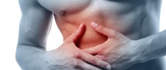
The formation of a cavity with pus in this organ is usually accompanied by pressing pain in the right hypochondrium, which often radiates to the shoulder blade or neck. Unpleasant sensations intensify when lying on the left side, and decrease on the right, especially if you pull the lower limbs to the chest. During examination, the liver is enlarged and protrudes from under the ribs.
Pain from under the ribs
The patient may experience signs of dyspepsia (loss of appetite, nausea, bloating, frequent loose stools, etc.). This occurs as a result of a disruption in the liver’s production of bile and its constituent bile acids, which are necessary for the complete digestion of food. Any inflammation is also accompanied by an intoxication syndrome: fever, sweating, increased heart rate, etc. However, often the only symptom is rapid weight loss.
The above complaints bother the patient in the initial stages of the disease. Later, when a capsule has already formed around the tissue melted by pus, a icteric coloration of the skin and mucous membranes appears. A liver abscess can also compress liver blood vessels or increase blood clots in them. In this case, the patient will accumulate fluid in the abdominal cavity, and the abdomen will increase in volume. This complication is called “ascites”.
Ascites
One of the features of this process in the liver is that the symptoms of the underlying pathology that led to the occurrence of the abscess often come first. That is why a lot of time can pass from the moment the first complaints appear to the identification of a formed focus.
Possibilities of ultrasound diagnosis of liver abscess (clinical observation)
Ultrasound scanner RS80
A benchmark for new standards!
Unparalleled clarity, resolution, ultra-fast data processing, and a comprehensive suite of advanced ultrasound technologies to solve the most challenging diagnostic problems.
Introduction
Liver abscess is a limited purulent-destructive suppuration of its parenchyma with a focus of lytic melting in the center, resulting from the introduction of infection by hematogenous, lymphogenous, cholangiogenic or contact routes, as well as infection of intraparenchymal and subcapsular hematomas. Liver abscess was first described by Hippocrates 400 BC. He suggested that the severity of the disease may depend on the nature of the abscess contents. At the beginning of the 19th century, R. Bright first suggested the possibility of the formation of liver abscesses in amebiasis, and in 1890, W. Osler was the first to discover amoebae in the liver abscess and feces of the same patient. P. Dieulafoy and H. Fitz considered suppurative diseases of the abdominal organs among the various causes of the formation of liver abscesses [1]. In 1926, pylephlebitic liver abscess was first described in a patient with diverticulitis. In 1903, L. Rogers noted the connection between purulent cholangitis, bile duct obstruction and liver abscesses. Before the advent of antibacterial drugs in the 30s of the last century, late diagnosed destructive appendicitis was the main cause of the development of pylephlebitic liver abscesses. In 1938, A. Ochsner and M. DeBakey, having analyzed surgical protocols and autopsy material, found that in 35% of cases the cause of the formation of liver abscesses was appendicitis [1].
Typically, a liver abscess is a secondary disease that develops against the background of existing inflammatory changes in the liver. But there are abscesses that are a manifestation of a primary disease (parasitic etiology). There are several classifications of liver abscesses; there is no single classification. The most comprehensive, in our opinion, is the classification of liver abscesses by B.I. Alperovich [2].
- Primary liver abscesses: bacterial (coccal, bacillary, mixed);
- parasitic (amoeba, roundworm, echinococcal, opisthorchiasis, giardiasis, etc.).
- suppuration of pathological neoplasms of the liver (non-parasitic liver cyst, decaying cancer, syphilitic or tuberculous granuloma);
- single;
The epidemiology of liver abscesses is not well understood. In different countries, incidence rates can vary greatly, ranging from 2.3 per 100 thousand population in North America to 275.4 per 100 thousand in Taiwan [3]. Before the era of antibacterial therapy, the main cause of liver abscesses was acute appendicitis. Mortality was extremely high and amounted to about 80%. With the introduction of antibiotics and surgical treatment methods into clinical practice, this figure decreased significantly, remaining, however, at the level of 10-40%. At the same time, the structure of etiological factors of liver abscesses has also changed. Diseases of the biliary tract and liver, malignant neoplasms and complications of invasive medical procedures began to come to the fore [4, 5].
The etiology of liver abscesses is heterogeneous. The main causes include infectious and inflammatory diseases, malignant neoplasms, iatrogenic conditions and blunt liver injuries (see table). In developed countries, abscesses of bacterial etiology predominate, while in Southeast Asia and Africa amoebiasis remains their most common cause [3].
Table
. The main causes of the development of liver abscesses [1].
| Hematogenous abscesses (portal and arterial) | Cholangiogenic abscesses | Abscesses in liver diseases | Iatrogenic causes and injuries |
| Sepsis | Cholangitis as a consequence of choledocholithiasis, strictures, neoplasms of the biliary tract, chronic opisthorchiasis | Primary malignant tumors of the liver (adenocarcinoma, etc.) and metastatic liver disease | Percutaneous ethanol injections Chemoembolization of the hepatic artery Radiofrequency ablation of liver tumors |
| Inflammatory diseases of the abdominal organs leading to pylephlebitis: - appendicitis; - acute cholecystitis; - pancreatitis; - diverticulitis; - Crohn's disease; - ulcerative colitis; - peritonitis | Biliodegestive anastomoses, fistulas | Liver granulomas | Surgical interventions and endoscopic procedures on the biliary tract and liver: - stenting; — papillosphinc-terotomy; — choledochoduodenostomy; - liver resection; – orthotopic liver transplantation |
| Violation of the integrity of the intestinal wall with the subsequent development of bacteremia: - endoscopic polypectomy; - non-metastatic colon cancer | Acute cholecystitis (hematogenous or direct spread of infection) | Liver hematomas | Blunt liver injuries |
| Amoebiasis |
The main risk factors for the development of liver abscesses are diabetes mellitus, liver cirrhosis, immunodeficiency states, old age, and male gender [4].
Despite significant advances in medicine, early and accurate diagnosis of liver abscesses remains critical. Difficulties in interpreting the clinical picture, the results of laboratory and instrumental research methods can cause late diagnosis, untimely initiation of treatment, the development of severe complications and, as a consequence, an unfavorable outcome of the disease. Thus, according to T. Pang et al., the diagnosis of liver abscess could be established on average a week after the appearance of its first symptoms [6].
The clinical manifestations of liver abscesses are nonspecific and may depend on their size, number and location. Most patients complain of pain in the right hypochondrium, fever and other symptoms of intoxication (chills, myalgia, weakness, sweating, tachycardia, nausea, anorexia). The pain often intensifies with a deep breath, changing body position, and can radiate to the right shoulder, scapula, and upper arm. An objective examination may reveal hepatomegaly. Sometimes jaundice and ascites occur. The clinical picture of the disease that led to the development of an abscess often comes to the fore [2, 5-8].
Laboratory indicators are not very specific and reflect the presence of an active inflammatory process. A general blood test reveals leukocytosis and accelerated ESR. Biochemical examination showed increased levels of C-reactive protein, bilirubin, ALT, and AST [1, 4].
The main methods for diagnosing liver abscesses are ultrasound and CT of the abdominal cavity. The sensitivity of these methods is 96-100%. At the same time, according to A. Lin et al., when contacting 25% of patients, ultrasound results give indeterminate results, and in 14% they are false negative [9].
To determine the etiology of liver abscesses, bacteriological examination of the contents of the abscess and blood is of great importance.
The ultrasound picture of a liver abscess has a number of features associated with the time of its existence. In the formation phase, in the presence of appropriate clinical and laboratory data in the liver parenchyma, it is possible to identify a zone of reduced echogenicity with a heterogeneous structure and unclear contours that transform into normal tissue. In the central part of this zone of reduced echogenicity (zone of intense parenchymal edema), an anechoic structureless area is usually detected, which is an area of tissue necrosis, so far without a liquid component. This picture can be observed for a short period of time (several hours). Subsequently, with a parallel increase in clinical manifestations, a fluid-containing anechoic cavity with internal echogenic contents is formed, due to the presence of pus and tissue detritus. In addition to the echographic signs characteristic of the liquid structure (the effect of enhancing the posterior wall, the effect of lateral shadows, the effect of distal pseudo-amplification of the echo signal), some special signs are observed: separation of the contents of the abscess cavity with the formation of a “liquid-liquid” boundary with a horizontal level, where the thicker part is located below ; possible appearance of air bubbles in the cavity (in the presence of gas-producing flora, for example the genus Klebsiella) in the form of hyperechoic structures near the upper wall, giving the “comet tail” reverberation effect; movement of all internal contents when the patient’s body position changes; the formation of a clear demarcation of the abscess cavity from the surrounding liver parenchyma in the form of a somewhat heterogeneous rim (pyogenic membrane) of increased echogenicity with a thickness of 0.5-1.5 cm [10].
In the literature, we came across a description of the ultrasound picture of a liver abscess depending on the predominance of infiltrative or destructive changes [11]. The authors identified two types of abscess images. Type I abscesses are located, as a rule, in the posterior segments of the right lobe (VI, VII) and represent areas of parenchyma of increased echogenicity of various sizes with unclear contours of a heterogeneous structure with the presence of foci of reduced echogenicity or fluid formations of irregular shape, corresponding to areas of necrosis. Such abscesses are a consequence of inflammatory infiltration of the parenchyma during exacerbations of opisthorchiasis cholangitis. As experience has shown, given the predominantly infiltrative nature of the lesion and a minor destructive component, these abscesses, with timely detection and adequate antibacterial therapy (preferably with intraportal administration), can be cured without surgery.
Type II abscesses are a consequence of purulent cholangitis and are unevenly dilated intrahepatic bile ducts with thickened and compacted walls, with the presence of heterogeneous echogenic content (pus, detritus) in the lumen. These abscesses are characterized by multiple localization, small size, connection with the bile ducts, the walls of which are the walls of the abscesses. Type II abscesses develop from cholangioectasis with prolonged biliary hypertension caused by obstruction of the ducts as a result of sclerotic changes, stenosis of the major duodenal papilla, and obstruction by opisthorchiasis detritus.
The echographic picture of a parasitic abscess in amoebiasis may have some differences [10]. When amoebas enter, inflammatory infiltration develops, followed by the formation of foci of necrosis and lysis of the liver tissue. Ultrasound initially shows moderate and then pronounced diffuse-focal heterogeneity of the liver parenchyma with mixed and predominantly reduced echogenicity. Then, against this background, hypoechoic areas of irregular shape of various sizes with uneven, unclear contours are formed. Subsequently, they form one or several cavities, almost similar in ultrasound signs to bacterial abscesses, but with a number of differences: the contours are usually uneven with the presence of “pockets”, the echogenic membrane around the abscesses is not clearly expressed, heterogeneous contents are visualized in the abscess cavity, up to the presence of sequestration of the liver tissue and a large amount of gases.
When abscesses break into the abdominal and sometimes pleural cavity, into the retroperitoneal space, accumulations of fluid similar to the contents of the abscess are detected extrahepatically, and a defect in the liver capsule may be visible.
The main treatment method for liver abscesses is their surgical or percutaneous drainage in combination with parenteral administration of antibiotics [12-14]. First-line therapy is usually a combination of a third-generation cephalosporin with metronidazole. For small abscesses (less than 3 cm), only systemic antibiotic therapy can be used. For amoebic abscesses, the drugs of choice are metronidazole and tinidazole [1, 2, 4].
The key to successful management of patients with liver abscesses is often timely diagnosis and treatment of the underlying disease.
To illustrate the above brief overview, we present a clinical observation.
Clinical observation
Patient A., born in 1971, instructor in therapeutic exercises. He complained of chills, fever, weakness, lack of appetite, and slight vague discomfort in the right hypochondrium.
I fell ill a week before going to the clinic, when, for no apparent reason, an acute “shooting” pain arose in the shoulder girdle and under the shoulder blade on the right, which intensified with breathing. The patient assessed the pain as “muscular.” Conducted a session of therapeutic exercises. The pain subsided. The next day, an aching and stabbing pain appeared in the right hypochondrium, radiating under the right shoulder blade, intensifying with breathing and movement. Body temperature increased to 37.8 °C.
On the 3rd day after the appearance of the first symptoms, the patient went to the clinic at his place of residence. Ultrasound of the abdominal organs revealed signs of moderate hepatomegaly, predominantly in the right lobe of the liver, and a general blood test revealed leukocytosis up to 14.6 x 10⁹ /l and acceleration of ESR up to 59 mm/h.
Over the next 4 days, there was no abdominal pain, but weakness, anorexia increased, and fever and chills were often bothersome. Low-grade fever persisted. I took paracetamol. I contacted our clinic.
From the life history it is known that the patient leads a healthy lifestyle and has no bad habits. Three months ago I went to Japan, and about three years ago I visited India. During the trips and after them I did not get sick.
At the time of treatment, the condition is of moderate severity. Body temperature 38.4 °C. The skin is pale pink. Subcutaneous fat is moderately developed. There is no swelling. Peripheral lymph nodes are not palpable. In the lungs, breathing is vesicular, weakened on the right in the lower parts, there is no wheezing. Heart sounds are rhythmic and clear. Blood pressure 120/75 mm Hg. Pulse 102 beats/min.
The abdomen is not enlarged, participates in the act of breathing, and is soft and painless on superficial palpation. The lower edge of the liver is percussion at the edge of the costal arch. On palpation, the liver has a soft-elastic consistency and is painless. Ortner's and Murphy's signs are negative. However, with deep palpation in the right hypochondrium, a stabbing pain occurred in the right shoulder girdle. Frenicus symptom is negative on both sides.
Stool once a day, formed, brown, without pathological impurities. Urination is free and painless.
To clarify the diagnosis, the patient underwent an ultrasound of the hepatobiliary zone with ultrasound angiography:
The liver is enlarged in size: the anteroposterior size of the right lobe is 14.3 cm (N up to 12.5 cm), the vertical size of the right lobe is 19.0 cm (N up to 15.0 cm), the anteroposterior size of the left lobe is 9.3 cm (N up to 7.0 cm). The diaphragmatic edge is smooth. The structure of the liver parenchyma is heterogeneous with the presence in the right lobe in the projection of the V segment of a formation of reduced echogenicity, with somewhat indistinct contours, dimensions up to 6.5 x 4.6 x 4.8 cm, a clearly heterogeneous structure with the presence in the central sections of an area of moderately increased heterogeneous echogenicity structure measuring 5.8 x 4.8 x 4.2 cm, avascular (Fig. 1). Along the periphery of the formation there is deformation of the vascular pattern (Fig. 2). The echogenicity of the liver is normal. Intra- and extrahepatic bile ducts are not dilated, their walls are compacted. The diameter of the portal vein is 1.2 cm.
Rice. 1.
B-mode. In the right lobe of the liver, in the projection of the V segment, a formation of reduced echogenicity with unclear contours of a clearly heterogeneous structure is visualized with the presence in the central parts of an area of moderately increased echogenicity of a heterogeneous structure.
Rice. 2.
Color flow mode. The described formation is avascular. Along its periphery there is a deformation of the vascular pattern.
The gallbladder is typically located, measuring 7.0 x 1.7 cm, with an inflection closer to the neck. The contours are clear and even. The wall is 0.3-0.4 cm thick, the echogenicity is moderately increased. The contents of the bubble are homogeneous. The common bile duct is not clearly visible, the diameter of the proximal part is 0.5 cm.
Pancreas: veiling of the pancreas is satisfactory, not enlarged in size: head 2.5 cm, body 0.9 cm, tail is not clearly visualized - 2.7 cm. The contours of the head and body are smooth and clear; the contours of the tail are somewhat indistinct. The structure of the parenchyma is somewhat heterogeneous. Echogenicity is moderately increased. The Wirsung duct is not visualized.
The spleen is not enlarged in size - 11.6 x 4.4 cm, area 47.66 cm² (N up to 50.0 cm²). The contours are smooth and clear. The structure of the parenchyma is diffusely heterogeneous. The echogenicity is average. The splenic vein is up to 0.6 cm in diameter. The retroperitoneal and para-aortic lymph nodes are not clearly visualized. No free fluid was detected in the abdominal cavity. Conclusion: suspicion of an abscess of the right lobe of the liver. Hepatomegaly. Moderate diffuse changes in the spleen.
Taking into account the data obtained, a preliminary diagnosis was established: “liver abscess.” The patient was urgently hospitalized at the Research Institute of Emergency Medicine named after. N.V. Sklifosovsky. In the hospital, a CT scan of the abdominal organs confirmed the diagnosis.
Percutaneous drainage of the abscess was performed. When inoculating the abscess contents, no growth of pathogenic flora was obtained.
Antibacterial therapy and washing of the abscess cavity with antiseptics were carried out. The symptoms of intoxication were completely stopped, the size of the abscess cavity was significantly reduced. The patient was discharged in satisfactory condition under the supervision of a polyclinic surgeon.
Conclusion
The presented clinical observation illustrates a case of acute cryptogenic liver abscess with a developing capsule in a young man who does not have significant risk factors for the development of this pathology. In the clinical picture, attention is drawn to the atypical onset of the disease, manifested by pain in the chest, and the spontaneous disappearance of pain with increasing symptoms of intoxication.
Difficulties in interpreting the clinical picture in this patient were also associated with the absence of significant pathology of the liver and gallbladder according to a previously performed ultrasound. However, the characteristics of the pain syndrome, the presence of intoxication, and objective examination data suggested an acute inflammatory disease of the liver or gall bladder. Repeated ultrasound allowed us to quickly and accurately establish the diagnosis on an outpatient basis, which ensured timely initiation of treatment and a favorable clinical prognosis.
Literature
- Akhaladze G.G., Tsereteli I.Yu. Liver abscesses // Annals of surgical hepatology. 2006; 11 (1): 97-105.
- Alperovich B.I. Liver surgery. M., 2010: 245-260.
- McKaigney S. Hepatic abscess: case report and review // West J Emerg Med. 2013; XIV (2): 154-158.
- Lebedev M.S. Experimental substantiation of minimally invasive combined surgical treatment of liver abscesses (experimental study): Diss. ...cand. honey. Sci. Saratov, 2015. 173 p.
- Roesch Dietlen F., Perez Morales A., Diaz Blanco F. Laparascopic surgical treatment of non-parasitic hepatic cyst // Rev Gastroenterol Mech. 1999; 64 (2): 56-60.
- Pang T.C., Fung T., Samra J. et al. Pyogenic liver abscess: An audit of 10 years' experience // Wld J Gastroenterol. 2011; 17: 1622-1630.
- Mavilia MG, Molina M., Wu GY The Evolving nature of hepatic abscess: a review // J Clin Transl Hepatol. 2016; 4 (2): 158-168.
- Lardiere-Degueltea S., Ragot E., Amrouna K. et al. Hepatic abscess: diagnosis and management // J Visc Surg. 2015; 152 (4): 231-243.
- Lin AC, Yeh DY, Hsu YH et al. Diagnosis of pyogenic liver abscess by abdominal ultrasonography in the emergency department // Emerg Med J. 2009; 26: 273-275.
- Practical guide to ultrasound diagnostics. General ultrasound diagnostics; Ed. Mitkova V.V. M.: Publishing house "Vidar-M", 2003: 106-108.
- Tskhai V.F., Brazhnikova N.A., Merzlikin N.V., Maksimov M.A., Saipov M.B., Eskov I.M., Khlebnikova Yu.A. Opisthorchiasis liver abscesses // Bulletin of Siberian Medicine. 2011; 3: 129-135.
- Chen C.-H., Wu S.-S., Chang H.-C. Initial presentations and final outcomes of primary pyogenic liver abscess: a cross-sectional study // BMC Gastroenterol. 2014; 14:133.
- Sleisenger and Fordtran's Gastrointestinal and Liver Disease. 9th ed. Elsevier, 2010.
- Boyko V.V., Tishchenko A.M., Maloshtan A.A., Skory D.I., Smachilo PM Treatment of solitary liver abscesses taking into account the stage of abscess formation // Xipyrgy of Ukraine. 2013; 1: 16-21.
Ultrasound scanner RS80
A benchmark for new standards!
Unparalleled clarity, resolution, ultra-fast data processing, and a comprehensive suite of advanced ultrasound technologies to solve the most challenging diagnostic problems.
Diagnostics
A correctly collected anamnesis can greatly facilitate making a correct diagnosis. In this case, we are interested in the presence of any inflammatory disease (especially in the abdominal organs), injury or tumor. You should also find out what the patient associates with the occurrence of complaints and how they changed over time.
The first laboratory test is a general blood test. It will show changes characteristic of the inflammatory process. Biochemistry will reflect an increase in indicators indicating damage to the liver parenchyma (AlAT, AST, alkaline phosphatase, bilirubin).
Among the instrumental diagnostic methods, radiography of the abdominal organs is used. In the image you can see a focus of clearing in the liver with a fluid level, a high position of the right dome of the diaphragm and other indirect signs. Also, an abscess cavity is often detected during an ultrasound. Under the control of this study, a fine-needle biopsy can be done to inoculate purulent contents on nutrient media and determine the microorganisms that caused this pathology. This analysis will play a leading role in the selection of drugs for antibacterial therapy.
Ultrasound of the liver
MRI
Well, we finally got to the main research method, which can confirm our diagnosis with a one hundred percent guarantee. Magnetic resonance imaging is highly informative. During the study, the number and location of purulent foci, their size and the degree of damage to the liver tissue are accurately determined.
The procedure is painless, because during its course the integrity of the skin is not compromised. The magnetic waves used during the examination are absolutely safe for the body. Thanks to this, MRI can be performed many times without fear of negative consequences for your health, as is the case with X-ray radiation. The scanning is performed in several planes, as a result of which a detailed image of the organs and tissues of the examined area in 3D format is projected onto the tomograph screen.
The examination is very comfortable for patients. All you need to do is remain motionless during the procedure so as not to reduce the quality of the image. Modern tomographs are equipped with ventilation, artificial lighting and two-way communication. If any problems arise, you can always inform the radiologist, and the examination will be temporarily suspended.
MRI
Almost anyone can undergo an abdominal MRI, with the exception of people with metal devices inside the body (artificial pacemaker, cochlear implant, etc.). Contraindications to such a procedure with contrast are pregnancy, breastfeeding and severe renal impairment.
Laboratory research
The results of laboratory tests indicate a pronounced inflammatory reaction and intoxication of the body:
- leukocytosis with a shift of the formula to the left;
- the appearance of reticulocytes in the blood;
- increase in ESR;
- drop in red blood cells and hemoglobin.
Damage to the liver tissue is indicated by elevated tests for bilirubin, alanine and aspartic transaminases, and alkaline phosphatase.
An increase in the pigment bilirubin is detected in the urine. Stool analysis reveals a lot of undigested food debris and blood.
Radiological signs of liver abscess are:
- areas of tissue clearing or one large formation with a fluid level;
- limited mobility of the right dome of the diaphragm;
- reactive pleurisy (fluid in the pleural cavity on the right).
Ultrasound examination (US) is the most convenient for diagnostic purposes. It is performed on patients for emergency indications and allows to identify:
- increased size of the liver and its individual sections;
- the presence of small and large cavities filled with fluid and pus;
- diameter and localization of ulcers.
The abscess has the appearance of a hypoechoic formation with rounded, even contours. Under ultrasound control, in the surgical department, material is taken from the suspected abscess with a thin needle for biopsy and bacteriological examination.
If possible, therapeutic drainage with evacuation of contents is carried out
Subsequent tank. the analysis allows you to accurately determine the pathological flora and its sensitivity to antibiotics. Experts consider it mandatory to study the histology of the abscess capsule. The diagnosis of tuberculous abscess and detection of tumor disintegration depend on the result.
If clarification is necessary and during preparation for surgery, magnetic resonance and computed tomography are performed. They detect even small lesions. Angiography and radioisotope scanning methods are required to clarify the characteristics of the blood supply and confirm the functional state of liver cells in the abscess area.
Diagnostic laparoscopy - insertion of an endoscope through an incision in the abdominal wall. It is carried out under anesthesia. Necessary for differential diagnosis with liver cancer, subphrenic abscess, purulent pleurisy and cholecystitis. It also allows you to drain the abscess and take material for analysis.
In the differential diagnosis of parasitic abscesses, a blood test for serological tests is used. It is positive if the patient’s body has antibodies to the supposed parasites (amoebiasis, echinococcosis, alveococcosis). Are used:
- indirect hemagglutination reaction;
- precipitation test;
- latex test.
Treatment
Treatment tactics are selected individually in each specific case. If there is one or more small abscesses, you can limit yourself to conservative treatment, which consists of long-term use of antibiotics and drainage of the pathological focus.
At the initial stages, drugs are prescribed by selection. To know exactly which microorganism is responsible for the development of inflammation, you need to wait for sowing, and this takes at least a week. Therefore, doctors prescribe an antibiotic that affects those pathogens that, according to statistics, most often cause this disease. When culture results come back, treatment is adjusted if necessary.
Medicines
Do not forget about symptomatic therapy. It includes detoxification of the body (Ringer's solution, saline solution), pain relief (ibuprofen), spasm relief (drotaverine), and intake of enterosorbents (smecta).
For multiple lesions, a combination of aminoglycosides and penicillins is usually used. Third generation cephalosporins can also be used. They have a wide spectrum of action and have proven themselves in this pathology. The drug of choice for amoeba infection is metronidazole.
If it is impossible to drain the abscess cavity, as well as in the presence of one or more foci of significant size, surgical treatment is performed. During surgery, the surgeon provides direct access (laparotomy), and then drains and sutures the cavity formed after the outflow of pus.
Surgical intervention
Surgical methods
Surgical treatment is preceded by puncture of the abscess under ultrasound control. Liver puncture is performed depending on the location through the intercostal spaces.
In case of viscous pus, a sodium chloride solution is first injected into the cavity, then sucked out with an aspirator
The procedure ends with inserting a conductor string into the needle, removing the needle and installing a drainage tube with side holes along the conductor. The drainage is attached to the skin with separate sutures. Through the tube, you can not only rinse the cavity, but also introduce a contrast agent. And from the photographs you can evaluate the size and quality of drainage.
The development of endoscopic surgery has made it possible to remove small abscesses. But large formations and lesions located in an area that is inconvenient for viewing are excised after an incision in the abdominal wall (laparotomy). Each abscess is carefully opened, and pus or other contents are removed using an aspirator. The empty membrane is washed with an antiseptic solution and then removed within healthy liver tissue.
With such an operation, the risk of pus releasing from the abscess when it is opened into the abdominal cavity and leaking between the intestinal loops increases. Therefore, special skills and experience of the surgeon are required. The patient's life depends on them. In case of cholangiogenic abscesses, after opening the abscess, the common bile duct must be drained for washing and subsequent rehabilitation of its inflammation (cholangitis).
Surgical treatment is necessarily accompanied by massive antibiotic therapy and other conservative methods of liver support.
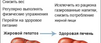
Diet for liver abscess
Do not underestimate the role of proper nutrition in the complex treatment of diseases of the abdominal organs. Patients with this pathology are recommended to adhere to diet No. 5.
Food must be taken in small portions, but often (6 to 8 times a day). Difficult to digest foods with a large amount of plant fiber should be excluded from the diet. You also need to forget about fatty, spicy, smoked and fried foods during treatment and limit the amount of salt consumed to 3 g per day.
Low-fat varieties of fish and meat, steamed, baked vegetables and all types of cereals are allowed. Soups made with vegetable broths are recommended as a first course. For breakfast, you can treat yourself to an unfried omelette.
Complications of liver abscess
The prognosis for life and ability to work in patients with this pathology is quite favorable with a timely diagnosis and adequate therapy. If these conditions are not met, the following complications may develop:
- Bleeding from the hepatic vessels.
- Formation of a purulent focus under the diaphragm.
- Ascites.
- Bleeding from the veins of the esophagus as a result of portal hypertension.
- Abscess ruptures in:
- Pleural cavity with the development of empyema (purulent inflammatory disease).
- In a nearby organ (for example, any part of the gastrointestinal tract).
- Bronchus.
- Abdominal cavity (leads to peritonitis).
- Pericardial sac with the development of tamponade (pus presses on the heart, preventing its normal functioning).
Urgent hospitalization
Conservative treatment
It is necessary to treat patients with suspected liver abscess only in a surgical hospital. The treatment plan is developed individually for each patient. If small single or multiple lesions are detected, then conservative measures are used. Among antibiotics, preference is given to drugs with a wide spectrum of effects:
- third generation cephalosporins;
- aminoglycosides;
- macrolides.
The drug is administered intravenously
A parasitic abscess requires the mandatory prescription of specific antiparasitic agents:
- for amoebic etiology - Yatren, Emetin, Diiodoquine, Hingamin, Chloroquine, Metronidazole, Tinidazole, Ornidazole are used according to indications;
- if alveococcosis is detected - Albendazole, Mebendazole;
- for echinococcosis - a group of benzolimidazoles.
If it is possible to drain the liver abscess after the procedure, a tube is installed through which antibiotics are injected directly into the cavity for several days and rinsed with an antiseptic solution.
Conservative treatment must be accompanied by:
- prescribing vitamins to improve immunity and support liver function;
- means that relieve intoxication (Hemodez, Ringer's solution, glucose);
- cardiac drugs and diuretics for ascites;
- hemostatic therapy for bleeding tendencies;
- prescription of antipyretics;
- sufficient pain relief;
- a course of enterosorbents to remove waste and toxins through the intestines (Smecta, Enterosgel);
- if necessary, use antiemetics.
It is imperative to treat the underlying disease that contributes to the formation of an abscess (sepsis, osteomyelitis, cholecystitis, peptic ulcer, tumors).
Consequences
With proper and timely treatment, the liver abscess resolves, and a scar forms in its place. The connective tissue that has replaced the cells of this organ does not perform their function. Consequently, with a large lesion volume, chronic liver failure may develop. In some cases, with a significant size of the abscess, the cavity may persist. Later it becomes overgrown with connective tissue from the periphery to the center, and over time it disappears completely.
An unfavorable outcome of the disease occurs only in 7-9% of patients. In many ways, mortality also depends on the underlying disease, which caused the development of focal inflammation in the liver. The healing process takes a long time, mainly due to the need for prolonged drainage of the abscess.
Frequency of distribution of different forms
Statistical records and studies of the etiology of liver abscesses revealed the following prevalence among patients:
- amoebic - 64.7% of patients;
- Echinococcal - 12.5%;
- bacterial - 6.5% (including the consequences of dysentery, phlegmonous appendicitis, putrefactive colitis, cholecystitis);
- cholangiogenic - 5.2%;
- traumatic - 1.8%;
- Giardiasis - 0.4%.
According to other data, the proportion of liver abscesses of appendicular etiology is up to 32%, and cholangiogenic - 39%. Rare cases of primary tuberculous liver abscess have been identified; only about 100 such cases have been described.