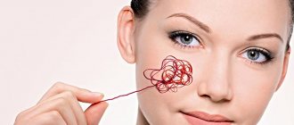
A birthmark is an acquired or congenital pigmented formation of the skin. It can be localized on the face, head, legs or arms. The degree of pigmentation varies - from brown and black to red and purple shades. There is a risk of malignancy of the formation, therefore, if it increases in size, changes in shape and relief, or is exposed to adverse external factors, treatment with a laser or other methods is necessary.
…
Pigment formations occur in people of any age and gender. The most common localization is the skin of the face and open areas of the body. It is believed that in newborns the number of birthmarks is limited or completely absent. Within 1 year of life, the first pigment spots appear, which are difficult to see.
What is the difference between a mole and a birthmark?
Dermatologists note that both formations are the same. However, people themselves often call large pigmented formations “birthmarks” - from 1 cm or more.
Causes
Doctors distinguish between congenital and acquired birthmarks. Separately, there are hemangiomas, which are associated with excessive formation of capillaries in the skin.
Purchased
It is known that as a person grows older, the number of nevi changes. Some of them disappear, but, more often than not, new ones appear, and old ones can change their color and clarity of outline. An important factor is heredity. If there are a large number of moles in one or both parents, there is a high probability of such a picture in the offspring.
The second most important factor is exposure to ultraviolet radiation. People who are exposed to excessive sun exposure or when visiting a solarium have a greater number of pigmented lesions on exposed skin.
The endocrine system has a great influence on the formation of melanin and the number of pigment cells in the skin. In this regard, the peak periods are puberty and pregnancy (changes in hormonal levels during pregnancy provoke the appearance of chloasma). Inflammatory diseases of the skin (dermatitis, acne, etc.) also provoke increased pigment formation.
Acquired nevi make up the bulk of all birthmarks. At the age of up to 1 year, they are observed in 5% of children, at the age of 16-17 years - already in 95%. In old age, their regression is often observed.
Congenital
The reasons for the appearance of birthmarks in newborns are associated with disturbances in the processes of embryonic development. Melanoblast cells, which normally give rise to pigmented melanocytes in the skin, must migrate into the skin from the primary neuroectodermal tube. When migration processes are disrupted and cell clusters form, visible foci of light brown color are formed.
Causes of formation of hemangiomas
A red birthmark found in a newborn or young child is a vascular formation - a consequence of excessive growth of capillaries inside the skin. This is due to a disruption in the process of cell division. Hemangioma can range in color from pale pink to red-purple.
Congenital hemangioma
Possible causes of impaired capillary formation include:
- viral diseases suffered by a pregnant woman - ARVI, influenza, cytomegalovirus infection, etc.;
- uncontrolled use of medications during pregnancy;
- exposure to adverse environmental factors during the gestation period (radioactive radiation, air pollution from industrial products, etc.).
In most cases, a specific cause cannot be determined.
Relationship between the number of moles and the risk of melanoma
There is a lot of research on this issue. If you start reviewing everything, you will end up with a long and boring article that you will not be interested in reading, and I will not be interested in writing.
I have selected some of the largest studies that will help us form our own opinion on this issue.
However, before considering research data, let’s move from the folk concept of “mole” to the medical concept of “nevus”.
Classification of birthmarks
All nevi are usually divided into two large groups:
- Pigmented, associated with the formation of a large amount of melanin in the skin, which has a brown color.
- Vascular, formed as a result of excessive growth of capillary vessels.
In addition, such formations are divided into congenital and acquired depending on the time of their appearance. Congenital ones are always assessed by size:
- small - from 0.5 to 1.5 mm;
- medium - no more than 1 cm;
- large - more than 1 cm;
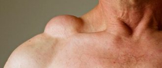
- gigantic - more than 10 cm or covering an entire anatomical area (cheek, buttock, etc.).
Such a division is necessary to determine the prognosis.
Are birthmarks dangerous or not?
Experts believe that the risk of malignancy is greatest if large and giant formations are identified. In this case, active surveillance and surgical removal are recommended.
Related articles: How to recognize malignant moles?
Pigmented birthmarks that appear after birth are divided into three types, depending on their location inside the skin:
- epidermal - accumulation of melanin is observed only within the epidermis;
- intradermal - accumulation of pigment cells occurs inside the dermis, i.e. under the epidermis;
- mixed or borderline - accumulation of melanin in both the epidermis and dermis.
There are a large number of types of birthmarks of vascular origin. They can be simple, cavernous, combined or mixed. The differences between them are associated with the morphological picture, which can be examined during a biopsy.
What are moles?
Every child is unique, and every mole is too. From the name you can easily guess that moles are spots that are on the baby’s skin from birth. There is also a theory that identical “marks” on the body were a sign of the same genus. Some moles form before the baby is born and are immediately visible. Others are present on the skin “in theory,” but only appear some time after birth.
Moles can differ significantly from each other in appearance: some can be flat, others can be convex, some can be regular in shape, others can have uneven edges. Colors can vary from yellowish and brown to red and blue.
Most often, moles on the skin are not dangerous. Some even shrink in size or fade completely over time. In rare cases, moles can be a symptom of an underlying disease. In any case, if your baby's moles cause you concern, consult your pediatrician.
Clinical manifestations
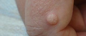
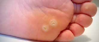
Pigmented and vascular nevi have characteristic external manifestations. Pigmented are represented by areas that have a color different from the unchanged skin. Sizes vary from a few millimeters to several centimeters. The appearance varies, but in most cases it is a round or oval lesion with clear boundaries, located at the level of the skin or rising above it.
Hemangiomas are flat or lumpy lesions that rise above the skin, from 1 mm to 10 cm or more in diameter, in the form of white (with insufficient blood supply to the vessels) or burgundy-bluish birthmarks.
With unchanged characteristics (color, relief, clarity of the border), a slight and gradual growth of the birthmark is the norm. If these indicators change, hair loss in the area of pigmentation, the formation of ulcers, peeling, inflammation, etc., the development of melanoma can be assumed.
In children, hemangiomas tend to increase in size. At the same time, they grow not only in width, but also in depth. This can lead to compression of soft tissues, neurovascular bundles and internal organs. When a vascular pathological focus is traumatized, bleeding may develop, which is difficult to stop using conservative methods.
It is possible to form a birthmark on the eye or mucous membranes. Normally, there are no accumulations of melanocytes on them. People with such symptoms should be under constant medical supervision and independently visually assess the condition to timely detect changes.
Diagnostic measures
When identifying small or large birthmarks, the main task is to promptly determine their transformation into malignant formations. Diagnosis of pigment and vascular formations includes similar activities:
- Determining the time of appearance of pigment formation - immediately at birth or during life. It is necessary to establish whether it has changed its shape, color and size. If there are changes, the dermatologist is interested in their connection with an adverse effect on the birthmark in the form of a burn, injury or scratching. If a person has previously consulted doctors, it is important to evaluate the completeness and effectiveness of the treatment.
- Physical examination - visual assessment of the formation and its palpation. In the case of a vascular birthmark, auscultation and determination of the area of the cutaneous hemangioma are necessary.
- Ultrasound examination of the latter makes it possible to assess the depth of its distribution, as well as structural features. Blood flow velocity is measured using ultrasound and Dopplerography. If it is necessary to accurately establish the boundaries and characteristics of the blood supply, angiography is possible.
- If the birthmark is located on the back of the head or other parts of the body near bone structures, an x-ray is taken.
Detection of malignant transformation is possible using epiluminescence microscopy. A special dermatoscope allows the doctor to evaluate the internal structure and boundaries of the formation inside the skin. If they are heterogeneous or there is ingrowth of pigment tissue deep into the skin, this indicates a malignant degeneration of the birthmark. A similar non-invasive diagnostic method is digital analysis on a computer. Photographs obtained using high-resolution cameras are analyzed by special computer programs that can identify signs characteristic of melanoma.
Malignant transformation of birthmarks
To make a diagnosis, histological analysis is recommended. It is known that any traumatic impact, for example, medical procedures in the form of cryodestruction or partial surgical excision, can cause malignancy. In this regard, material for histological analysis is obtained using a smear by applying a glass slide to the surface of the nevus. Non-invasive examination is possible if there are cracks or ulcers on it.
In most cases, if surgical excision of a pigment spot is necessary, the resulting material is necessarily sent for morphological analysis. Depending on the conclusion about the benignity or malignancy of the process, further treatment tactics are selected.
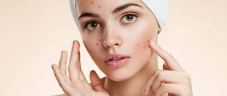
Why do red moles appear on the body?
Red moles are formed by a large number of microscopic blood vessels (hemangiomas) and lymphatic vessels (lymphangiomas). The main reason for the appearance of red moles is dysfunction of the circulatory and lymphatic systems. Angiomas are divided according to type:
| Types of angiomas | |
| Types of angiomas depending on the morphological structure | hemangiomas that develop from blood vessels |
| lymphangiomas, which form from lymphatic vessels | |
| Types of angiomas depending on the cellular structure | monomorphic, consisting of the same type of cells |
| polymorphic, which combine several cell types | |
| Types of angiomas depending on type | simple (capillary) angioma - occurs due to the proliferation of capillaries |
| branched angioma - a branched network of dilated capillaries | |
| cavernous angioma - wide spongy cavities filled with blood | |
| combined angioma, combining cavernous and simple types | |
| Types of angiomas depending on location | spine - predominant location in the thoracic and cervical regions, affecting one or more vertebrae |
| skin - red moles on the body | |
| brain - vascular angiomas in the brain substance | |
| liver - single or multiple vascular neoplasms located in different parts of the organ | |
| kidneys - angiomas are formed from the veins and arteries that supply the organ | |
The most common causes of angiomas on the body are:
- hormonal changes in women (during pregnancy, taking oral contraceptives);
- skin injuries;
- genetic characteristics of the organism;
- diseases of the gastrointestinal tract, liver and pancreas;
- long exposure to sunlight and frequent visits to the solarium.
Treatment approaches
Therapy for vascular and pigmented formations has a number of differences. In this regard, after establishing the nature of the birthmark, the immediate treatment tactics for a particular patient are determined.
Treatment of pigmented formations
Any mole is a potential melanoma, and therefore it is necessary to minimize its damage during normal life activities.
Is it possible to get a tattoo on a birthmark?
Definitely no. Such manipulation can trigger malignant transformation and the development of skin cancer.
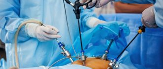
Removal is carried out for 2 indications: oncological alertness and for cosmetic problems. In the case of surgical treatment for suspected melanoma, the pathological focus is removed by excision within healthy tissue. After the operation is completed, the material must be submitted for histological analysis.
If it is localized on the neck, where the collar constantly rubs, or on the face, leading to a cosmetic defect, it is possible to remove even a small birthmark without medical indications for this. In this case, wide surgical excision is not necessary. It is possible to use limited surgical removal, electrocoagulation, cryodestruction or laser treatment. Each of the methods has differences in its mechanisms and indications for implementation:
- Surgical removal is the standard treatment method for large lesions or transformations located deep in the skin. Excision of the birthmark should be 2-6 cm in diameter, depending on its location. The main disadvantage of surgical removal is associated with a cosmetic defect in the form of a scar.
- Electrocoagulation involves excision by thermal exposure to high-frequency current. Removal is carried out over healthy tissue, which allows the surgical material to be sent for histological examination. Unlike conventional surgery, cicatricial changes on the skin are not typical during electrocoagulation.
Additional information in the article “Surgical removal and electrocoagulation of skin tumors”
- Laser removal is a modern procedure recommended by most doctors for cosmetic reasons. The laser acts locally without damaging surrounding tissue. A crust remains at the site of laser treatment, protecting the wound from infection and cicatricial deformation. However, this method completely destroys pathological tissues, without the possibility of their histological examination.
- If the lesion is small, does not protrude above the skin and is located on the back and other closed areas of the body, cryodestruction can be performed.
The choice of the specific type of intervention is made by the oncologist. If there is a suspicion of malignancy, traditional surgical removal is necessary.
Before and after electrocoagulation of a birthmark
Therapy for vascular spots
Vascular birthmarks differ in their prognosis. In this regard, the question arises: is it possible to remove them in childhood? Experts recommend early surgical treatment for children with hemangiomas on the scalp and neck, as well as in the perineal area and oral mucosa. Removal is necessary in case of frequent bleeding, bacterial infection, aggressive growth and the presence of necrotic changes. Surgical intervention can be postponed if it has a capillary structure, the absence of a cosmetic defect and the onset of spontaneous regression.
With superficial localization and small sizes, destruction can be carried out using laser exposure, cryodestruction or electrocoagulation. In case of cavernous growth, sclerotherapy is effective, which consists of introducing sclerosing substances into the vessels that form pathological tissues. Surgical excision within healthy tissue is necessary if the birthmark is deep or it is impossible to perform minimally invasive procedures. If a large vessel feeding it is identified in the neoplasm, then embolization can be performed during endovascular surgery.
If localized in the orbital area or large in size, removal using the described methods cannot be performed. For treatment, radiation therapy or exposure to an ultra-high-frequency magnetic field is performed. Such an effect leads to stopping the growth of the vascular neoplasm and allows it to be partially removed through surgery or cryodestruction.
Laser removal of hemangiomas
It is not possible to remove hemangioma with a laser in all cases. Although with the help of a laser the tumor is removed layer by layer, in several “passes”, if the modified tissue has deep proliferation or the tumor is located in the internal organs, it is impossible to remove the tumor in one procedure. In addition, with a large volume of modified tissue, complex therapy is necessary.
Despite all the advantages, laser procedures have many contraindications.
Among the advantages of laser removal of red spots are the non-invasiveness of the procedure, it is carried out under local anesthesia, a short period of postoperative rehabilitation, as well as a minimal cosmetic defect remaining after removal.
Any procedures using a laser require preliminary examination for the presence of contraindications. Thus, laser surgery is not used if the patient has:
- problems of the cardiovascular system and rheological properties of blood;
- oncological neoplasms;
- hypertension and atherosclerosis;
- disorders of the kidneys and liver;
- nervous system disorders;
- elevated body temperature.
Laser surgery is also not used during pregnancy and lactation. If there are no contraindications, postoperative rehabilitation takes up to two weeks, during which redness and swelling may be observed in the affected area. They do not require any additional treatment and soon go away on their own.
Possible complications
A serious complication is malignancy with the development of melanoma. This is one of the most malignant tumors, which can quickly lead to the appearance of metastases and damage to internal organs. As a result of multiple organ failure, the patient dies.
Vascular or light-colored birthmarks do not have a tendency to become malignant. However, they are able to quickly increase in size and compress anatomical formations located under the skin. This can cause damage to neurovascular bundles, bones, etc. Their trauma leads to bleeding, which is difficult to respond to conservative therapy. Against the background of such injuries, a bacterial infection may develop, accompanied by necrosis, general intoxication, sepsis, etc.
Forecast
Pigmented nevi are divided into 2 types: simple and melanoma-dangerous. The latter include formations that have an uneven structure and are characterized by a high risk of progression to melanoma. In the first case, the prognosis is favorable - the person needs to independently monitor the condition of the pigment formation. The need to visit an oncologist or therapist arises when changes in its shape, relief or contours are detected. A sign of malignancy may be a complaint that the birthmark itches and hurts. The appearance of pain indicates the beginning of invasive growth into healthy soft tissue.
In the second case, the prognosis depends on the morphology of the lesion, its location and the person’s lifestyle. With regard to the latter, the type of work, the presence of bad habits, concomitant diseases, family history of cases of melanoma, etc. are important.
What does the appearance of red moles on the body mean?
If red moles appear on the body, this may be a simple cosmetic defect, or it may indicate pathological processes occurring in the body. In some cases, the occurrence of angiomas indicates poor nutrition, prolonged exposure to the sun, and a sludge buildup in the body.
Red dots like moles may indicate increased estrogen levels in the body, liver dysfunction and increased insulin in the blood. In addition, in some cases, angiomas appear as a response of the body to a lack of iodine, magnesium, chromium, vitamins C and K.
Prevention of development
Specific prevention has not been developed. Regarding congenital moles, doctors recommend that women during pregnancy eliminate unfavorable factors (bad habits, working with chemical reagents), normalize diet and lifestyle, and are also under constant supervision by obstetricians and gynecologists.
The level of insolation should be reduced - avoid prolonged exposure to the sun, including in winter. The development of acquired vascular formations is associated with bad habits (smoking, drinking alcohol and drug addiction), exposure to ultraviolet radiation on the skin and chronic diseases of internal organs, primarily the endocrine system.
It is necessary to follow a number of medical recommendations:
- Reduce the amount of time spent in the sun during the day. This is especially true for the period from 11 a.m. to 5 p.m. At this time, the maximum amount of ultraviolet radiation is observed.
- When you are near open water or in winter, you must remember that ultraviolet exposure in this case increases.
- Sunscreens, including those with a high protective factor, do not reduce the risk of developing melanoma. They only reduce the risk of sunburn.
- It is necessary to avoid excessive insolation in the solarium.
- If you have moles on the neck, wrists or other areas of the body that are exposed to mechanical impact from clothing, bracelets or watches, you should consult a doctor about their removal.
- The condition of pigmented lesions should be constantly monitored. If the changes described above occur, you should consult a dermatologist.
- Under no circumstances should you practice self-removal using cauterization or any other methods. Such damage contributes to the development of melanoma.
It is recommended to visit an oncologist once every six months.
Preventing the appearance of red moles
It is impossible to prevent the occurrence of congenital angiomas. A congenital red mole in a child is most often a short-term formation and disappears by the age of 5-7 years. Therefore, if the child does not complain of pain or itching in the area of the angioma, there is no need to worry.
In order to avoid the occurrence of angiomas after birth, it is necessary to follow these recommendations:
- avoid prolonged exposure to direct sunlight and frequent visits to the solarium;
- follow a sleep and nutrition schedule;
- lead a healthy lifestyle.
If you have a red mole, it is important to monitor its development and contact a dermatologist at the first signs of injury or change in size. With timely consultation with a doctor, it is possible to prevent complications of angiomas and, if necessary, begin therapy on time.
5
2
8
Article rating:
4.08 out of 5 based on 40 ratings
Author: Christopher Vasily Alekseevich
Dermatologist, pediatric dermatologist. Highest category. Work experience 24 years.