Table of contents
- First question: what is a bezoar?
- Classification of bezoars
- Symptoms of the presence of a bezoar in the stomach
- Diagnosis of bezoar
- Possible complications
- Treatment for the presence of a bezoar in the stomach
A feeling of heaviness in the stomach up to severe pain, belching with an unpleasant odor, vomiting, weakness, etc. Such complaints from the patient should make the doctor think twice. Perhaps this is due to a rather rare, but still occurring problem: the formation of a bezoar in the stomach.
First question: what is a bezoar?
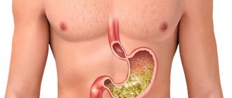
A bezoar is a foreign body that forms over a certain period of time, mainly in the stomach. Most often, bezoars are found in ruminants, but there are many known cases of their formation in the human body. The trouble in the form of a bezoar occurs due to substances entering the stomach that are not digested in it, but accumulate and thus form a foreign body. In addition, bezoars can form due to the proliferation of Candida fungi in the stomach. Bezoars have a rather interesting classification.
Classification of bezoars
Phytobezoar
Phytobezoars are the most common. Help for its formation in the stomach is a decrease in the secretory function of the stomach, as well as a violation of the removal of contents from it, poor chewing of food, etc. These bezoars are formed from plant fibers of wild persimmons, grapes, wild plums, figs, bird cherry, etc. The rate of their formation can vary from 1 day to 25 years. Rapid formation occurs from unripe persimmons containing many astringent and resinous substances. Phytobezoars may have
the consistency is soft and loose, and can reach the density found in natural stones. These bezoars can be either single or multiple. The color may be dark brown or green, and the odor may be foul. The dimensions of this type of bezoar vary from a few millimeters to tens of centimeters. As a rule, they are formed in patients who have undergone surgery to remove part of the stomach (resection) or surgery associated with the intersection of the main trunk (or branch) of the vagus nerve passing to the stomach (vagotomy). This happens due to the rapid and unhindered movement of undigested foods into the small intestine. But it is not possible to say for sure about the frequency of occurrence of bezoars in the stomach, because Not all patients who have previously undergone these operations are examined using endoscopic and radiological methods.
Trichobezoar
Trichobezoars form when hair enters the stomach. Most often, this type of bezoar is found in people with a disturbed psyche, those suffering from an irresistible addiction to biting hair, as well as in those whose work involves hair. Trichobezoars often form in children suffering from schizophrenia. Their weight can reach 3.5 kg or more.
Shellacobesoar
Shellacobesoars are formed as a result of the abuse of alcohol varnish, nitro varnish, and polish by persons suffering from alcohol addiction. The thing is that shellac is a natural resin used in the production of varnishes. Polish is an alcohol solution of shellac used in finishing work. So, with the regular consumption of all these liquids, shellac stones are formed in the stomach, which are found mainly in the stomach and never enter the duodenum. This type of bezoar has a brownish-white color and a smooth or slightly rough surface. In cross-section, shellac bezoar has a layered structure and black-brown color. It is also known that such a bezoar can burn, be cut with a knife, and its weight can reach 500 g or more.
Sebobezoar
Sebobezoars occur when animal fats become compacted. Their formation is due to the fact that the melting point of fats (beef, lamb and goat fat) is higher than the temperature inside the stomach. As a result, crystallization of triglycerides occurs with the formation of fat stones.
Pixobesoar
Pixobesoars are found in people who have the habit of chewing var and resin.
Lacto- and hemolactobezoar
Lactobesoars are formed in premature babies who are on a high-calorie artificial diet, which contains lactose and casein. Their formation occurs during the first 2 weeks of a child’s life. Hemolactobezoars disintegrate on their own after gastric lavage, diet correction, and use of breast milk.
Trichobezoar of the stomach
Return to section:
Therapy
Patient V., 19 years old, was examined and treated at City Hospital No. 40 from November 19, 2009 to December 10, 2009. From the anamnesis it is known that several years ago, due to psycho-emotional stress (death of close relatives), she began to eat her own hair, "to have something to do." She was operated on several times for gastric trichobezoar.
In September 2009, hypochromic anemia was detected. She was treated on an outpatient basis with iron supplements. Due to the progression of anemia, she was referred to the therapeutic department of City Hospital No. 40.
Upon admission, the patient complained of lack of appetite, weakness, and pain in the epigastric region. Palpation of the abdomen reveals a round formation in the epigastric region measuring 15x5 cm, mobile, painless.
Clinical blood test: hemoglobin 89 g/l, red blood cells 3.4×10¹²/l. Serum iron – 3.8 mmol/l.
She was consulted by a psychiatrist and diagnosed with obsessive-compulsive disorder (trichotillomania).
Based on complaints, anamnesis, objective and laboratory examinations, it was impossible to exclude a trichobezoar of the stomach.
Fibrogastroduodenoscopy revealed that the lumen of the stomach is 2/3 filled with trichobezoar (Fig. 1).
In addition, an ulcerative defect of the pyloric part of the stomach was revealed (Fig. 2).
Conclusion: Trichobezoar of the stomach. Ulcer of the pyloric stomach.
Thus, the final diagnosis was made: Trichobezoar of the stomach. Ulcer of the pyloric stomach. Moderate chronic iron deficiency anemia.
Comprehensive treatment was carried out:
- therapy for obsessive-compulsive disorder;
- surgical removal of the trichobezoar;
- therapy for peptic ulcers and chronic iron deficiency anemia.
During treatment, the psycho-emotional state improved, the pain syndrome was relieved, and the hemoglobin concentration increased to 101 g/l. The patient was discharged on December 10, 2012 in satisfactory condition with recommendations for further treatment in the neurosis clinic.
Commentary on the clinical case
Bezoars (bezoar stones) are foreign bodies that are often found in the gastrointestinal tract of ruminants and are used for medicinal purposes, including in traditional Chinese medicine. The name comes from the Arabic word "bedzehr" or the Persian word "padzhar", which means "prevents poison".
The first description of a bezoar in a human was made in 1779 during an autopsy of a patient who died of gastric perforation and peritonitis.
Trichobezoar of the stomach is a rare disease that occurs predominantly in young people (80% of patients under 30 years of age), females (90%) with the presence of neurosis or mental illness.
Despite their long history, bezoars are rare, with just over 500 cases described to date. Trichobezoar develops in only 1% of patients with trichophagia.
There are several types of bezoars:
- phytobezoars, consisting of plant fiber that is not digested under the influence of gastric juice (fiber from persimmons, dates, wild dogwood, less often apples, bananas);
- trichobezoars (a ball of hair, sometimes wool mixed with mucus and food); Trichobezoars are caused by trichotillomania (pulling out scalp hair, eyebrows and eyelashes) and trichophagia (swallowing hair). In addition to their own hair, children can also pull out and eat the “hair” of dolls and the bristles of toothbrushes;
- lactobezoars (contain undigested milk);
- sebobezoars (animal fat);
- pixobesoars (consist of resinous substances and are found in people who use alcohol substitutes, BF glue, polish);
- shellac-bezoars (bitumen or tar stones among the inhabitants of Central Asia, who sometimes have the habit of chewing and swallowing tar and bitumen);
- anthracobezoars (coal stones);
- medicinal (magnesium) bezoars.
The rate of formation of bezoar stones depends on their nature and ranges from 1-5 days to several years. Their sizes and shapes are also different. Bezoars are localized most often in the stomach, less often in the esophagus and small intestine.
The clinical picture of gastric bezoars is varied. The onset of the disease is usually asymptomatic; As the bezoar grows, periodic pain and a feeling of heaviness in the abdomen, nausea, vomiting, and loss of appetite appear; laboratory tests often reveal chronic iron deficiency anemia. With large sizes, bezoars can be palpated through the anterior abdominal wall. Complications of bezoars are erosions and ulcers of the stomach with bleeding, perforation of the stomach wall with the development of peritonitis, obstruction of the small intestine with subsequent perforation. Treatment is usually surgical. Less dense bezoars (phytobezoars, piscobezoars, sebobezoars) can be removed endoscopically.
The clinical picture of trichobezoars does not differ from that of bezoars of other origins. An extremely rare occurrence is Rapunzel syndrome (named after the princess from the fairy tale of the Brothers Grimm) - a trichobezoar that constantly increases in size due to the accumulation of non-absorbable dietary fiber and reaches the loops of the small intestine.
Treatment of trichobezoars is surgical.
Thus, our observation represents a typical case of trichobezoar that developed in a young woman with neurosis.
Literature
- Sidorov P.I., Mikheeva V.V. Trichobezoar of the gastrointestinal tract in the clinic of childhood neurosis // Journal of Neurology and Psychiatry. - 2000. - No. 2. - P. 59–60.
- Gonuguntla V., Joshi D.-D. Rapunzel syndrome: a comprehensive review of an unusual case of trichobezoar // Clin. Med. Res. - 2009. - Vol.7, N 3. - P. 99–102.
- Kement M. et al. Synergistic effect of multiple predisposing risk factors on the development of bezoars // World J. Gastroenterol. - 2012. - Vol. 18, N 9. - P. 960–964.
- Oh SH et al. Bezoar-induced small bowel obstruction // J. Korean Soc. Coloproctol. - 2012. - Vol.28, N2. — P. 89–93
Bogdanov A.N., Anisenkova A.Yu., Belokopytov I.Yu., Efendiev A.O., Esinkina M.V., Savilova I.V., Azarenko S.V.
← Back
Symptoms of the presence of a bezoar in the stomach
The patient complains of stomach pain, nausea, general weakness, belching with an unpleasant odor, vomiting, decreased appetite, and weight loss. Sometimes a movement in the stomach may be felt, reminiscent of a ball rolling. Children with a bezoar sometimes suffer from edema and have hypoproteinemia, which occurs due to metabolic disorders, insufficient absorption of vitamin B12 and folic acid, as well as the rapid proliferation of bacteria in the small intestine (its upper parts).
However, it should be said that symptoms in the presence of a bezoar in the stomach may be completely absent. Moreover, they are not characteristic. Also, the need depends largely on the type, size, location and duration of bezoar formation, as well as on the complications caused by its presence.
How is the diagnosis confirmed?
The diagnosis is confirmed using special diagnostic methods. X-ray examination methods (X-ray, fluoroscopy), fibrogastroduodenoscopy (FGDS) and ultrasound examination of the abdominal organs. All of these methods confirm the presence of a bezoar in the stomach or small intestine.
Photo: https://pixabay.com/photos/hypertension-high-blood-pressure-867855/
When performing an X-ray examination, a special contrast agent is used. The images reveal a round formation against the background of a filled cavity (stomach cavity) with contrast. When using fibrogastroduodenoscopy (FGDS), it is possible to study the formed formation in more detail and differentiate it from a tumor in the stomach. FGDS also makes it possible to take material (pinch off a piece of stone) for a more detailed study.
CT (computed tomography) and MRI (magnetic resonance imaging) methods can be used to determine in more detail the location and structure of the bezoar.
Diagnosis of bezoar
Due to the fact that the symptoms in the presence of a bezoar are not characteristic, in some patients the exacerbation of this problem is mistaken for food poisoning. In this case, the doctor prescribes plenty of water, mainly mineral water, and gastric lavage, which somehow gives relief as a result of removing bezoar masses from the stomach. The exacerbation itself can occur as these bezoar masses accumulate and subside after they enter the duodenum or are excreted through vomiting.
If the size of the bezoar is sufficiently dense, it can be palpated in the epigastric region, but there is a possibility that a bezoar in a stationary state may be mistaken by the doctor for a tumor. For this reason, X-ray, echographic and endoscopic research methods are of great importance in diagnosing a bezoar. A laboratory blood test may reveal anemia.
In any case, to establish the correct diagnosis, the doctor must conduct a thorough analysis of the anamnestic data obtained from the patient, namely: previous stomach surgeries, consumption of food of plant or animal origin and the presence of bad habits that can provoke the formation of bezoars, as well as the condition of the teeth, nature of chewing and swallowing food.
Chew and drink!
The only way to get rid of any bezoars is surgery. Therefore, it is important to prevent their formation.
Eating rough, plant-based foods that contain a lot of fiber is healthy, but in moderation. It cannot be abused. It is also important to wash down such food with enough water to help digest the food. The diet should be varied. Eating only meat or only vegetables and fruits is wrong.
In addition, food must be chewed thoroughly. If this is not done, not only does the risk of bezoars increase, but also the risk of simply choking increases. In the warm season, when the barbecue season begins, the number of calls to endoscopists regarding the removal of unchewed pieces of meat from the esophagus sharply increases.
Important
Do not forget to undergo preventive gastroscopy and colonoscopy every two years, especially if you are over 40 years old. Remember: cancer in the initial stages does not manifest itself in any way, it can sometimes be detected only with these examinations, while the successful outcome of treatment depends precisely on early diagnosis.
Possible complications
If the bezoar is a large stone moving towards the small intestine, it can cause recurrent intestinal obstruction up to its complete blockage. For this complication, surgical treatment is indicated.
Another complication caused by the formed bezoar may be perforation of the stomach, as well as ulceration of its mucous membranes and bleeding. The same thing can happen with the duodenum.
Treatment for the presence of a bezoar in the stomach
In the absence of complications, treatment begins with conservative methods. It should be said that in each specific case it is not the same. For example, with phytobezoars that have a soft consistency, you can drink warm mineral waters (Borjomi) on an empty stomach for 1–2 weeks, limit the consumption of vegetables and fruits for a while, take laxatives, perform a light massage of the stomach, etc. These kinds of measures help destruction and removal of such a bezoar.
If the bezoar is small, it can sometimes come out on its own (like any foreign body). By the way, in children, compared to adults, the movement of foreign bodies occurs faster. One way or another, to speed up this process, you can use mechanical removal of the bezoar using an endoscope or use the method of applying jets of solutions under pressure to divide the bezoar into smaller parts. Subsequently, prokinetics (drugs that speed up the process of moving food through the intestines) and enveloping drugs are used to get rid of individual small fragments.
As for dense and (or) voluminous bezoars (we are talking here mainly about tricho-, shellac- and pixobesoars), the stomach is opened (gastrotomy), and the problem is solved by removal. Also, surgical intervention is necessary if conservative treatment methods are ineffective, regardless of the type of bezoar.
Author: Ekaterina Solovyova
Please note that the information presented on the site is for informational and educational purposes only and is not intended for self-diagnosis and self-medication. The selection and prescription of medications, treatment methods, as well as monitoring their use can only be carried out by the attending physician. Be sure to consult a specialist.
From what, from what?
In addition to persimmons, phytobezoars can be formed by excessive consumption of figs, plums, dates, grapes, nuts and sunflower seeds, especially poorly peeled ones and with seeds. The risk increases if you do not drink these foods and chew them poorly. Pathology is also facilitated by various problems with the gastrointestinal tract - increased viscosity of mucus in the stomach, impaired evacuation function, decreased secretion of gastric juice, and others.
Much less common than phytobezoars, but other formations are still found. For example, sebobezoars are formed by consuming goat, lamb and beef fat without sufficient heat treatment; anthracobezoars are an accumulation of undissolved residues of activated carbon and other medications; Pixobesoars appear when drinking low-quality alcohol or swallowing chewing gum.
Trichobezoars, which appear when hair is swallowed, are, oddly enough, a fairly common phenomenon. It turns out that many people under stress not only bite their nails, but also bite the ends of their hair, and this can lead to the formation of giant “hairpieces” in the abdominal cavity, which disrupt the functioning of the entire digestive tract and can lead to the most serious complications.
A spoon of tar. What is the harm from healthy food? More details


