Inflammation of the pancreas
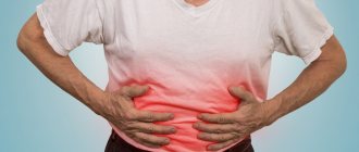
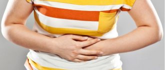
Pancreatitis is inflammation of the pancreas and can be acute or chronic. Causes – poor diet, stones in the bile ducts, poisoning, smoking, chronic alcoholism, disruption of the gastrointestinal tract (stomach, duodenum), viral diseases, infections, etc. Symptoms of acute pancreatitis – nausea, vomiting, heartburn, aching, monotonous pain in abdomen, in the left hypochondrium, persisting for several days. As well as spasmodic pain in the lower abdomen, flatulence, abnormal bowel movements (diarrhea or constipation), chills or excessive sweating, an unpleasant taste in the mouth, and changes in skin color.
Causes of pain
Pancreatitis
Inflammation of the pancreas is the most common factor causing pain on the left side under the ribs. A number of reasons can cause inflammation of an organ:
- alcohol abuse;
- binge eating;
- cholelithiasis;
- viral lesions;
- injuries of the upper floor of the abdominal cavity;
- Helicobacter pylori infection;
- complications after operations and manipulations (endoscopy).
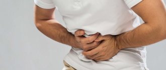
An attack of pancreatitis is characterized by severe pain under the left rib, radiating to the back and shoulder. Often people complain of a sharp girdling pain, combined with uncontrollable vomiting.
With chronic inflammation, the pain syndrome has a dull, aching character, intensifying against the background of physical activity, overeating, alcohol abuse or excessive consumption of fresh fruit. This form of the disease is a logical continuation of untreated acute pancreatitis.
Relief of pain in acute pancreatitis consists of therapy aimed at eliminating its cause - inflammation of the pancreas. In addition, for symptomatic purposes, analgesics are prescribed and novocaine blockades are carried out. The introduction of a local anesthetic to the organ bed in the acute phase of the inflammatory process significantly relieves its painful manifestations.
Intercostal neuralgia
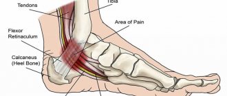
Osteochondrosis, herniated intervertebral discs, and compression fractures of the vertebrae due to osteoporosis often serve as a trigger for sharp pain in the left hypochondrium. Unpleasant stabbing sensations begin behind the spine with a sudden attack. The mechanism of this pathology is compression of the intercostal nerves by the structures of the spinal column. Neuralgia can simulate some diseases of the abdominal organs. Its distinctive features:
- sharp pain under the left rib behind, starting from the vertebrae;
- the beginning is associated with a sudden movement;
- the pain worsens when turning or simply bending the body;
- may disappear when stationary;
- there are no dyspeptic symptoms (nausea, vomiting, bloating, changes in stool character).
Another feature of the pain syndrome is its rapid relief after an injection of an anti-inflammatory drug (diclofenac, dicloberl) or a manual therapy session. In the absence of proper care and elimination of the cause, attacks of neuralgia recur periodically.
Peptic ulcer
Aching pain under the left rib in the front is provoked by a stomach or duodenal ulcer. It is localized below the xiphoid process or, popularly, “in the pit of the stomach.” There is a clear connection with food, thanks to which the presumptive localization of the ulcerative defect is established:
- pain immediately after eating corresponds to an ulcer of the entrance (cardiac) section and body of the organ;
- symptoms appear a few hours later with an ulcer of the exiting section;
- night pain is predominant for a defect in the duodenal mucosa.
In addition, peptic ulcer disease is characterized by dyspeptic symptoms. Similar symptoms and aching pain under the left rib can also be observed with a diaphragmatic hernia. A distinctive feature of a hernia is early vomiting after eating, which brings relief. It is also characterized by a specific symptom - lying down immediately after eating causes regurgitation of gastric juice into the esophagus, manifested by an unbearable sour taste in the mouth.
The pain of a peptic ulcer disappears or is significantly reduced by diet and taking medications that reduce acidity.
Injury
The lateral walls of the abdomen and chest are rich in nerve endings, which, if traumatically damaged, cause pain. After a rib fracture, it persists for several months. It is relieved after taking analgesics and local anti-inflammatory compresses.
The main danger of injury to the left costal arch is not considered to be a fracture of the ribs, but damage to the internal organs. The etiology of severe pain under the left rib may be a rupture of the spleen - a condition that threatens massive intra-abdominal hemorrhage.
When an organ ruptures, pain radiates to the lower abdominal wall, where blood flows, and the left shoulder, which is associated with irritation of the fibers of the phrenic nerve. There is also a specific sign for this injury. When a person tries to lie down, the pain in the abdomen and shoulder increases sharply, causing him to quickly get up. In the medical literature, this is called the “vanka-vstanka” symptom.
A splenic rupture is a serious condition that requires immediate surgical intervention. In most cases, the essence of the operation is the complete removal of an organ that no longer performs vital functions in adults. In extremely rare cases, when the patient’s condition is stable and the degree of organ destruction is insignificant, it is possible to perform organ-saving surgery.
Heart diseases
Dull pain under the left rib is a manifestation of angina pectoris and myocardial infarction, when delay and incorrect diagnosis have a poor prognosis. Treatment of pancreatitis, neuralgia or ulcers during an acute heart attack is worse than absolute inactivity. It is advisable to remember the symptoms accompanying the abdominal form of myocardial infarction:
- chest pain;
- numbness of the left hand;
- dyspnea;
- cardiopalmus;
- pallor.
For these symptoms, an electrocardiogram should be the leading diagnostic procedure, without which treatment cannot be started. Pain relief for a heart attack involves the administration of narcotic analgesics and the use of nitroglycerin tablets. If this diagnosis is suspected, an ambulance should be called immediately.
Pneumonia and pleurisy
Inflammation of the lung or pleura covering this organ is a potential cause of pain in the area of the left costal arch. The stabbing pain intensifies with deep inhalation and finger pressure on the intercostal spaces. These diseases occur against the background of a cold and, in addition to pain, are accompanied by a number of symptoms:
- febrile temperature;
- dyspnea;
- dry cough;
- lag of the left half of the chest in the act of breathing;
- smoothing of intercostal spaces.
Pain relief occurs virtually independently after the start of treatment for the underlying problem. For this purpose, antibacterial drugs, anti-inflammatory, antitussive and expectorant drugs are used. In cases of exudative pleurisy, when fluid accumulates in the pleural cavity, it may be necessary to perform puncture and aspiration of inflammatory exudate.
Colon diseases
On the left in the hypochondrium is the splenic angle of the colon. When it is inflamed, torsion, or a tumor, pain may occur in this area. It is often accompanied by some of the symptoms:
- pathological impurities in feces (mucus, blood, pus);
- diarrhea or constipation;
- bloating;
- increased gas formation;
- dyspepsia;
- absence of gas and stool.
Delay in such a case threatens surgical intervention. Therefore, if you have such symptoms, you should immediately go to the hospital. Pain syndrome in pathology of the colon is rarely the leading symptom, so it quickly stops after eliminating the root cause of the disease.
Renal colic
Stones or sand in the urinary tract are a likely cause of obstruction of the flow of urine into the bladder from the kidneys. In such situations, increased pressure occurs in the pelvis of the latter, the tissue and capsule of the organ stretches, and severe pain occurs. Acute piercing pain is predominantly localized under the ribs at the back. Such an attack is accompanied by a disturbance in the nature of urination in the form of retention, frequent urge or bloody urine. Patients are always restless, it is difficult for them to find a place or a comfortable position in which the attack would not be so pronounced.
Emergency care for renal colic involves administering a combination of various anti-inflammatory, analgesic and antispasmodic drugs. More often than not, the pain subsides after this. To prevent a recurrent attack, it is recommended to take a hot bath, place a heating pad on the painful area, or drink a small amount of strong alcohol. These activities lead to relaxation of the smooth muscles of the urinary tract, the passage of small stones and sand. Subsequently, the patient needs a mandatory scheduled consultation with a urologist to determine the tactics for further treatment of urolithiasis.
Shingles
The chickenpox virus, after the symptoms of the disease subside, does not leave the human body, but is sent to the ganglia of the spinal nerves, where it remains in an inactive state. In case of weakened immunity, hypothermia and other aggravating circumstances, the virus can be activated. But in these cases, it no longer causes chickenpox, but a disease called herpes zoster.
The clinical picture is characterized by burning pain spreading from the spine along the intercostal space. After a few days, small vesicles appear in this place, reminiscent of herpes rashes on the lips. Even later, the vesicles burst and wounds appear, causing a lot of inconvenience to the person. The pathology is characterized by malaise and low-grade fever.
Treatment of pancreatic inflammation
Treatment of acute pancreatitis should take place in a hospital.
First aid - you need to apply an ice bag to the left hypochondrium, avoid eating any food. Drug treatment includes taking antispasmodics, painkillers (opioid analgesics), and antiemetics. In the acute phase, infusion drugs are administered intravenously. For complications, antibiotics may be prescribed. Fasting is observed for three to five days or parenteral nutrition is used.
Therapy for the chronic form of the disease is based on quitting smoking, drinking alcohol, diet therapy with a small amount of fat in the diet, using painkillers, taking enzyme preparations; antidepressants (pregabalin, gabapentin) can be prescribed to relieve pain, as well as treatment of complications.
If there are stones in the gland ducts, then it is rational to perform lithotripsy and endoscopic therapy.
Surgical treatment is performed when drug therapy is impossible or the body is tolerant to their action.
The duration of treatment depends on the form and complications of the disease, and on the body’s susceptibility to therapy.
Treatment
For treatment, in addition to analgesics, antiviral agents are used to eliminate the cause of the disease.
As already mentioned, pain in the left hypochondrium is just a symptom and not an independent disease. When it first appears, a mandatory comprehensive examination is necessary to help identify the cause of the painful condition and prevent its occurrence in the future. Otherwise, the process may become chronic, threatening constant pain, which leads to apathy, depression and other psychological disorders.
Call our contact center at 8 (495) 230 03 09 and we will help you make an appointment with a specialist!
Pancreatic tumors
A pancreatic tumor can be of two types – cancer and a hormonally active tumor.
Men are more often affected; the average age is 55 years. Most often the tumor is localized in the head of the gland. Factors that provoke the development of pancreatic cancer include smoking, chronic pancreatitis, high body mass index, and male gender.
Symptoms of cancer depend on the part of the gland that is affected. If the head of the gland is affected, jaundice may develop, because compression of the bile duct occurs. Skin itching appears. When the body and tail are affected, diabetes mellitus, varicose veins of the esophagus and stomach, and the risk of bleeding develop.
Treatment is surgical or chemotherapy.
Causes of pain in the intestines on the right and left lower abdomen
Experts identify several reasons that could cause pain in the intestines on the left and right side of the lower abdomen.
- Problems of the small intestine.
If the patient feels severe pain in the lower right abdomen, then these symptoms indicate problems with the small intestine. They can be caused by metabolic disorders, microflora, poor diet, long-term use of medications, and alcohol. Pain may indicate the development of dangerous diseases - enteritis, cancer, celiac disease, dyskinesia, ischemia, dysbacteriosis, the appearance of neoplasms, ulcers, polyps, cracks.
- Overeating food.
If a few minutes after eating any food a person begins to belch, acute pain in the intestines in the lower abdomen, these symptoms indicate a weakness of the digestive process. Stomach pain and gastrointestinal disorders are associated with low acidity. The human body does not have enough gastric juice. As a result, the body does not properly digest food, which over time begins to ferment and cause pain.
- Severe stress.
Cutting pain in the lower abdomen may be associated with problems of the nervous system. Nervous tension causes the body to produce nonspecific ulcerative colitis. The pain goes away after a few days. If a person does not feel improvement, the disease enters the chronic stage.
- Pain in the intestines on the right and left lower abdomen in women is associated with the genital tract.
During menstruation, blood flow into the pelvis increases, so this process is accompanied by pain.
- This can provoke stagnation in girls who are prone to developing varicose veins.
Pain in the intestines can appear with decreased vascular tone and observed weakness of connective tissue. The patient begins to experience painful sensations, pain during bowel movements, an increase in the size of hemorrhoids, and gastrointestinal disorders.
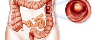
- Diverticulitis.
The disease is accompanied by painful sensations in the intestines in the lower abdomen. Diverticulitis occurs due to inflammation in areas of protrusion of the colon. Painful sensations occur when feces and food stagnate in these areas.
- The appearance of neoplasms.
The appearance of tumors in the human body is accompanied by severe pain in the lower abdomen on the left and right. The pain syndrome increases with changes in the size of the tumor.
- Irritable bowel syndrome.
Irritable bowel syndrome is characterized by severe acute pain in the lower abdomen, and the patient has no appetite. This disease is accompanied by gastrointestinal disorders, diarrhea and constipation, diarrhea, and flatulence. Sometimes the patient notices weakness, an increase in the size of the abdomen, bloating and discomfort in the intestinal area.
- Problems with the genitourinary system
Stitching pains in the lower abdomen on the left and right indicate problems with the kidneys and bladder. The patient should visit a urologist and get tested.
- Pregnancy.
In pregnant women, the size of the uterus increases. This process can affect the appearance of pain in the intestines in the lower abdomen. The enlarged uterus puts pressure on the intestinal walls, which can be accompanied by severe cutting pain, nausea, vomiting and flatulence.
- Pancreatitis.
The next cause, which is characterized by pain in the intestines in the lower abdomen and nausea, is pancreatitis. With pancreatitis, the patient experiences severe cutting pain in the upper abdomen. In some cases, the size of the abdomen increases. Gastrointestinal disorders, including constipation, accompany pancreatitis.
- Duodenal ulcer.
With a duodenal ulcer, the patient is bothered by severe sharp pain in the intestines in the lower abdomen. They are accompanied by vomiting and upset.
Pancreatic stones
Pancreolithiasis is the formation of stones in the pancreatic ducts.
Often stones form if there is a cyst or tumor; this is one of the complications of chronic pancreatitis. Stagnant processes occur in the organ, the secretion of the pancreas thickens, and the protein precipitates. The ducts dilate, the pressure in them increases, and symptoms of pancreatitis appear. Symptoms of pancreolithiasis:
- burning pain in the abdomen radiating to the back and shoulder blade;
- nausea, vomiting with bile;
- steatorrhea – “fatty” stool;
- diabetes;
- increased salivation;
- obstructive jaundice.
Chronic abdominal pain and irritable bowel syndrome
Abdominal pain has been and remains a serious problem in internal medicine and gastroenterology. The greatest difficulties arise when identifying the causes of chronic abdominal pain syndrome. Understanding the mechanisms underlying the formation of pain can partly help in establishing its cause and choosing a way to relieve it [2].
The appearance of pain is associated with activation of nociceptors located in the muscular wall of a hollow organ, in the capsules of parenchymal organs, in the mesentery and peritoneal lining of the posterior wall of the abdominal cavity, stretching, tension of the wall of a hollow organ, and muscle contractions. The mucous membrane of the gastrointestinal tract (GIT) does not have nociceptive receptors, so its damage does not cause pain. Inflammation and ischemia of the gastrointestinal tract through the release of biologically active substances (BAS): bradykinin, serotonin, histamine, prostaglandins, etc. lead to a change in the sensitivity threshold of sensory receptors or directly activate them. The same processes can provoke or aggravate spasm of intestinal smooth muscles, which in turn causes irritation of nociceptors and a sensation of pain. Signals from the intestine are transmitted along afferent fibers through the spinal ganglia and reach the anterior parts of the brain, where the sensation of pain is realized in the postcentral gyrus. Efferent fibers travel to the periphery and cause contraction and relaxation of smooth muscles and vasodilation. A large number of different neurons modulate pain perception and response.
In general, there are four main mechanisms for the formation of abdominal pain: visceral, parietal, radiating and psychogenic.
One of the variants of abdominal pain due to organic causes may be parietal pain arising from involvement of the peritoneum. It is mostly acute, clearly localized, accompanied by tension in the muscles of the abdominal wall, and intensifies with changes in body position and coughing.
The most common mechanism of abdominal pain is visceral pain, which is caused by increased pressure, stretching, tension, circulatory disorders in the internal organs and can be the result of both organic and functional diseases. The pain is usually dull, spastic, burning, and has no clear localization. It is often accompanied by a variety of vegetative manifestations: sweating, anxiety, nausea, vomiting, pallor. Due to the large number of synapses between neurons, double innervation often occurs, which underlies the radiating nature of pain. The latter is understood as the reflection of pain during an intense visceral impulse in the area of zones of increased skin sensitivity, at the site of projection of other organs innervated by the same segment of the spinal cord as the involved organ.
At the initial stages, organic diseases (appendicitis, diverticular disease, etc.) may be accompanied by visceral pain, then, in the case of inflammation of the peritoneum, parietal pain.
Psychogenic pain occurs in the absence of somatic causes and is caused by a deficiency of inhibitory factors and/or an increase in normal incoming afferent signals, due to damage to central control mechanisms and/or a decrease in the synthesis of BAS. The pain is constant, sharply reducing the quality of life, and is not associated with impaired motor skills, food intake, intestinal motility, defecation and other physiological processes.
In functional diseases, the mechanisms of pain formation are different and can be isolated or combined: visceral genesis is often combined with radiating and/or psychogenic mechanisms. The pain is mainly of a daytime nature and rarely occurs during sleep [4].
In practice, the likelihood of an organic cause underlying visceral pain is much greater in the presence of “anxiety” symptoms, which include: predominantly nocturnal pain, waking the patient from sleep; onset of symptoms after the age of 50; presence of cancer in the family; the patient has a fever; unmotivated weight loss; changes identified during direct examination of the patient (hepatomegaly, splenomegaly, etc.); changes in laboratory parameters of urine, feces and blood; changes identified through instrumental studies (stones in the biliary tract, colon diverticula, dilated common bile duct, etc.).
The attempt to differentiate abdominal pain using the least number of examinations, often traumatic for the patient, can be well illustrated by irritable bowel syndrome (IBS). Despite the presence of the term “syndrome” in the name, this pathology refers to independent nosological forms. According to the World Gastroenterology Organization (WGO), IBS is a functional bowel disorder in which abdominal pain or discomfort is associated with bowel movements or changes in intestinal transit [20]. Associated symptoms may include bloating, rumbling, and defecation disorders. To make this diagnosis, according to the Rome III criteria, pain must be recurrent, present at least three days per month for the last three months or more, and be associated with at least two of the following three features: change after defecation, its occurrence must be associated with a change in frequency or stool shape. Symptoms must have bothered the patient for the last three months and first appeared six or more months ago [12]. IBS, like most other functional gastrointestinal diseases, is characterized by an increased level of depression, anxiety, and a tendency to hypochondria.
Abdominal pain in IBS is necessarily present, but depending on the prevailing disorders of intestinal passage, the following options are possible: IBS with diarrhea (frequency of loose stools more than 25% of the time, and dense stools less than 25%, more often in men), IBS with constipation (hard stool more than 25% of the time and, accordingly, liquid less than 25%, women are more likely to suffer), mixed or cyclic IBS (liquefied and solid stool more than 25%) [12, 20]. According to the WGO recommendations, it is possible to divide into subgroups depending on which symptoms dominate: IBS with a predominance of intestinal passage disorders, IBS with a predominance of pain, IBS with a predominance of bloating. And finally, according to the provoking factor, it is possible to subdivide the pathology into post-infectious IBS, food-induced IBS (or certain foods), and stress-induced IBS.
The algorithm for the actions of a practicing physician was developed by WGO and published in 2009 (
). If a patient is under 50 years of age with typical symptoms, no signs of alarm, a low incidence of parasitic infections and celiac disease in the population and no diarrhea, and no changes in the results of routine routine tests (complete blood count), the likelihood of IBS in this patient is so high that there is no need to conduct other examinations [20].
In the presence of persistent diarrheal syndrome, a high incidence of celiac disease or parasitic diseases, it is necessary, respectively, to conduct tests for celiac enteropathy, stool analysis to detect parasitic diseases and colonoscopy (for chronic diarrheal syndrome). In the absence of deviations from normal values, the diagnosis of IBS will be most likely.
Chronic abdominal pain syndrome with gastrointestinal transit disturbances, characteristic of IBS, is similar to the symptoms that occur with enteropathies (gluten, lactase, parasitic), colorectal cancer, microscopic, parasitic colitis, diverticulitis and some gynecological diseases: endometriosis, ovarian cancer. This is due to a single visceral mechanism of pain, which is often accompanied by its radiating genesis, which makes it even more difficult to determine the localization of the pathological process [17].
Relieving chronic abdominal pain is a serious independent problem, since not only elimination, but even an attempt to establish the main cause of its occurrence is not always possible. Considering that pain is often combined, in real practice it is often necessary to use a combination of various means.
One of the approaches to relieving visceral pain is the removal of muscle spasm, which is a universal mechanism of smooth muscles to respond to any pathological influences, which inevitably leads to excitation of nociceptors located in the muscular layer of the gastrointestinal tract [1–4, 18].
The group of antispasmodic drugs is diverse and quite heterogeneous in terms of the mechanism of action and point of application, since a rich receptor apparatus takes part in the contraction of muscle fiber, and this process itself is complex and multicomponent. Thus, drugs that suppress muscle fiber contraction can exert their effect in the following way:
- block the transmission of nerve impulses to muscle fibers (M-anticholinergics - atropine, platiphylline, hyoscine butyl bromide (Buscopan));
- suppress the opening of Na+ channels and the entry of Na+ into the cell (sodium channel blockers - mebeverine);
- suppress the opening of Ca+ channels and the flow of Ca+ from the extracellular space into the cytoplasm and the release of K+ from the cell - the initial stage of repolarization (calcium channel blockers - pinaveria bromide, otilonium bromide);
- suppress the activity of phosphodiesterase, the breakdown of cAMP, thereby blocking the energy processes of the muscle cell (phosphodiesterase blockers - alverine, drotaverine, etc.);
- act through serotonergic receptors, disrupting the regulation of ion transport;
- act on opioid receptors (trimebutine);
- influence oxidases (nitroglycerin and nitrosorbide).
The prescription of each drug must be justified from the standpoint of effectiveness and safety. The more selective the drug, the fewer systemic side effects it has.
Of all the selective antispasmodic drugs, the anticholinergic quaternary ammonium compound hyoscine butyl bromide (Buscopan) has been used the longest in Europe. The drug was first registered in Germany in 1951, and currently it is one of the most studied experimentally and clinically and selective antispasmodic drugs for the gastrointestinal tract. The most important pharmacological properties of hyoscine butylbromide are its dual relaxing effect through selective binding to muscarinic receptors located on the visceral smooth muscles of the gastrointestinal tract, and the parasympathetic effect of blocking nerve ganglia through binding to nicotinic receptors, which ensures selectivity of suppression of gastrointestinal motility.
Hyoscine butyl bromide, due to its high affinity for muscarinic and nicotinic receptors, is distributed mainly in the muscle cells of the abdominal and pelvic organs, as well as in the intramural ganglia of the abdominal organs. Because the drug does not cross the blood-brain barrier, the incidence of systemic anticholinergic (atropine-like) adverse reactions with hyoscine butyl bromide is very low and similar to placebo. Therefore, the feasibility of using this drug is obvious and proven to relieve pain of the visceral component of any origin [1, 3, 18, 19].
The onset of effect when taking Buscopan orally is approximately 30 minutes; duration of action is 2–6 hours. After a single oral administration of hyoscine butyl bromide in doses of 20–400 mg, average peak plasma concentrations are reached after approximately 2 hours. The half-life of the drug after a single oral dose of 100–400 mg ranges from 6.2 to 10.6 hours. Recommended oral dose: 10–20 mg 3–5 times daily. There is also a dosage form of Buscopan in rectal suppositories.
Published in 2006, a comparative placebo- and paracetamol-controlled study of the effectiveness and tolerability of hyoscine butylbromide in the treatment of recurrent cramping abdominal pain, conducted at 163 clinical centers under the leadership of such famous gastroenterologists as S. Müller-Lissner and G. N. Tytgat , included 1935 patients. It showed high efficacy and safety of hyoscine butyl bromide for recurrent abdominal pain [14].
Evidence of the antispasmodic effect of hyoscine butylbromide is the improvement in the results of instrumental examination of the intestine during endoscopic and x-ray examination, which is demonstrated by both an increase in the intestinal lumen and visualization of polyps, diverticula, as well as less pain during manipulations [11, 15].
An example of effective relief of abdominal pain with antispasmodics is their use in IBS [12].
A meta-analysis conducted by T. Poynard et al demonstrated that many antispasmodic drugs individually (mebeverine, cimetropium bromide, trimebutine, otilonium bromide, hyoscine butyl bromide, pinaveria bromide) and the entire group of antispasmodics (OR 2.13; 95% CI 1 .77–2.58) is more effective than placebo in the treatment of IBS pain [16]. Thus, the likelihood of improvement with the use of hyoscine butylbromide in the treatment of IBS is 1.56 times higher (95% CI 1.14–2.15) than with placebo. A number of studies have shown that, in addition to the antispasmodic effect, the good analgesic effect of Buscopan may also be associated with a decrease in the threshold of visceral hypersensitivity, which plays an important role in the pathogenesis of IBS [10].
Antispasmodics with proven effectiveness in the treatment of IBS from the point of view of the special American Gollege Gastroenterology (ACG) are hyoscine butyl bromide, cimetropium bromide, pinaveria bromide and peppermint oil. These drugs can relieve pain or discomfort associated with IBS [5].
Antispasmodics not only relieve pain, but also help restore the passage of contents and improve blood supply to the organ wall. Their administration is not accompanied by direct interference with the mechanisms of pain sensitivity and does not complicate the diagnosis of acute surgical pathology.
Of course, an important place in the relief of pain not only of parietal origin, but also of visceral and psychogenic origin is given to analgesics. The World Health Organization has proposed the following step-by-step approach to pain relief: 1st step - non-opioid analgesics, 2nd step - adding soft opioids, 3rd step - opioid analgesics. Among non-opioid analgesics, it is preferable to prescribe paracetamol due to fewer side effects on the gastrointestinal tract. A number of studies have shown a good effect for pain relief when combining the antispasmodic hyoscine butyl bromide with the analgesic paracetamol [13].
Sometimes it is necessary to use direct analgesics in case of functional diseases, in particular in IBS. The prescription of opiates should be avoided in every possible way, since with such chronic conditions there is a high risk of developing addiction and dependence. Such cases are described in the literature and are called “narcotic bowel syndrome” (intestinal syndrome caused by narcotic drugs). The criteria for this condition include chronic or often recurrent pain that progresses over time, which cannot be explained by a specific pathology, the relief of which requires large doses of narcotic drugs, which increases with the abolition of opiates and is quickly relieved with their use [8, 9].
The effect of antidepressants to potentiate and enhance the analgesic effect of other drugs is well known and proven. Taking into account the presence of a psychogenic mechanism of pain in functional diseases, clinically identified psycho-emotional characteristics of patients (tendency to depression, high levels of anxiety), the interest in psychotropic drugs for IBS is understandable. A recently published systematic review, although highlighting flaws in some study designs, provided evidence to support the use of antidepressants (both tricyclics and selective serotonin reuptake inhibitors) for IBS (amitriptyline 10–75 mg/day at bedtime; selective serotonin reuptake inhibitors: paroxetine, 10–60 mg/day, citalopram, 5–20 mg/day) [6, 20].
Explaining the genesis of symptoms and, above all, abdominal pain, taking into account the level of education, social status of the patient, and establishing a trusting, empathetic relationship between the doctor and the patient is effective in relieving symptoms [7].
Nutritional adjustments to reduce pain and relieve other symptoms should be used with some caution so as not to cause nutritional problems in the patient (deficiency of microelements, vitamins, other nutritional ingredients).
There is no convincing connection between abdominal pain and other symptoms of IBS. The use of drugs effective for relieving various disorders in IBS did not affect the severity of pain. In the presence of IBS with constipation, various classes of laxatives, fiber and other volume-forming drugs are used. A good evidence base exists for osmotic laxatives (lactulose preparations, polyethylene glycol in individual dosages). To speed up the normalizing effect on passage through the gastrointestinal tract in IBS with constipation, irritating laxatives (Dulcolax, etc.) can be prescribed in short courses. For the treatment of IBS with constipation in women, it is possible to use a selective activator of C-2 chloride receptors - lubiprostone [6]. There is evidence of the advisability of using the probiotic strain Bifidobacterium lactis DN-173 010 in order to accelerate intestinal transit.
The main drug for the treatment of IBS with diarrhea is loperamide, which requires individual dosage selection. For severe diarrheal syndrome in women, the serotonergic receptor antagonist (5-HT3)-alosetron has been registered for use in a number of countries [6, 20]. To reduce gas formation, sorbents and other defoamers are used, and some recommendations also call the antibiotic rifaximin (400 mg 3 times a day).
Some effects on improving the general condition and reducing pain were demonstrated by the antagonist of serotonergic receptors (5-HT3) - alosetron (in IBS with diarrhea syndrome), the selective activator of C-2 chloride receptors - lubiprostone (in women with constipation) and the probiotic strain Bifidobacterium infantis 35624 .
To relieve pain and other symptoms in functional pathology, in particular in IBS, a variety of therapy methods are used, including psychological ones: cognitive/behavioral therapy, relaxation methods, hypnosis. The ACG states that psychological therapies including cognitive therapy, dynamic psychotherapy and hypnotherapy are more effective in relieving common IBS symptoms than standard treatments. The attitude towards herbal medicine and acupuncture in general today is optimistically restrained.
The course of any pathology and especially IBS is largely influenced by both the patient’s personal characteristics (attitude to treatment, level of anxiety and degree of trust/distrust in medical manipulations, the presence of chronic traumatic situations, individual emotional characteristics, as well as mental illness), and the behavior of the medical professional. staff (the ability to establish contact and trust, the ability to provide psychological support to the patient). An important point that always increases the patient’s confidence in the doctor is the rapid relief of pain. Therefore, the choice of drugs must be made competently and in a timely manner.
Literature
- Baranskaya E.K. Abdominal pain: clinical approach to the patient and treatment algorithm. The place of antispasmodic therapy in the treatment of abdominal pain // Farmateka. 2005. No. 14.
- Wiley J. Assessment and meaning of abdominal pain. Chapter 1. In the book: J. Henderson. Pathophysiology of the digestive organs. St. Petersburg: Nevsky Dialect, 1997. 275 p.
- Livzan M.A. Pain syndrome in gastroenterology - treatment algorithm // Medical advice. 2010. No. 3–4. pp. 68–70.
- Shulpekova Yu. V., Ivashkin V. T. Symptom of visceral pain in pathology of the digestive organs // Doctor. 2008. No. 9. pp. 12–16.
- Brandt LJ, Chey WD, Foxx-Orenstein AE, Schiller LR, Schoenfeld PS, Spiegel BM, Talley NJ, Quigley EM American College of Gastroenterology Task Force on Irritable Bowel Syndrome An evidence-based position statement on the management of irritable bowel syndrome // Am J Gastroenterol. 2009, Jan; 104, Suppl 1: S1–35.
- Camilleri M. Review article: new receptor targets for medical therapy in irritable bowel syndrome // Aliment Pharmacol Ther. 2010, Jan; 31 (1): 35–46.
- Camilleri M. Evolving concepts of the pathogenesis of irritable bowel syndrome: to treat the brain or the gut // J Pediatr Gastroenterol Nutr. 2009, Apr; 48, Suppl 2: S46–48.
- Drossman DA Severe and refractory chronic abdominal pain // Clin Gastroenterol Hepatol. 2008, vol. 6, no. 9, pp. 978–982.
- Grunkemeier DMS, Cassara JE, Dalton CB, Drossman DA The narcotic bowel syndrome: clinical features, pathophysiology and management // Clin Gastroenterol Hepatol. 2007; No. 5; 1126–1139.
- Khalif IL, Quigley EM, Makarchuk PA, Golovenko OV, Podmarenkova LF, Dzhanayev YA Interactions between symptoms and motor and visceral sensory responses of irritable bowel syndrome patients to spasmolytics (antispasmodics) // J Gastrointestin Liver Dis. 2009, Mar; 18 (1): 17–22.
- Lee JM, Cheon JH, Park JJ, Moon CM, Kim ES, Kim TI, Kim WH Effects of Hyosine N-butyl bromide on the detection of polyps during colonoscopy // Hepatogastroenterology. 2010, Jan-Feb; 57 (97): 90–94.
- Longstreth G., Thompson W., Chey W., Houghton L., Mearin F., Robin C. Spiller. Functional Bowel Disorders // Gastroenterology. 2006; 130:1480–1491.
- Mertz H. How effective are oral hyoscine butylbromide and paracetamol for the relief of crampy abdominal pain // Nat Clin Pract Gastroenterol Hepatol. 2007, Jan; 4 (1): 10–11.
- Mueller-Lissner S., Tytgat GN, Paulo LG, Quigley EM, Bubeck J., Peil H., Schaefer E. Placebo- and paracetamol-controlled study on the efficacy and tolerability of hyoscine butylbromide in the treatment of patients with recurrent abdominal cramps pain // Aliment Pharmacol Ther. 2006, Jun 15; 23 (12): 1741–1748.
- Misra SP, Dwivedi M. Role of intravenously administered hyoscine butyl bromide in retrograde terminal ileoscopy: a randomized, double-blinded, placebo-controlled trial // World J Gastroenterol. 2007 Mar 28;13(12):1820–1823.
- Poynard T., Regimbeau C., Benhamou Y. Meta-analysis of smooth muscle relaxants in the treatment of irritable bowel syndrome // Aliment Pharmacol Ther. 2001; 15: 355–361.
- Spiegel B., Bolus R., Harris LA, Lucak S., Naliboff B., Esrailian E., Chey WD, Lembo A., Karsan H., Tillisch K., Talley J., Mayer E., Chang L. Measuring irritable bowel syndrome patient-reported outcomes with an abdominal pain numeric rating scale // Aliment Pharmacol Ther. 2009, Dec 1; 30 (11–12): 1159–1170.
- Tytgat GN Hyoscine butylbromide: a review of its use in the treatment of abdominal cramping and pain // Drugs. 2007; 67(9):1343–1357.
- Tytgat GN Hyoscine butylbromide — a review on its parenteral use in acute abdominal spasm and as an aid in abdominal diagnostic and therapeutic procedures // Curr Med Res Opin. 2008.
- WGO Practice guideline - Irritable bowel syndrome: a global perspective. April 2009.
M. F. Osipenko , Doctor of Medical Sciences, Professor S. I. Kholin , Candidate of Medical Sciences, Associate Professor A. N. Ryzhichkina , Candidate of Medical Sciences, Associate Professor Novosibirsk State Medical University, Novosibirsk
Contact information for authors for correspondence
Diagnosis of pancreatic diseases
To diagnose diseases of the pancreas, the doctor examines and palpates the abdomen and prescribes laboratory and instrumental methods.
A biochemical blood test examines the level of pancreatic enzymes (amylase, lipase), blood glucose, and liver function tests.
A stool analysis is carried out to assess the deficiency of enzyme production, the presence of undigested food, fat, and certain enzymes (pancreatic elastase) is examined.
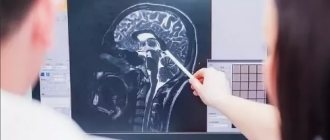
Visual instrumental examinations are carried out using transabdominal ultrasound (ultrasound of the abdominal cavity), CT, radiography, magnetic resonance cholangiopancreatography.
In the structure of radiology diagnostics of the abdominal organs, visualization of the spleen occupies a special place due to the low incidence of pathological processes. However, we must not forget about the variations in the normal anatomy of the spleen and the possibility of regenerating its tissue after removal (reparative regeneration).
Clinical example
In an outpatient patient K., 70 years old, an ultrasound examination on January 28, 2013 revealed a mass formation in the left hypochondrium; there was a history of splenectomy (automobile trauma in 2002); multislice computed tomography (MSCT) with contrast enhancement was recommended as a follow-up examination.
An MSCT study with intravenous contrast enhancement on January 31, 2013 revealed an additional volumetric formation in the bed of the removed spleen with smooth, clear contours, dimensions 38×51×40 mm, the nature of the accumulation of the contrast agent was similar to the spleen tissue with the presence of a feeding vessel extending from the splenic arteries, most likely ectopic tissue of the spleen (splenosis; Fig. 1, a-c).
Figure 1. MSC tomogram of the abdominal organs with intravenous contrast enhancement, arterial phase in patient K. a - hypervascular formation in the bed of the removed spleen (arrow)
Figure 1. MSC tomogram of the abdominal organs with intravenous contrast enhancement, arterial phase in patient K. b - maximum intensity projection (MIP): a feeding vessel extending from the splenic artery to the formation in the bed of the removed spleen (arrow)
Figure 1. MSC tomogram of the abdominal organs with intravenous contrast enhancement, arterial phase in patient K. c - multiplanar reconstruction (MPR; coronal projection): hypervascular formation in the bed of the removed spleen (arrow).
A control CT study with intravenous contrast after 6 months did not reveal any growth dynamics of the previously detected spleen tissue.
Discussion
Normal anatomy and developmental variants of the spleen
Spleen
- an unpaired parenchymal organ of the hematopoietic and lymphatic systems, which is located in the upper floor of the abdominal cavity, deep in the posterior part of the left hypochondrium. The dimensions of the spleen are variable: length 12-14 cm, width 8-10 cm, thickness 3-4 cm, density characteristics of the parenchyma in MSCT studies 40-50HU. The main blood supply to the spleen is carried out by the largest branch of the celiac trunk - the splenic artery. It is important to be aware that during the arterial phase of contrast enhancement, a characteristic “marbled” (pallisading) pattern of enhancement of the splenic parenchyma is detected, which is likely due to differences in pulp perfusion and should not be confused with a focal lesion.
Normally, 10% of adults have, in addition to the normal spleen, additional spleens called accessory lobules of the spleen, or spleens. They are usually found in the hilum of the spleen or near the tail of the pancreas, but can also be found in the mesentery of the small intestine or greater omentum [1].
Anatomical asplenia (absence of the spleen) and polysplenia (several small vestigial bodies of the spleen) are associated with congenital heart disease and isomerism (bilateral right or left-sided anatomy of the heart, bronchi and abdominal organs). This is influenced by the fact that the development of the spleen and heart occurs during the same period of intrauterine development.
Splenosis
Splenosis is an autotransplantation of splenic tissue, usually as a result of traumatic rupture of the spleen or splenectomy. The average interval between injury and the onset of abdominal or pelvic splenosis is 10 years (range 5 months to 32 years). On average, the period of onset of thoracic splenosis is 21 years (3-45 years) [2]. Thoracic splenosis usually occurs with splenic rupture accompanied by a simultaneous diaphragmatic rupture and is therefore less common [3]. Subcutaneous splenosis is a rare condition, the pathogenesis of which may be explained by mechanical implantation. In all cases where he was observed, there was surgery or post-traumatic scarring.
It is important to distinguish splenosis from accessory lobules of the spleen, since both conditions are manifestations of ectopic splenic tissue. In the first case we are dealing with an acquired process, and in the second with a congenital condition. The accessory lobule of the spleen is histologically represented by unchanged splenic tissue, in contrast to splenosis, in which the structure is changed due to poorly formed white pulp, normal content of red pulp and the absence of trabeculae. Also, splenic tissue with splenosis has fewer elastic fibers, the absence of a hilum, and a poorly formed capsule [4]. In addition, accessory spleens are located near the gastrosplenic ligament, left hypochondrium, while splenosis can be in any floor of the abdominal cavity or even have an extraperitoneal localization. Autografts of splenic tissue after splenic trauma are often numerous, have variable size and shape, and are located along the peritoneum and pleura, retroperitoneum, pericardium, lungs, and even subcutaneously.
Pathogenesis
Splenosis begins at the time of splenic rupture or splenectomy, when the splenic pulp enters the abdominal cavity [5]. The number of ectopic splenic tissue nodules that develop in the peritoneal cavity is hypothesized to correlate with the severity of splenic injury.
Another mechanism of splenic tissue transplantation is splenic vein embolism or hematogenous spread of splenic pulp, which occurs in intrahepatic and intracranial splenosis.
Splenosis has rare clinical manifestations. Occasionally, patients experience nonspecific abdominal pain (likely due to tissue infarction), intestinal obstruction due to external mechanical compression by ectopic splenic tissue, gastrointestinal bleeding, or hydronephrosis. Pleurisy and hemoptysis may be symptoms of thoracic splenosis.
A particular difficulty in diagnosis is splenosis, which simulates a primary disease of other organs (liver, pancreas).
Patient A., 48 years old, was routinely admitted to the surgical department of the Federal State Budgetary Institution Central Clinical Hospital with a polyclinic for additional examination and surgical treatment. On an outpatient basis, ultrasound and MSCT with intravenous contrast were used to diagnose her as a “formation of the tail of the pancreas with focal liver damage of a secondary nature.”
However, during a preoperative examination during ultrasound of the abdominal organs and retroperitoneal space, a volumetric formation was revealed in the bed of the removed spleen (splenectomy in 1998, torsion of the elongated pedicle of the spleen) measuring 31×33 mm, comparable in echogenicity and echo structure to spleen tissue (Fig. .2).
Figure 2. Ultrasound data of the abdominal organs. a — isolated scanning mode: in the bed of the removed spleen a formation is visualized, comparable in echo structure and echogenicity to spleen tissue (arrows)
Figure 2. Ultrasound data of the abdominal organs.
b — energy mapping mode, longitudinal scanning: single vessels are visualized in the formation. No focal liver damage was detected by ultrasound. A routine MSCT examination of the abdominal cavity with intravenous contrast enhancement revealed an additional structure with smooth, clear contours in the bed of the removed spleen measuring 30×33×32 mm, closely adjacent with its lateral surface to the tail of the pancreas, the nature of the accumulation of the contrast agent is similar to the spleen tissue, without signs of hypervascularization, the liver parenchyma is homogeneous (Fig. 3).
Figure 3. MSC tomogram of the abdominal organs with intravenous contrast enhancement, arterial phase in patient A. a - hypervascular formation at the level of the tail of the pancreas, according to the density characteristics and type of contrast enhancement corresponding to the spleen tissue (arrow)
Figure 3. MSC tomogram of the abdominal organs with intravenous contrast enhancement, arterial phase in patient A. b - maximum intensity projection (MIP; arterial phase): a feeding vessel extending from the splenic artery to the ectopic tissue of the spleen, simulating the formation of the tail of the pancreas ( arrow)
Figure 3. MSC tomogram of the abdominal organs with intravenous contrast enhancement, arterial phase in patient A. c - multiplanar reconstruction (MPR; coronal projection): a formation in the bed of the removed spleen at the level of the tail of the pancreas (arrow).
Thus, taking into account the anamnesis, the absence of a clinical picture, and the results of radiation imaging methods, the incoming diagnosis was changed to “splenosis of the left subdiaphragmatic region.”
Diagnostics
Since most patients do not complain, the presence of ectopic splenic tissue is often an incidental finding on ultrasound, CT, or MRI. If a mass similar to normal splenic tissue is found during diagnosis in a patient with a history of splenic trauma or splenectomy, the diagnosis of splenosis should be considered.
According to ultrasound data
a well-demarcated (for example, with perihepatic splenosis), hypoechoic formation with clear contours and single arterial and venous vessels is determined.
With MSCT
A hypodense round formation with smooth, clear contours is determined, possibly multiple. The density and features of contrast enhancement of the formation are similar to those of the spleen tissue: ~50HU, hyperdense in the arterial phase, isodense with the liver parenchyma in the portal phase (with the perihepatic variant of splenosis), and hypodense in the parenchymal phase.
With MRI
Normal splenic tissue is hypointense on T1-weighted images (WI) and hyperintense on T2-weighted images. Occasionally, the spleen may be hypointense on T2-weighted images due to excess iron deposition. Typically, splenic tissue implants for splenosis have the same signal intensity and contrast enhancement characteristics on MRI. Typically, heterogeneous enhancement of splenic tissue occurs in the arterial phase of MR studies, while in the delayed phase this ectopic tissue becomes homogeneous.
In splenosis simulating a liver mass, a hypointense rim around the mass may be noted on T1-weighted images. The presence of a peripheral rim of the lesion is characterized by low signal intensity on T1-weighted images and T2-weighted images and represents a thin layer of fat or fibrous capsule around the lesion and is described as a characteristic feature of splenosis. This sign would be nonspecific for primary liver disease.
Scintigraphy
A widely used radionuclide diagnostic method for identifying additional splenic tissue after ineffective splenectomy is scanning with the introduction of colloidal radiopharmaceuticals labeled with 99mTc and 111In. Their spectrum is quite wide, but colloidal sulfur labeled with 99mTs has the greatest sensitivity. Scintigraphic isotopes, such as 99mTc, are detected in reticuloendothelial tissue (liver, spleen, bone marrow) [6]. Using this method, even small areas of spleen tissue can be visualized.
However, a more specific method is scanning with the introduction of 99mTc-labeled red blood cells that have been heat-treated. The physiology of this method lies in the fact that damaged red blood cells will selectively accumulate in the spleen tissue. Few radioisotope laboratories are equipped for this technique, so its use is limited.
MRI with intravenous iron oxide
Another specific method for diagnosing splenosis is MRI with the introduction of nanoparticles as an alternative to traditional contrast agents.
For this purpose, intravenous administration of superparamagnetic iron oxide nanoparticles is used, which are subject to nonspecific capture by cells of the reticuloendothelial system.
In this regard, iron oxide nanoparticles are used for imaging the liver, spleen and lymph nodes. Superparamagnetic iron oxide nanoparticles are highly detectable
even at very low concentrations. There is evidence of imaging of individual cells containing these nanoparticles and even individual nanoparticles. Therefore, this type of nanoparticles
actively used for tagging individual
cells and tracing their migration routes in vivo [7].
Differential diagnosis
- metastatic lesion;
- enlarged lymph node (abdominal lymphadenopathy);
- depending on the location, it can imitate malignant formations of other organs and tissues;
- endometriosis.
Conclusion
Splenosis is a benign condition that is often misdiagnosed as a neoplastic process, and therefore splenosis should be included in the differential diagnosis for neoplasms, single or multiple, in the abdominal cavity or even in atypical locations (intrahepatic, pleura, lungs, pericardium, retroperitoneum , or subcutaneously) in patients with a history of splenic trauma or after surgical removal of the spleen.
It is necessary to know the normal anatomy of the spleen, variants of the anatomical structure and be able to interpret data from various imaging methods for the correct diagnosis of splenosis.
Diet for diseases of the pancreas
The diet should include soft, well-chopped food.
Steam, bake, boil, stew foods. Include puree soups and cream soup in your diet. Bread can be consumed dried, yesterday's bread made from first and second grade flour. Meat - chicken, turkey, lean parts of beef, low-fat fish is allowed. Eggs should be in the form of omelettes, steamed in the oven. You can eat non-acidic types of fruits, baked fruits. You can drink rosehip decoction, sugar-free compote, herbal teas, water. Of the fats in the diet, butter with 82.5% fat content and sunflower oil should be present, but limited. Food should be taken warm. It is necessary to exclude sausages, smoked meats, fatty, fried, canned and pickled foods.
Forbidden:
- alcohol;
- spicy food;
- coffee;
- energy, carbonated sweet drinks;
- chocolate;
- ice cream;
- duck, goose meat, pork, lard, bacon;
- legume products;
- garlic, cabbage;
- grapes, bananas.
The severity of the diet depends on the disease of the pancreas. In the acute period, fasting is prescribed for three to five days; in case of exhaustion, parenteral and special enteral nutrition is used.
1.General information
As you know, ribs are not only part of the musculoskeletal system, but also serve as mechanical protection for vital internal organs: heart, lungs, liver, gall bladder, etc. Therefore, any pain behind or under the ribs should alert you. If pain discomfort in the hypochondrium has become recurrent or chronic, you should definitely consult a doctor: there are simply no problems in this area that can be neglected. But it is, on the contrary, very easy to waste precious time and miss the development of a dangerous pathology, and this happens, unfortunately, all the time in clinical practice.
On the left under the ribs there are many potential sources of pain: individual parts of the intestines, stomach, spleen, pancreas. Heart pain often radiates to the left hypochondrium, but it would be wrong to assume that “any pain on the left is from the heart.” Possible causes are far from limited to cardiac problems.
A must read! Help with treatment and hospitalization!