Author Peter Deryabin
03/06/2016 14:00 (Updated: 09/16/2021 14:58)
Health » Healthcare
Why is modern man increasingly faced with colon diseases? How to avoid the development of malignant tumors? And what intestinal diseases are most common today in Russia and the world? The director of the Federal State Center for Coloproctology, Doctor of Medical Sciences Yuri Shelygin, spoke about this.
Why is inflammation of the intestinal walls dangerous?
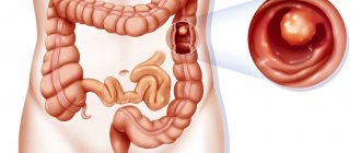
Recently, the number of Russians diagnosed with IBD has increased annually by an average of 10–12%. Most often, residents of modern megacities suffer from ulcerative colitis and Crohn's disease. In the first case, the mucous membrane of the colon is affected, and in the second, the end of the small intestine and the beginning of the large intestine. Inflammation leads to the formation of bleeding ulcers and progresses rapidly, leading to irreversible tissue damage if undiagnosed and treated.
- Patients with Crohn's disease have a significantly increased risk of intestinal obstruction and, as a result, fistula formation.
- Massive bleeding in ulcerative colitis can cause intestinal perforation and life-threatening peritonitis.
- Finally, IBD is recognized as one of the factors that significantly increases the likelihood of tumor formation in the intestine.
Even at the initial stage, intestinal diseases manifest themselves with unpleasant symptoms that significantly reduce the quality of life.
Anatomy and physiology
The large intestine is separated from the overlying small intestine by a muscular valve, the so-called. ileocecal sphincter or bauhinian valve. The large intestine is located in the lower part or floor of the abdominal cavity and includes the following sections:
- Caecum (lat. – caecum). It is 3-8 cm long and 4-7 cm wide. This section of the large intestine is equipped with a vermiform appendix, the appendix. The cecum with the appendix is projected onto the right middle and lower part of the anterior abdominal wall.
- Colon (lat. – colon). The longest section of the small intestine. The colon, about 4 cm in diameter, borders the entire lower floor of the abdominal cavity with loops of the small intestine and other organs. Hence the name. In the colon, its initial ascending part is distinguished. 12-20 cm long from the cecum to the right bend, the so-called. hepatic angle. The transverse colon is its middle part, 50 cm long, from the hepatic angle to the splenic angle, the left bend. The descending part, 22-23 cm long, runs from the splenic angle to the sigmoid colon. Due to the fact that the transverse part sags slightly, the configuration of the colon resembles the letter M.
- Sigmoid colon (lat. - colon sigmoideum). The same applies to the colon, and in the form of two loops 54-55 cm long and 4 cm in diameter is located between the descending part and the rectum. It is projected onto the anterior abdominal wall in its lower left part.
- Rectum (lat. - rectum). Located in the pelvic cavity. The length of the rectum is 14 cm. The width in the initial sections is 4 cm, then it expands into the ampulla of the rectum 7.5 cm, and ends with a narrow opening of the anus.
Compared to other parts of the gastrointestinal tract , the large intestine has its own characteristics. Unlike the pink small intestine, the large intestine has a grayish color. On the outside it is covered with a serous connective tissue membrane. Parts of the colon, and in some cases the cecum, are fixed to the walls of the abdominal cavity by the mesentery. This mobile anatomical formation is formed by two layers of peritoneum. Vessels and nerves pass through the mesentery to the intestines.
The middle muscular layer is represented by an inner circular or circular layer and an outer longitudinal layer. Peristaltic (wave-like) contractions of these muscles ensure the movement of the food bolus through the intestinal tube.
But, unlike other parts of the gastrointestinal tract, the outer muscle layer here is continuous only at the junction of the cecum with the appendix , and in the rectum. And for the most part it is represented by three ribbons. These tapes seem to tighten the intestinal tube. As a result, protrusions and haustra are formed on its surface. The haustrae are separated from each other by transverse grooves. These grooves correspond to the semilunar folds of the mucous membrane.
There are no villi on the mucous membrane, like in the small intestine. It contains secretory glands that secrete digestive juice. Colonic juice is secreted at rest, but its amount is small.
Food contents enter the large intestine from the small intestine in small portions at the moment the ileocecal sphincter opens. In response to mechanical irritation, juice secretion increases many times over. It contains: amylase, lipase, and a number of other digestive enzymes. But in general, the content of enzymes in the juice of the large intestine is low, 20 times less than in the juice of the large intestine.
The amount of absorbed food components is also small. Here, those food components that are not absorbed in the small intestine are absorbed in small quantities. The main function of the large intestine is to absorb water from the bolus of food and form feces in a daily amount of 150 g or more.
When eating plant foods, more feces are formed than when eating foods of animal origin. Plant foods contain a large amount of indigestible fibers that absorb intestinal toxins. The final section of the gastrointestinal tract, the rectum, serves as a reservoir for feces. The act of defecation is controlled by muscle valves - the internal and external sphincter.
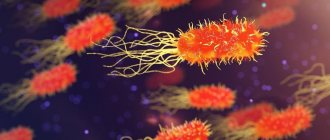
The functioning of the large intestine is largely influenced by the microflora it contains, represented by bifidobacteria, lactobacilli, and bacteroides.
The main functions of physiological intestinal microflora:
- The final breakdown of undigested food components.
- Inactivation of bile components and digestive juices that enter the large intestine from the overlying sections of the gastrointestinal tract.
- Neutralization of toxic products formed during the digestion of food.
- Synthesis of vitamin K and some B
- Destruction of pathogenic intestinal microflora.
- Participation in other nonspecific immune reactions.
Along with microflora, immune protection is provided by accumulations of lymphoid tissue located in the mucous membrane, lymphatic follicles.
IBD in children: growth and development of the body is at risk
Ulcerative colitis in children can appear even before the age of one year and very quickly lead to total damage to the colon. If left untreated, the disease can result in a serious disruption of the physiological process of development of the child’s body: growth retardation or retardation in physical development. If endocrine pathology is not detected, a good pediatrician will definitely prescribe a gastrointestinal tract diagnosis for the child. In addition, this pathology causes serious psychological problems in both children and schoolchildren.
From clinical practice
Liver damage, although without obvious clinical manifestations, is quite common in chronic intestinal diseases. In adults, pericholangitis with the portal triad develops in 70% of cases. 50% have fatty liver changes. 10% acquire liver disease. Liver cirrhosis develops in 5%.Other, more severe and rare complications are chronic active hepatitis, sclerosing cholangitis and biliary tract carcinoma.
It should be noted that in children, chronic active hepatitis is a common complication, while pericholangitis and sclerosing cholangitis are very rare and are asymptomatic.
We present a clinical case of ulcerative colitis in a three-year-old girl. The initial phase of the disease was asymptomatic. Subsequently, pronounced hepatomegaly developed.
The girl was hospitalized at the age of three. 8 months before hospitalization, irritability, loss of appetite, intermittent abdominal pain, and stool mixed with scarlet blood were noted. With the symptoms described above, the girl was hospitalized. On examination, significant hepatomegaly was noted (+4 cm from below the costal arch along the right midclavicular line). In the blood test, ESR is 104 mm per hour; total blood protein 9.9 g%; albumin 2.5 g%; a1-globulin 0.3; a2-globulin 1.1; b-globulin 1.0; g-globulin 5.0; IgG 4810 mg%; alkaline phosphatase 1273 IU; Antinuclear and anti-smooth muscle antibodies are absent.
During hospitalization, the girl complained of frequent abdominal pain. There is an admixture of mucus and scarlet blood in the stool. Analysis of stool mucus revealed many neutrophilic leukocytes. Endoscopically revealed an extensive inflammatory process throughout the colon with swelling of the mucous membrane. Upon contact with the instrument, the mucous membrane bled easily.
With multiple biopsies, histological analysis was performed, which revealed thinning of the glandular ducts with mono- and polynuclear infiltration.
Percutaneous liver biopsy revealed inflammatory and granulomatous changes predominantly in the portobiliary spaces. Moreover, an inflammatory process such as pericholangitis and initial cholangitis in the interlobular ducts was noted.
Initial treatment with prednisone and subsequent salazopyridine (SAZP) resulted in complete remission of intestinal symptoms. Clinical remission with complete regression of hepatomegaly (the liver was palpated at the costal arch) was confirmed histologically. Laboratory parameters were also normalized (IgG 997 mg%).
It is known that ulcerative colitis is often accompanied by cow's milk protein intolerance, so a skin test and a test to determine specific IgE were performed. IgE levels were significantly increased.
Rapid clinical remission was due to two factors: the administration of salazopyridine (SAZP) and the simultaneous exclusion of cow's milk from the diet.
References 1. Walker Smith J., Crohn's Disease. In: Diseases of the small intestin in children. 1989. 2. Booth IW Chronic inflammatory bowel disease // Arch. Dis. Chid. 1991; 66: 742. 3. Elewaut D., De Keyser F., Culvelier C., Lazarovits AI, Mielants H., Verbruggen G., Sas S., Devos M., Veys EM Distinctive activated cellular subset in colon from patients with Chron's disease and ulcerative colitis 1998. 4. Casini-Raggi V., Kam L., Chong YJF, Fiocci C., Pizarro TT, Cominelli F. Mucosal imbalance of IL-1 and IL-1 receptor antagonist in inflammatory bowel disease. A novel mechanism of chronic intestinal inflammation // J. Immunol. 1995; 154: 2434-2440. 5. Olafsdottir E. J., Fluge G. and Hanf K. Chronic inflammatory bowel disease in children in Western Norway // J. Ped. Gastroen. North. 1989; 8: 454. 6. Castro M., Ansaldi N., Bianchi E. et al. Malattie inflammatorie chroniche intestinali: studio multicentrico italiano // Riv. Ital. Ped. 1991; 17:137. 7. Blaser MJ, Miller RA, Singleton J. W: Patients with active CD have elevated serum antibodies of seven enteric bacterial pathogens // Gastroenterology 1984; 87: 88-95. 8. Shanahan F., Duerr R., Rotter J. et al.: Neuthrophil autoantibodies in ulcerative colitis: familial aggregation and genetic heterogenety // Gastroenterology. 1992;103:456-461. 9. Halstensen T., Mollnes T., Garred et al. Surface epithelial related activation of complement differs un Crohn's disease and ulcerative colitis // Gut. 1992; 33: 902-908. 10. McNeish AS La diagnosi e la terapia della m. Di Crohn in eta pediatrica // Prospettive in Pediatria. 1988; 71: 245. 11. Shurmann G., Bretzler M., Meuer S. et al. Soluble IL-2r, IL-6 and IL-1 beta in patients with CD and US: preoperative levels and postoperative changes in serum concentrations // Digestion. 1991; 51: 51-59. 12. Sciumert R., Towner J., Zipser R.: Role of eicosanoids in human and experimental colitis // Dis. Sci. 1988; 33: 58-64. 13. McLain B.I., Diridson P.N., Stokes K.B. et al. Growth after gut resection for Crohn disease // Arch. Dis. Child. 1990; 65: 760. 14. Davies G., Evans CM, Shand WS, Walker Smith J. Surgery for Crohn's disease in childhood: influence of sites of disease and operative procedure on outcome // Br. J. Surg. 1990; 77: 91. 15. Booth IW The nutritional concequences of gastrointestinal disease in adolescence // Acta Pediatr. Scand. Suppl. 1991; 373: 91. 16. Layden T., Rosenberg J., Neuncharnsky B. Reversal of growth arrest in adolescents with Chronic disease after parental alimentation // Gastroenterology. 1979; 70: 101. 17. Hunter JO Nutritional factors in inflammatory bowel disease. 18. Morin CL, Rulet M., Roy CC, Webwn A. Contnous elemrntal enteral alimentation in children with Crohn's disease and growth failure // Gastroenterology. 1980; 79: 1205. Belli DC, Seiduman E, Bouthiller L et al. Chronic intermittent elemental diet improves growth failure in children with Crohn's disease // Gastroenterology. 1989; 97: 905. 19. Aiges H, Mancovitz J, Rma J et al. Home nactural sullplemental naso-gastric feedings in growth retarded adolescents with Crohn's disease // Gastroenterology. 1989; 97: 905. 20. Sanderson IL, Udeen S, Davies PSW et al. Remission induced by elemental diet in small bowel Crohn disease // Arch. Dis. Child. 1987; 62, 1223. 21. Giaffer MN, Nath G., Holdsworth CP Controlled trial of Polvmenic versus elemental diet in the treatment of active Crohn's disease // Lancet. 1990; 335: 816. 22. Seidman E., Leleiko N., Ament M. et al. Nutritional issues in pediatric inflammatory bowel disease // J. Ped. Gastroenterol. Nutr. 1991; 12: 424. 23. O'Sullivan M. A, O'Mora CA Nutritional therapy in Crohn's disease. 1998. 24. Zoli G., Care M., Parazza M., Spano C., Biagi PL, Bernardi M., Gasbarrini G. A randomized controlled study comparing elemental diet and steroid treatment in Crohn's disease. 1997. 25. Malchow H., Ewe K., Brandes JW, Coebell H., Ehms H., Sommer H., Jesdinsky H. European cooperative Crohn's disease study (ECCDS): results of drug treatment // Gastroenterology. 1984; 86: 249-266. 26. Prantera C., Pallone F., Brunetti G., Cottone M., Meglioli M. The Italian IBD study group: Oral 5-Aminoswlieye acid (Asacol) in the maintenance treatment of Crohn's disease // Gastroenterology. 1992; 103: 363-368. 27. Steinhart AH, Hemphill DJ, Greenberg GR Sulphasalazine and mesalazine for the maintenance therapy for Crohn's disease. A meta-analysis. DDW Abstract Book. 1994. A-2842. 28. Tremaine WJ Maintenance of remission in Crohn's disease: is 5-fminosalicylic acid the answer? // Gastroenterology. 1992; 103: 697-704. 29. Bernstein IH, Frank MS, Brandt LG et al.: Healing of perineal Crohn's disease with metrondazolo // Gastroenterology. 1980; 79: 357-365. 30. Markovitz J., Grancher K., Mandel F., Daum F. Immunosopressive therapy in pediatric inflammatory bowel disease: results of a survey of the North American Society of Pediatric Gastroenterology an Nutrition // A J. Gastroenterol. 1993; 88(1): 44-48. 31. Lloyd-Still JD Azathioprine and the treatment of chronic inflammatory bowel disease // J. Pediatr. 1990; 117(5):732-735. 32. Verhave M., Winter HS, Grand RJ Azathiopine in the treatment of children with inflammatory bowel disease // J. Pediatr. 1990; 117(5): 809-814. 33. Feldman M., Achord JM, Chang EB Cyclosporine in Crohn's disease - low doses won't do it // Gastroenterology. 1990; 98 (5): 1383-1384. 34. Balfour RB Cyclosporine therapy for inflammatory bowel disease // N. Eng. J. Med. 1994; 330(26): 1897-1898. 35. Sachar DB Cyclosporine treatment for inflammatory bowel disease. A Step Backward or a Leap forward? // N. Eng. Med. 1995; 321(13):894-896. 36. Geagan BG, Rochon J, Fedorac RN, Irvine EJ, Wild G. et al. Metrotrexate for the treatment of Crohn's disease // N. Eng. J. Med. 1995; 332(5):292-297. 37. Martin LW, Torres AL, Fisher JE, Alexandre F. The critical level for preservation of continuity in the ileo-anal anastomosis // JPS 1985; 20:664
Symptoms of intestinal diseases: when should you see a doctor?
The disease often manifests itself with intimate symptoms, which the majority of patients try to fight on their own, without rushing to a paid appointment with a proctologist or gastroenterologist. In addition, intestinal inflammation is often accompanied by “masking” symptoms: for example, only stomatitis, as in Crohn's disease, or only inflammation around the anus - perianal dermatitis, or lesions of the eyes or joints. Therefore, the patient often comes to the doctor with a severe form, when the path to recovery turns out to be both longer and more financially expensive.
The following signs should alert anyone:
- frequent diarrhea - up to 15–20 times a day;
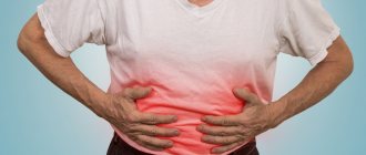
- pain and/or cramping in the abdominal area;
- weakness, increased fatigue;
- loss of appetite and noticeable weight loss;
- temperature increase;
- the appearance of traces of blood in the stool.
The likelihood of damage to the large and small intestines is increased in people who regularly and in large quantities consume dark varieties of meat and processed products (sausages, sausage, ham, bacon, etc.). But you should know that adherence to a diet with a predominance of vegetables, fruits, and foods made from whole grains is not a panacea.
A genetic factor plays a certain role: the tendency to intestinal inflammation can be inherited. Among the sick people, people who abuse smoking predominate. Also important are the lack of vitamin D in the body and frequent and uncontrolled self-administration of antipyretic medications.
An appointment with a proctologist in Moscow is recommended not only for people with unstable stools. In order not to fill the army of colorectal cancer patients, even a generally healthy person after 45 years of age should visit a doctor at least once every five years for preventive purposes. The absence of visible signs of inflammation does not exclude the possibility of a hidden disease. Symptoms of intestinal cancer detected at an early stage allow doctors to save the patient and achieve stable remission.
How are inflammatory diseases and bowel cancer diagnosed?
The specialists of the multidisciplinary clinic “Trit” are proficient in all the techniques that allow them to identify even the smallest changes in the intestinal mucosa.
- An ultrasound examination is performed to detect thickening of the walls of the large and small intestine and narrowing of their lumen.
- Ultrasound with Doppler function allows you to check blood flow parameters.
- If necessary, an MRI OR CT scan is prescribed, as well as an x-ray examination.
- To detect blood in the stool, indicating the presence of bleeding in the gastrointestinal tract, laboratory tests are performed (including determination of fecal transferrin and hemoglobin).
The “gold standard” of research is a modern endoscopic technique, intestinal colonoscopy under anesthesia. If the patient has extraintestinal manifestations of the disease, doctors of relevant specialties - an ophthalmologist, dermatologist or rheumatologist - are also involved in the diagnosis.
Diagnostics
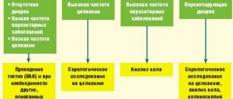
Diagnosis of the large intestine begins with a digital examination of the anus. Next, laboratory and instrumental studies begin. , endoscopy of the large intestine is diagnostically valuable - sigmoidoscopy and colonoscopy . During sigmoidoscopy, the doctor examines the rectum and sigmoid colon, and during colonoscopy, the entire large intestine.
Irrigoscopy is an X-ray diagnosis of the large intestine after filling through the anus with a contrasting barium suspension. Plain radiography of the abdominal organs without the use of contrast is informative in case of intestinal obstruction. The so-called Kloiber cups, horizontal fluid levels in the intestinal lumen. This is a symptom specific to intestinal obstruction.
Of the laboratory tests, the main place is occupied by the diagnosis of stool. Depending on the tasks assigned, feces are taken for:
- general analysis (coprogram);
- occult blood;
- worm eggs;
- bacterial culture on nutrient media.
A supporting role is played by a general and biochemical blood test, immunodiagnostics and blood culture on nutrient media.
Colonoscopy of the intestine: are patients’ fears justified?
An illness diagnosed at an early stage creates the least amount of problems for the patient. Therefore, highly informative screening colonoscopy is included in government programs to combat colon cancer in the USA, Israel and European countries. Citizens of developed countries are constantly informed about the need for regular examinations for the prevention and early diagnosis of colorectal cancer.
The specifics of such an intimate procedure do not evoke the most positive emotions among Russians. Pain is a major deterrent even for those people who are fully aware of the need for testing. But is it possible to compare the sensations that arise during a colonoscopy with the torment experienced by patients suffering from already developed oncology!
It should be understood that gentle diagnostic technology, which can be performed under anesthesia, will allow you to avoid dangerous symptoms, frightening diagnoses and a solid set of diagnostic procedures in the future.
Advantages of endoscopic examination in a paid clinic
Colonoscopy under anesthesia is carried out after appropriate preparation - following a special diet and complete cleansing of the intestines with the help of potent laxatives.
- In a European-style clinic, the patient will receive detailed recommendations on balancing the diet, which will help them calmly, without weakness or discomfort, undergo the stage of fasting and cleansing the intestines.
- Modern equipment allows a highly qualified specialist to conduct an examination with pinpoint precision without damaging the intestinal mucosa.
- The systematic process will take no more than 20 minutes, after which the patient will wake up in the room without experiencing any negative sensations.
- Patients who have already undergone intestinal endoscopy note that their expectations of something painful and terribly unpleasant were absolutely not justified.
Be attentive to your health! If sudden intestinal problems do not go away within 2-3 weeks, consult a specialist.
| +7 (495) 134-06-66 Write to WhatsApp | Moscow, st. Kantemirovskaya 53, k1 | Mon-Sat: from 9 to 21, Sun: from 9 to 20 |
| +7 (495) 134-06-66 Write to WhatsApp | Moscow, Kashirskoe highway, 51, k3 | Mon-Sat: from 9 to 21, Sun: from 9 to 20 |