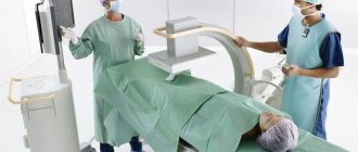
X-ray of the stomach
X-ray of the stomach - radiation examination with barium or other contrast agent. Contrast is used so that the contours and defects of the walls are clearly visible in the pictures. Using gastrography, the abdominal organs and duodenum are examined.
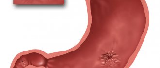
What does a barium stomach x-ray show?
- foreign bodies;
- internal bleeding;
- phlebeurysm;
- ulcers;
- protrusion of the mucous membrane;
- polyps and tumor formations;
- obstruction.
The disadvantage of x-rays is the rather high dose of radiation, so a repeat examination can only be done a year after the first procedure. Therefore, it is impossible to assess the effectiveness of the prescribed treatment over time.
If a malignant tumor is suspected or before surgery, it is recommended to undergo an MRI.
What does the study show?
X-ray of the stomach is used not only to visualize this organ, but also neighboring anatomical structures:
lower third of the esophagus;- diaphragms;
- duodenum;
- transverse colon;
- upper abdominal cavity;
- pancreas;
- right lobe of the liver;
- lymph nodes of the upper abdominal cavity;
- spleen.
When performing a targeted X-ray of the stomach, the doctor should pay attention to the following characteristics of the organ:
- location in the abdominal cavity;
- organ shape and presence of developmental anomalies;
- surface of the mucous membrane;
- wall thickness and its structure;
- contours of the greater and lesser curvature of the stomach;
- volume of the gas bubble of the stomach;
- presence/absence of air under the diaphragm dome;
- activity of gastric peristalsis (during fluoroscopy).
Help The use of contrasts based on Barium preparations significantly increases the information content of diagnostics. However, in this case, it is necessary to prepare the patient.
Conditions for performing fluoroscopy of the gastrointestinal tract
X-rays are taken at intervals of twenty minutes to an hour. The process may take several hours. The subject periodically needs to change position on the table (lying, standing, on his side) and wait from half an hour to an hour for the contrast to move to the next section of the digestive canal.
Depending on the areas studied, there are several types of fluoroscopy of the stomach and intestines :
- irrigoscopy - x-ray of the colon (the suspension is administered by enema);
- fluoroscopy – images of the upper gastrointestinal tract (pharynx, esophagus, stomach, first part of the small intestine);
- esophagography – pictures of the esophagus, pharynx.
During intestinal fluoroscopy, general photographs and targeted ones are taken from different angles for a better examination of pathologies or disorders of the examined area. A suspension of pure barium sulfate is used as a contrast.
Contrast in intestinal fluoroscopy
X-ray diagnostics can be conventional (contrast), with double and air contrast.
- The patient takes part of a pre-prepared barium suspension orally. To better distribute the solution throughout the gastric mucosa, the patient’s abdomen is thoroughly massaged.
- Double contrast - taking the solution through a perforated tube. X-ray of the intestines and stomach with 2nd contrast allows you to study in detail the disturbances in the relief of the organ lining.
- Air contrast - taking a solution of crystalline soda instead of barium.
In what cases is a tomography necessary?
MRI allows you to obtain layer-by-layer series of images in different planes, giving doctors information about changes in cell metabolism. The examination of the gastrointestinal tract includes: stomach, intestines, esophagus, pancreas, and, if necessary, other abdominal organs.
Experts recommend undergoing an MRI of the pancreas and stomach in the following situations:
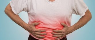
- chronic digestive disorders;
- pain in the stomach;
- suspicion of tumors or mass process;
- control of the therapy performed.
With an inflammatory process in the gastrointestinal tract, it is quite difficult to recognize neoplasms. MRI helps to avoid a biopsy when determining the diagnosis. Tomography also allows you to detect metastases, and even compactions less than 1 centimeter.
When gadolinium-based contrast is used, ulcers and bleeding may be visible.
Unlike X-rays, MRI is performed without quantitative restrictions, since ionizing radiation is not used during magnetic resonance examination. It is tomography that is used to evaluate the effectiveness of treatment and control in oncological processes.
Pathologies and changes in the esophagus
X-ray examination allows us to detect various inflammatory, oncological, degenerative pathologies, as well as their complications:
| Disease | X-ray signs |
| Esophageal hernia (hiatal hernia) | The subphrenic part of the esophagus is missing, and it may be significantly narrowed. When contrast is used, this area is cleared of it more slowly, as peristalsis activity in this segment decreases |
| Peptic ulcer | A mucosal defect of various sizes is visualized, which is not filled with contrast. Possible deformation of the stomach wall and its thinning |
| Perforation of gastric ulcer | When the patient is in an upright position, air appears under the diaphragm |
| Developmental anomalies | There are changes in the shape of the stomach, the presence of narrowing in the antrum (lower) section, partitions of connective tissue in the cavity, underdevelopment of the organ |
| Ingress of foreign objects | Areas of darkening in the lower esophagus, stomach or duodenum of the corresponding shape |
| Malignant neoplasms (stomach cancer and metastases of other tumors) | The presence of deformation of the stomach wall, violation of the homogeneity of its structure, defects of the mucous membrane, enlargement of regional lymph nodes. When contrast is used, a filling defect with an unequal external contour and a lack of peristalsis in the affected segment are visible |
| Esophageal stenosis | Narrowing of the lumen and change in the contour of the esophagus before entering the stomach |
| Benign tumor | Deformation of the stomach wall with protrusion and thickening of the wall is detected. When using contrast, an equal external contour of the tumor is visible, peristalsis in the affected segment is preserved |
| Esophageal achalasia | The expansion of the esophagus is visible, in which food masses and ingested liquid accumulate |
| Acute intestinal obstruction | They find “Kloiber cups” - in the transverse intestine, areas of air accumulation with clear lower boundaries are observed, which resemble inverted cups |