If you've recently watched a crying baby or the latest sentimental Hollywood movie, you're well aware that tears run down your face.
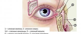
However, we also have a tear duct (also called the "nasolacrimal duct" and "tear duct"). through which tears pass through the nose. These drainage tubes cause a runny nose when crying or an allergic reaction in the eyes.
When the tear duct becomes blocked, problems arise. Let's look at the causes of blocked tear ducts, how it happens and how to get rid of it.
Reasons for appearance
Dacryocystitis occurs in the presence of physiological pathologies, namely congenital narrowing of the duct (stenosis). Sometimes doctors detect a complete blockage of the tear duct.
- Trauma to the eyes or paranasal sinuses.
- Inflammatory process of the nose, which provokes swelling of the tissues around the eye.
- An infectious process caused by bacteria and viruses, which leads to blockage of the duct.
- Getting foreign particles into the eye or working in dusty and smoky rooms. As a result, the channel becomes clogged.
- Allergy to exposure to an irritant.
- Reduced protective properties of the body.
- Overheating and hypothermia.
- Presence of diabetes mellitus.
Very often this pathology occurs in newborn babies. This is due to the structural features of the tear ducts. When the baby is in amniotic fluid, the tear duct is closed with a special membrane, which must rupture during or after childbirth. This process does not occur if pathology occurs. Tears collect in the canal and this provokes an inflammatory process. It mainly develops in women. Men are also no exception, but this pathology is detected very rarely in them. The reason is differences in the structure of the lacrimal canal. Women use cosmetics, most of which cause inflammation.
Sources
- Grassiri B., Zambito Y., Bernkop-Schnürch A. Strategies to prolong the residence time of drug delivery systems on ocular surface. // Adv Colloid Interface Sci - 2021 - Vol288 - NNULL - p.102342; PMID:33444845
- Zhu WX., Zhang YY., Sun ZP., Gao Y., Chen Y., Yu GY. Differential diagnosis of immunoglobulin G4-related sialadenitis and Kimura's disease of the salivary gland: a comparative case series. // Int J Oral Maxillofac Surg - 2021 - Vol - NNULL - p.; PMID:33384237
- Wang Y., Zhuo W., Hei Y., Tong QZ., Li T.Y. . // Zhonghua Yan Ke Za Zhi - 2021 - Vol56 - N11 - p.832-838; PMID:33152841
- Tao H., Wang YS., Wang F., Wang HB., Dong WL., Bai F., Wang P., Zhou XB., Wang LH., Liu C. Diagnosis of lacrimal punctum lesions using optical coherence tomography: a preliminary study. // Int J Ophthalmol - 2021 - Vol13 - N6 - p.902-906; PMID:32566500
- Nagendran S., Alsamnan M., Strianese D., Malhotra R. Ectopic Lacrimal Gland Tissue: A Systematic Review. // Ophthalmic Plast Reconstr Surg - 2021 - Vol36 - N6 - p.540-544; PMID:32205779
- Singh S., Sharma A., Mittal V., Ali MJ. Lacrimal drainage anomalies in Tessier cleft 3 with unilateral anophthalmos. // Eur J Ophthalmol - 2021 - Vol - NNULL - p.1120672119891475; PMID:31771345
- Jang JK., Lee SM., Lew H. A histopathological study of lacrimal puncta in patients with primary punctal stenosis. // Graefes Arch Clin Exp Ophthalmol - 2021 - Vol258 - N1 - p.201-207; PMID:31713749
- Tanaboonyawat S., Idowu OO., Copperman TS., Vagefi MR., Kersten RC. Dacryops - A review. // Orbit - 2021 - Vol39 - N2 - p.128-134; PMID:31512543
- Kubota T., Katayama M., Nishimura R., Moritani S. Long-term outcomes of ocular adnexal lesions in IgG4-related ophthalmic disease. // Br J Ophthalmol - 2021 - Vol104 - N3 - p.345-349; PMID:31272957
- Kojima T. Contact Lens-Associated Dry Eye Disease: Recent Advances Worldwide and in Japan. // Invest Ophthalmol Vis Sci - 2018 - Vol59 - N14 - p.DES102-DES108; PMID:30481813
Symptoms of the disease
Tears are necessary for the normal functioning of the visual organs. They moisturize the cornea of the eye, protect against mechanical irritants, and perform an antibacterial function.
Sometimes tears stop flowing, this is the first sign of tear duct obstruction. Treatment is one way to cope with the problem and prevent the development of canaliculitis. Sometimes massage of the tear duct helps.
- painful and unpleasant sensations in the eye area;
- redness of the skin around the eye;
- feeling of squeezing and bursting;
- swelling of the skin;
- lacrimation;
- edema;
- vision problems;
- increased secretion of mucus, which smells bad;
- formation of pus;
- high body temperature;
- intoxication of the body.
The acute stage of dacryocystitis appears as an inflammatory process affecting one eye. In the chronic stage, the tear duct swells, the eye turns red and the number of tears increases.
If you experience such symptoms, you should consult a doctor. Blockage can occur in both acute and chronic stages. The accumulation of tear fluid increases the likelihood of infectious processes.
Lacrimal gland disorders
Disorders of the lacrimal gland manifest themselves in increased tear production ( hyperfunction ) or insufficient production of tear fluid ( hypofunction ).
Increased lacrimation may occur due to bright light, strong wind, cold and other external irritants, or as a result of disturbances in the innervation of the eye. A characteristic sign of lacrimal duct pathology is increased lacrimation (epiphora).
Hypofunction of the lacrimal glands (or “dry eye syndrome”) is one of the manifestations of Sjögren’s syndrome. It also occurs in endocrine and autoimmune diseases, in patients on hormone replacement therapy, in people who work for a long time behind a monitor, in smokers.
Diagnostics
Dacryocystitis is detected without much difficulty. At the appointment, the doctor performs a visual assessment of the eye and palpation of the lacrimal sac.
- Test using paint. The eye is instilled with a solution containing a dye. If pigment appears in the eye after a few minutes, this indicates a blockage of the tear ducts.
- Probing. Using a probe with a needle, the ophthalmologist penetrates the duct, which helps to expand it and get rid of the problem.
- Dacryocystography. Carrying out an X-ray examination with the introduction of a dye. In the picture you can see the structure of the eye system and identify the problem.
- Patency can also be checked with the West test. A cotton swab is placed in the nasal passage, on the affected side. Collargol is instilled into the eye. The condition is considered normal when after 2 minutes the tampon turns dark. If the swab remains clean or stained after 10 minutes, there is a problem.
Treatment
Eyes are the mirror of the soul. When an eye problem arises, there is no need to take risks. Treatment should be prescribed by a doctor after preliminary diagnosis. The treatment method is selected depending on the form and cause of the pathology that provoked it, and age characteristics.
- Rinsing the eyes with antibacterial and disinfectant solutions.
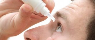
- The use of special drops and ointments.
- Massage procedures and compresses to help clean the canal.
Eye rinsing with antiseptic solutions is carried out several times a day. The procedure is performed by an ophthalmologist in a hospital setting.
Ointments and drops that have an antibacterial effect:
- Phloxal . Antibacterial drug with a wide spectrum of effects. Fights the inflammatory process. The course of treatment is 10 days, two drops twice a day.
- Dexamethasone . Drops with an antibacterial effect. Effective against infectious processes. Instill 5 times a day. The required dosage and course of treatment are selected by the doctor individually for each patient.
- Levomycetin is a hormonal drug. Used for allergic reactions and inflammations.
- Ciprofloxacin . Prescribed for infections of the lacrimal duct. Buried every three hours.
The drugs have contraindications and side effects. Drug therapy is carried out under the supervision of the attending physician.
If the treatment does not have a positive effect, bougienage is performed - cleaning the lacrimal canal from purulent contents;
You can quickly cope with the disease only if treatment is started in a timely manner. If symptoms are negative, you should visit an ophthalmologist.
Radical ways to fight
If there is no positive effect from drug treatment, or if the cause is a tumor or cyst, surgical treatment is performed.
Surgical intervention happens:
- Endoscopic dacryocystorhinostomy I. A device with a camera is inserted into the duct. A puncture or incision is made using an endoscope. A special valve is created, the main purpose of which is drainage. The recovery period is 7 days. In parallel, antibiotic therapy is carried out to prevent the risk of developing an inflammatory process. The main advantage is the absence of visible marks after the operation.
- Balloon dacrycytoplasty is an intervention that, due to its safety, is performed even on newborns. A conductor with a reservoir filled with liquid is inserted into the channel. Allows you to expand the area, thereby breaking through it. The procedure is carried out under local anesthesia. During the rehabilitation period, special drops and antibacterial drugs are prescribed.
What are tears for?
With the help of tears, you can not only express your emotional state, first of all, we need them to protect our eyes. A thin layer of tear film covers the surface of the cornea and makes it perfectly transparent and smooth, protecting the eyes from drying out.
At the heart of tears is an antibacterial enzyme - lysozyme , which helps cleanse the conjunctival sac of small foreign bodies and microorganisms.
In normal conditions, a small amount of tears is required to moisturize the eye - 0.4-1 ml per day; it is produced by additional conjunctival glands. Large lacrimal glands begin to work when additional irritants appear: severe pain, emotional stress, foreign body contact with the conjunctiva or cornea. And also in too bright light, exposure to smoke and toxic substances.
Massage
Prepare your hands before the procedure: wash, disinfect or wear gloves.
Massage scheme:
- Press on the outer corner of the eye, turn your finger towards the bridge of the nose.
- Gently press and massage the lacrimal sac, removing purulent masses from it.
- Instill a warm solution of furatsilin and remove the discharge.
- Carry out pressing and massaging movements along the tear ducts.
- Jerky movements along the nasolacrimal sac with some force to open the canal and extract the discharge.
- Instill a solution of chloramphenicol.
Folk remedies
After prior approval from a doctor, traditional medicine is successfully used at home.
Folk remedies:
- Aloe. For inflammation, it is good to instill freshly prepared aloe juice, half diluted with saline solution.
- Eyebright. Cook in the same way. Use for eye drops and compresses.
- Chamomile. Has an antibacterial effect. You need to take 1 tbsp. l. collection, boil in a glass of boiling water and leave. Use to wash eyes.
- Thyme . Due to its anti-inflammatory properties, the infusion is used for dacryocystitis.
- Kalanchoe. Natural antiseptic. Cut off the leaves and keep in the refrigerator for two days. Next, extract the juice and dilute it in a 1:1 ratio with saline. This remedy can be used to treat children. Adults can instill concentrated juice into the nose, 2 drops each. The person begins to sneeze, during which the tear duct is cleared of pus.
- Leaves from a rose . Only those flowers that are grown on your own plot are suitable. You will need 100 gr. collection and a glass of boiling water. Boil for five hours. Use as lotions.
- Burda ivy-shaped . Brew a tablespoon of herb in a glass of boiling water, simmer for 15 minutes. Use for rinsing and compresses.
- Bell pepper . Drink a glass of sweet pepper fruit every day. adding a teaspoon of honey.
Consequences and complications
Dacryoadenitis can be complicated by:
- Orbital phlegmon.
- Abscess of the upper eyelid.
- Formation of an external or internal fistula with spontaneous resolution of the abscess.
- A long-existing chronic form can develop into a lacrimal gland cyst - it forms gradually.
In the chronic course of inflammation of the lacrimal tubules, pronounced structural changes in the tubules are observed. Its narrowing (stricture) or obliteration (complete occlusion of the tubule) often develops, as a result of which the outflow of tears is significantly impaired or stops altogether. It is possible that surrounding tissues may be involved in the process with the development of orbital cellulitis and subperiosteal abscess.