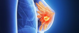
A beautiful bust is the dream of many women!
The owner of beautiful breasts radiates confidence and sexuality. However, it is extremely important that not only external beauty, but also the health of the mammary glands, gives a woman joy of life and confidence in the future. Every representative of the fair sex needs to avoid pathological changes in the breasts that adversely affect the general condition of the female body. After all, breasts are nothing more than mammary glands - a paired organ of the reproductive system responsible for the secretion of milk - the main food product of future generations.
Evolutionary purpose of the mammary glands
The mammary gland in women is an important organ of the reproductive system, which begins to function at a certain period of life. Its main task is to produce milk to feed the baby after birth. This feature places a person in the class of mammals - according to the scenario planned by nature, the cub is fed milk for several months, receiving all the most useful things from the mother.
The gland can fully function only after the birth of the child; its cells “mature” and begin to produce milk. This feature depends on the hormonal background, the variability of which affects the qualitative composition of its tissue.
Interesting: the structure of the mammary gland does not differ between boys and girls before puberty. When puberty occurs, under the influence of female hormones, its tissue begins to grow and develop. In men, the gland remains in an atrophied state for life.
Prevention
The main preventive measure is regular and systematic breast examination by a mammologist or gynecologist:
- women 30-35 years old - ultrasound (annually), mammography (every 2-3 years);
- after 40 years – ultrasound of the mammary glands, mammography (annually);
- after 50 years - ultrasound, mammography (2 times a year).
The following measures will help prevent the disease:
- timely and complete treatment of inflammation of the genital organs;
- maintaining hormonal balance;
- timely implementation of childbearing function;
- absence of abortions;
- maintaining an active and healthy lifestyle;
- correct selection of underwear;
- avoiding stress, nervous tension and negative emotions;
- moderate physical activity;
- maintaining weight within recommended limits;
- balanced diet;
- complete rest;
- moderate passion for solarium and sunbathing;
- careful and attentive attitude towards your body.
Each woman is independently responsible for her health. It is always worth remembering that it is easier to prevent a disease than to start it and subsequently undergo hormonal therapy or, what is much worse, surgical intervention.
If you find an error, please select a piece of text and press Ctrl+Enter
Features of anatomy
In an adult woman, the mammary gland has a structured structure, consisting of several types of tissue, each of which performs its own function. Let's take a closer look at the anatomical features:
- The outside of the gland is covered with skin, on which the juice area is located. It is rich in pigment cells, which plays an important role in feeding the baby - the baby instinctively finds the nipple, since its surface is slightly different from smooth skin. The areola also contains a large number of nerve endings.
- The parenchyma of the breast has a segmental structure - dense strands extend from the skin, which form large cells (15-20), surrounded by fatty tissue. They contain lobules of the mammary gland, separated by connective tissue.
- Each lobule consists of glands, inside of which there are many alveoli that produce milk. Convoluted tubules extend from them, they gradually unite and form the nipple opening.
- The entire anatomical structure is supported by a special cord called the ligament that supports the mammary gland. Underneath it there is a layer of fat that prevents soft tissue injuries.
Normally, there should be no lumps in the breast; the entire structure is soft and can be painlessly palpated. The mobile ligament provides slight displacement without noticeable discomfort. Proportional sizes mainly depend on the volume of adipose tissue that fills the free space between the cells.
Size and shape
Women's breasts are generally larger in size than the mammary gland. The size of the bust largely depends on the content of fat cells in it. It’s not for nothing that if a girl loses weight, her breasts “lose weight” along with her - after all, fat leaves the body evenly.
As a rule, the breast size of thin petite women is small, while larger or fuller ladies have a solid bust. However, there are exceptions - sometimes it is the glandular tissue that develops intensively. In this case, a thin woman may have large or even very large breasts - this is called macromastia. Accordingly, underdevelopment of the glands is micromastia. It is expressed by the practical absence of a bust, regardless of the build of its owner.
Breast asymmetry occurs quite often - this is a normal variant. The difference between the right and left breasts can be up to two sizes.
The tendency to ptosis (sagging) depends on the elasticity of the connective tissue of the chest capsule, the shape and size of the bust. The heavier it is, the more it will stretch even the elastic capsule and, accordingly, sag. In most cases, appetizingly protruding breasts of size 5+ are the result of the use of implants. The tendency to ptosis depends not only on the size, but also on the shape of the bust.
Shape and size do not affect the health of the breast and its ability to lactation - this is a purely aesthetic issue.
Fact! The “chest exercise” recommended in women’s magazines is aimed at developing the anterior pectoral muscles on which it is attached. But these muscles have little effect on the fit of the bust and its shape, since it itself does not have muscle tissue. Therefore, if a woman is not satisfied with the appearance of her breasts, this can only be eliminated surgically.
Breast surgery can be performed no earlier than 18 years of age. And preferably after the first lactation, since surgery can affect the ability of the gland to produce milk.
The structure of the mammary gland during puberty
In girls, the mammary gland is in a embryonic state. There is skin on the outside and a small layer of fat on the inside.
Characteristic age-related features appear during the production of female sex hormones:
- milky tubes appear in the lobules of the mammary gland, which begin to grow and branch;
- when the ducts finish growing, alveoli form at their ends;
- cells of adipose and connective tissue begin to actively divide, as a result of which the mammary gland increases in size.
The result of all these processes is breast growth. The body prepares for pregnancy, and subsequently the glandular and adipose tissue undergoes certain cyclic changes.
Oleogranuloma
Oleogranuloma is a benign tissue compaction altered by inflammation as a result of a foreign body entering the mammary gland. As a rule, it occurs as a result of surgery. Silicone implants, synthetic threads, etc. can act as a foreign body.
The symptoms of oleogranuloma and cancer are similar to each other, so it is often mistaken for oncology. The final verdict can be made by the doctor after a thorough examination.
There are the following types of breast oleogranuloma:
- injection (artificial) – formed as a result of the introduction of various fats and oils;
- post-traumatic – occurs after trauma to the chest (blow, fall, improper massage). A frequent companion is necrosis of fatty fibers;
- periinflammatory – location near the source of the inflammatory process, gradual movement to fatty tissue, which turns out to be destructive;
- spontaneous (cutaneous) – causes unknown.
Oleogranuloma can be identified by the following signs:
- the appearance of voluminous nodules under the skin of the breast;
- lumpy and dense consistency of the skin surface;
- redness in the area of formation;
- pain and discomfort in the area of compaction;
- retraction of the skin over the tumor;
- increasing education over time;
- formation of ulcers, discharge of pus through fistulas (in advanced forms of the disease).
Causes of oleogranuloma:
- breast injuries;
- surgical interventions (removal of the mammary gland or its lobe, breast augmentation by introducing implants);
- a sharp decrease in body weight;
- ingress of foreign bodies (silicone, gels, ointments, synthetic threads);
- infectious and inflammatory diseases;
- hormonal imbalance;
- radiation exposure.
The effect of menstruation on breast anatomy
In an adult woman, breast tissue is constantly exposed to hormones, their concentration varies depending on the phase of the cycle. Clinically, two periods can be distinguished during which changes occur at the cellular level:
- Temporary hypertrophy - in the second phase of the cycle, progesterone is produced in high concentrations. Under its influence, milk tubes begin to grow and alveoli appear. By the end of the month, the described changes reach their peak, the breasts may increase slightly in size, and sometimes pain occurs.
- Reduction - if fertilization does not occur, against the background of a decrease in progesterone concentration, the alveoli undergo reverse development. The body takes a short break to prepare for the next phase of the cycle.
Fibroadenoma
Fibroadenoma (fibrosis) is a benign neoplasm that develops from connective tissue. The tumor occurs against the background of the production of hormones - estrogens. The disease is predominantly observed in women 20-30 and 40-50 years old, sometimes occurring at a young age (12-20 years). It does not pose a threat to the health and life of a woman. In most cases, fibroadenoma is not prone to progression and development of complications.
Sometimes it can reach large sizes and thereby deform the mammary glands. Only fibrosis of gigantic size is prone to malignancy. As a rule, the method of treating a tumor is surgical, but if it is small in size (up to 5-8 mm), therapeutic treatment is carried out aimed at its resorption.
Pathology is of the following types:
- Nodular - deformation of connective tissues near the glandular ducts or their ingrowth into the ducts;
- Leaf-shaped - characterized by an extreme speed of development and an increased threat of degeneration into sarcoma. This is a terrible disease that leads to breast amputation and even death.
Fibroadenoma is asymptomatic for a long time. The general symptoms are not pronounced. It is manifested by compactions in the body of the mammary glands, which have the following features:
- painlessness, density and sometimes mobility of the formation;
- clearly defined boundaries;
- variation in tumor size;
- lack of direct dependence of size changes on the phase of the menstrual cycle;
- intensive growth of education (sometimes).
In medicine, the exact causes of fibroadenoma are still unknown. There are a number of factors contributing to the development of pathology:
- genetic predisposition;
- puberty;
- diseases of the endocrine system and thyroid gland;
- pregnancy;
- frequent abortions;
- nervous and physical exhaustion;
- severe frequent stress;
- obesity;
- incorrect selection of oral contraceptives or their uncontrolled use;
- mammary gland injuries;
- overheating of the mammary glands;
- abuse of solarium and sunbathing.
Changes in the breasts during pregnancy
The mammary gland can function fully only during pregnancy and after childbirth. Natural physiological mechanisms are launched that ensure milk secretion.
The structural features are determined by three hormones:
- Progesterone - under its influence, the alveoli reach their final development and are ready for secretion. The milk ducts grow in the lobules, the glands prepare to produce milk.
- Prolactin - produced immediately after childbirth, triggers secretion in the alveoli and the release of colostrum through the ducts. It contains a large amount of vitamins, minerals and nutrients that a child needs.
- Oxytocin - supports the secretion and production of mother's milk for several months. Under its influence, the alveoli remain in a hypertrophied state until the end of feeding. When the hormone titer decreases, all structures of the mammary gland undergo reduction.
Against the background of all these changes, the mammary glands are exposed to a dozen hormones. The whole system works like a clock; even a small malfunction can lead to disruption of cell division in the breast and the appearance of problems.
Top 11 most frequently asked questions from women about breast health.
ANSWERS OF OUR MAMMOLOGIST, EKATERINA NIKOLAEVNA MAKAROVA TO THE TOP 11 MOST FREQUENT QUESTIONS OF WOMEN ABOUT BREAST HEALTH.
QUESTION #1 – WHY CAN ONE BREAST BE BIGGER THAN THE OTHER?
Asymmetry of the mammary glands can be detected in healthy women.
A slight difference in the volumes of the right and left breasts is observed quite often and is considered a normal variant. This asymmetry occurs in many women and should not be a cause for concern.
Fear and alarm should be caused by a sudden increase in the volume of one of the breasts, which occurred in a short time. When such an increase occurs, a woman needs to undergo an examination such as an ultrasound of the mammary glands (on the 5-7th day from the start of menstruation) and an examination by a mammologist. This way you can exclude a severe pathological process (including oncopathology), which can provoke an increase in the volume of one of the mammary glands.
QUESTION No. 2 – WHY CAN THE MAMMY GLANDS BE THICK BEFORE MENSTRUGGLES?
Before the onset of menstruation, women may experience pain (in the nipples or breasts as a whole) and engorgement of the mammary glands for several days or 1-2 weeks. In most cases, the appearance of such symptoms is associated with mastopathy, which is provoked by a hormonal imbalance that occurs due to various reasons.
To identify this common pathology of the mammary glands, a woman needs to undergo a number of standard examinations:
- examination by a mammologist;
- examination by a gynecologist;
- Ultrasound of the mammary glands, uterus, ovaries and thyroid gland;
- blood tests for hormones (FSH, progesterone, estradiol, TSH, T3 and T4) and analysis for tumor markers;
- cytological analysis of nipple discharge (if any);
- puncture biopsy (if necessary).
One month before the examination, it is advisable to stop taking oral contraceptives - this issue should be discussed with your doctor.
After analyzing all the data received, the mammologist will be able to make a diagnosis. As a rule, such symptoms are characteristic of one of the forms of mastopathy (fibrous, nodular, mixed, cystic, etc.). After this, the woman can be prescribed the necessary treatment.
QUESTION #3 – WHY DOES HAIR GROW AROUND NIPPLES?
Throughout their lives, women may experience periods of hormonal surges, which may manifest as hair growth around the nipples. This symptom is considered a variant of the norm, because normally there are hair follicles in the areola, which in some women can be activated.
However, we should not forget that the symptoms of hirsutism should be a cause for concern; this is a sudden excess growth of male-pattern hair (on the chest, back, chin or upper lip). This disease indicates a significant imbalance in hormones and requires clinical observation and treatment.
QUESTION No. 4 – WHY DO SERUS OR BLOODY DISCHARGE APPEAR FROM THE NIPPLES?
The presence of transparent yellowish or brown discharge indicates a pathological process occurring in the mammary gland. Typically, such a symptom indicates the development of one of the forms of mastopathy, but sometimes discharge from the breast can be a sign of more dangerous diseases. If such a symptom appears, a woman should definitely contact a mammologist to clarify the diagnosis.
QUESTION #5 – WHAT ARE THE MOST COMMON SYMPTOMS OF BREAST DISEASES?
The most common signs of pathological processes in the mammary glands are the following symptoms:
- feelings of tension;
- painful sensations;
- skin changes in the breast, nipple or areola area;
- nipple discharge;
- determination by palpation of areas of compaction of gland tissue.
QUESTION No. 6 – HOW AND WHEN TO CONDUCT A BREAST SELF-EXAMINATION?
Breast self-examination can help detect cancer in the early stages.
Every woman should know that once a month (about the same days, 6-7 days after the end of her period) she should conduct a breast self-examination: this is the best way to early detect any changes in the condition of the mammary gland tissue .
The procedure should be carried out as follows:
- Inspect the bra in those places where it comes into contact with juices and make sure there are no stains.
- Examine the nipples and areola, making sure there are no changes: redness, retractions, peeling, rashes or ulcerations.
- Stand in front of the mirror, raise your hands behind your head and pay attention to the shape of your breasts and the presence of any irregularities, protrusions or depressed areas in certain parts of the chest.
- Examine the skin of the breast for changes in its color or changes in the appearance of “lemon peel”.
- Perform alternate palpation of the mammary glands. To do this, you need to lie down on the bed and place a bolster or small pillow under the shoulder blade (on the side of the gland being examined) so that the chest is slightly elevated and the mammary gland is most spread out. The right mammary gland can be felt with the fingertips of three or four fingers of the left hand. The fingers should be placed flat and moved in a circle (from the nipple to the outer edge of the gland), moving them centimeters and feeling the entire area of the gland. In the same way (only with the right hand) the left breast is felt.
- If you are unable to palpate your breasts this way, you can do it in the bathroom - wet fingers with soapy suds will glide over the skin better, and it will be easier for you to feel the lumps in the gland.
Remember that identifying any lumps, lumps or painful areas should always be a reason to immediately contact a mammologist. And don’t put off visiting your doctor until later, because malignant tumors of the mammary glands can be rapidly progressing and extremely aggressive, and you may miss your chance for recovery.
QUESTION No. 7 – HOW OFTEN DO YOU NEED TO HAVE A PREVENTIVE EXAMINATION WITH A MAMMOLOGIST?
Remember that even regular breast self-examination does not free a woman from the need to undergo a preventive examination once a year with a mammologist or breast oncologist.
Only a specialist who has experience in diagnosing diseases of the mammary glands and works in a medical institution with sufficient technical equipment for professional examination of the mammary glands will be able to professionally assess the condition of the breast and give you the necessary recommendations.
It is better to schedule a visit to the doctor in the first days after menstruation: approximately from 1 to 10 days.
QUESTION No. 8 – WHAT DIAGNOSTIC METHODS ARE USED TO STUDY THE BREAST GLANDS?
The main methods for diagnosing breast pathologies include the following types of studies:
- examination and palpation of the mammary glands;
- Ultrasound of the mammary glands;
- x-ray mammography;
- blood test for hormones (prolactin, FSH, progesterone, estradiol, TSH, T3 and T4);
- blood test for breast tumor markers CA 15-3, CA 27-29 and CEA;
- microwave radiothermometry (RTM research);
- electrical impedance tomography (EMM);
- CT;
- MRI;
- biopsy of tumor tissue followed by cytological or histological examination;
- axillography;
- ductography;
- scintigraphy.
The scope of breast examinations is determined by a mammologist individually for each patient.
For the purpose of preventive examination, women are recommended to conduct the following regular studies:
- Ultrasound of the mammary glands (for women under 40 years old - once a year, and for people with an increased risk of the disease, mammography is recommended);
- mammography (for women over 40 years old - 1-2 times a year, over 50 years old - annually).
Such preventive instrumental methods for examining the mammary glands are the gold standard for screening breast diseases and are recognized throughout the world. They make it possible to identify various diseases at the earliest stages and significantly reduce the risk of uncontrolled progression of tumor diseases of the mammary glands.
QUESTION No. 9 – WHICH EXAMINATION IS MORE RELIABLE – BREAST ULTRASOUND OR MAMMOGRAPHY?
Ultrasound of the mammary glands is a non-invasive, safe (in terms of dose load), painless and informative procedure, which is usually prescribed to young women and pregnant or nursing mothers. However, most doctors, when treatment is necessary, prefer to rely on mammography data, because they consider this diagnostic method to be more informative and accurate.
Some mistrust of the data obtained from ultrasound is due to the fact that the female breast is one of the most difficult objects for examination, since the structure and density of gland tissue constantly changes throughout a woman’s life and depends on age, body weight, physiological periods life, phase of the monthly cycle and the presence of pathological foci.
QUESTION #10 – WHAT IS THE PROBABILITY OF GETTING BREAST CANCER?
The risk of malignant neoplasms in the mammary glands is individual and depends on various factors. Taking into account the fact that the number of patients with breast cancer is steadily increasing from year to year, it should be remembered that, starting from the age of 40, every woman should regularly visit a mammologist and undergo preventive mammography.
The main risk factors for the development of breast cancer are:
- age – the risk increases after 40-45 years and reaches its peak at 65 years;
- heredity - especially if there are cases of illness in the mother or siblings;
- presence of pathologies of the mammary glands in the anamnesis;
- earlier onset of menstruation – before the age of 11-12 years;
- late onset of the first pregnancy (or complete absence of childbirth) - after 30-35 years;
- the onset of menopause is too early - before 45 years of age;
- too late menopause - after 55 years;
- refusal to breastfeed;
- prolonged contact with carcinogenic substances or ionizing radiation;
- Frequent chest X-ray examinations;
- obesity.
Research by scientists to study other factors that contribute to the degeneration of breast tissue cells into cancer cells continues to this day. These include: taking hormonal oral contraceptives and hormone replacement therapy for perimenopausal disorders, smoking, drinking alcohol and eating high-fat foods.
QUESTION No. 11 – HOW TO PREVENT THE DEVELOPMENT OF BREAST CANCER?
Despite the fact that scientists have not yet been able to create an effective vaccine for breast cancer, a woman can take care of its prevention by following simple rules:
- regularly conduct breast self-examination and contact a mammologist if any changes in its tissue are detected;
- regularly conduct preventive ultrasounds of the mammary glands and mammography;
- wear a properly selected bra - it must fit perfectly, properly hold the breasts in a normal anatomical position (it is not recommended to choose underwire or push-up models);
- to refuse from bad habits;
- eat right - introduce more vegetables and cereals into your diet, eat fresh fruits and vegetables that have antioxidant properties, do not abuse fatty and fried foods;
- maintain normal weight and get rid of extra pounds;
- do not give up breastfeeding;
- strengthen the chest muscles with gymnastics and lead an active lifestyle;
- to be a self-sufficient person - according to the observation of many scientists, doctors and psychologists, breast cancer often develops in those women who neglect themselves, deny themselves care and attention to themselves and are often offended by the people around them who treat them badly.
Dear women and girls, please take care of your health, love, take care of yourself! We are waiting for you at our Medical Center!
Types of mammary gland structure
In women, the structure of the breast changes at different periods of life, sometimes the anatomy is influenced by the individual characteristics of the body. There are many classifications, we will note the gradation by type of fabric:
- Glandular type - occurs in young women and is considered a normal variant. Adipose tissue is present, but its proportion is small.
- The fat type of structure is typical for overweight women. It also occurs with a normal physique, when after 35-40 years the processes of reverse development begin in the chest and the glandular tissue is replaced by adipose tissue.
- Mixed type - intermediate. Characterized by an equal ratio of adipose and glandular tissue.
Lipoma
Breast lipoma is a benign disease characterized by the growth of fatty tissue in the breast area. The appearance of such tumors is typical for women after 40 years of age. Lipoma itself does not pose a threat to the health and life of a woman. It creates only a cosmetic defect: due to its ability to reach a large size, the lipoma deforms the mammary gland.
The only treatment option is surgery. Malignancy is extremely rare.
Breast lipoma can be either single or multiple (lipomatosis). It can include not only fat cells, but also other tissues. The following types of disease are distinguished:
- lipofibroma – predominance of adipose tissue;
- fibrolipoma – the presence of fibrous (connective) tissue;
- angiolipoma - riddled with small blood vessels;
- myxolipoma – the presence of mucus cells;
- myolipoma - muscle fibers are detected.
Typically, lipoma does not manifest itself with characteristic symptoms. It can be detected independently by palpation (palpation). It is inactive, painless and has a dough-like consistency. Often the lipoma reaches up to 2 cm in diameter, and can also be large in size - up to 10 cm. In some forms of the disease, a woman may feel pain and discomfort in the chest.
The following causes of the disease are identified:
- blockage of the outlet duct of the sebaceous glands;
- metabolic disorders;
- excess body weight;
- hereditary predisposition.
The following factors are predisposing:
- sedentary lifestyle (lymph movement is impaired);
- unbalanced diet, which is dominated by fats and carbohydrates;
- changes in hormonal levels;
- mechanical injuries of the mammary gland;
- long-term use of hormonal contraceptives;
- the appearance of stretch marks on the chest due to pregnancy and lactation;
- bad habits (smoking, drinking alcohol, drugs);
- X-ray and ultraviolet irradiation;
- incorrectly selected underwear;
- unfavorable environmental conditions.
Pathologies of the mammary glands
If hormonal imbalances or abnormalities in physiology occur in the body, local diseases may develop. We list the pathologies in descending order of frequency of occurrence:
- mastopathy – the appearance of lumps in the breast;
- cancer - usually occurs as a complication from a previous disease;
- mastitis – inflammation in the breast tissue due to congestion;
- infections - often bacterial or fungal in nature.
It is very important to prevent the listed pathologies - these are regular examinations and palpation of the breast. Timely delivery and breastfeeding play an important role. You should also choose a comfortable bra made from natural materials.
Lactostasis
Lactostasis is the name given to milk retention, which occurs when there is significant formation of milk or when its outflow is disrupted. The disease predominantly develops in primiparous women in the first 10 days after birth.
Lactostasis does not pose a threat to the life and health of a woman. However, ignoring the disease or its illiterate elimination can lead to dangerous consequences - the development of mastitis (inflammation of the mammary gland). Hypothermia (feeding in a draft) or overheating (compresses, hot baths) can also lead to complications.
Lactostasis manifests itself with a number of symptoms:
- breast lump;
- pain on palpation;
- feeling of heaviness and fullness of the chest;
- expansion of the saphenous veins on the chest in the area of stasis;
- feeling of heat and local hyperemia;
- increase in body temperature to 38°C.
Symptoms may either improve after emptying the mammary glands or persist. During the feeding process, a woman may experience severe pain. The stagnation zone can shift and increase.
The main reasons for the development of lactostasis:
- insufficient emptying of the breast;
- compression of the mammary glands by tight clothing or fingers during feeding;
- infrequent feedings (excess milk forms in the mammary glands);
- incorrect position of the woman or child during breastfeeding;
- aggressive pumping (provokes the production of additional
- milk);
- premature introduction of artificial formulas into the child’s diet (the baby may refuse breastfeeding, which will lead to stagnation of unused milk);
- refusal to breastfeed;
- stress and physical exhaustion;
- sagging of the mammary glands (impedes the flow of milk from the lower part of the large breast).
Lactostasis is easily eliminated in the first stages, so it is important to contact a gynecologist immediately.
Intraductal papilloma
It is a growth several millimeters in size that occurs inside the milk ducts of the mammary gland. Intraductal papilloma does not cause pain, but it causes discharge from the nipples: clear, bloody and even black. Danger: Intraductal papilloma often causes the development of a malignant tumor. Treatment: The tumor is treated with surgery, which does not change the appearance of the breast in any way. Risk group: Intraductal papilloma often occurs in older women before menopause and indicates disruptions in the hormonal system.